. 2023 Apr 24;17(4):e0011254.
doi: 10.1371/journal.pntd.0011254. eCollection 2023 Apr.

A recombinant rabies virus chimera expressing the DC-targeting molecular MAB2560 shows enhanced vaccine immunogenicity through activation of dendritic cells

Affiliations

A recombinant rabies virus chimera expressing the DC-targeting molecular MAB2560 shows enhanced vaccine immunogenicity through activation of dendritic cells

Zhiyuan Gong et al. PLoS Negl Trop Dis. .

Abstract

Background: Rabies, caused by the rabies virus (RABV), is an ancient and neglected zoonotic disease posing a large public health threat to humans and animals in developing countries. Immunization of animals with a rabies vaccine is the most effective way to control the epidemic and the occurrence of the disease in humans. Therefore, the development of cost-effective and efficient rabies vaccines is urgently needed. The activation of dendritic cells (DCs) is known to play an important role in improving the host immune response induced by rabies vaccines.

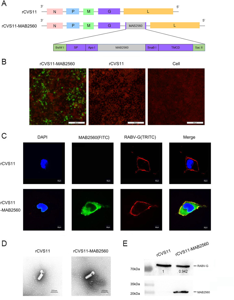
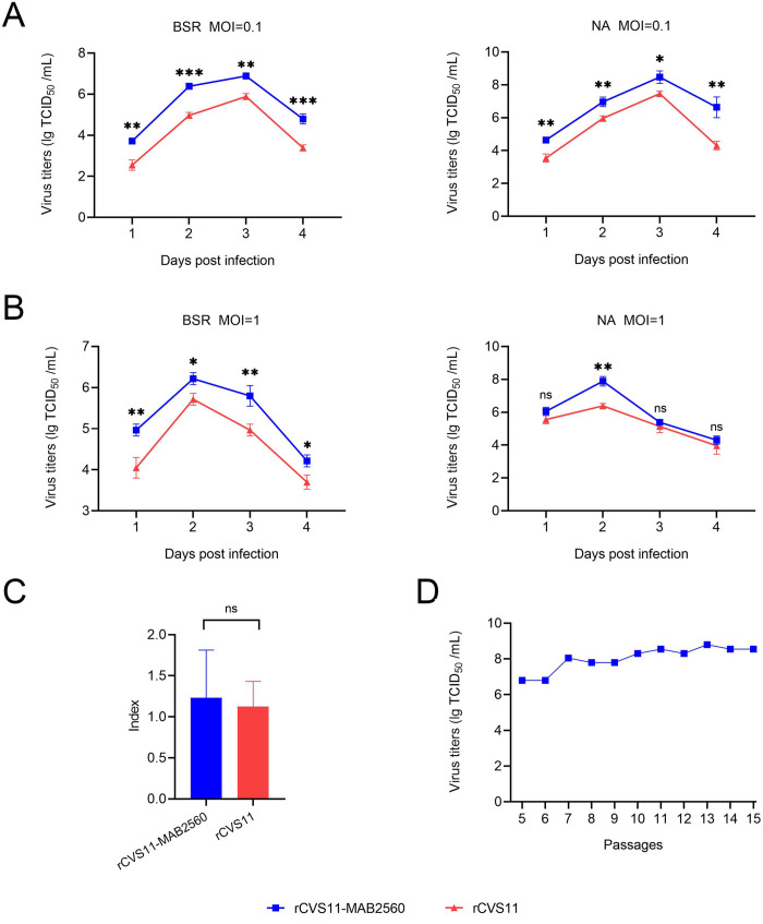
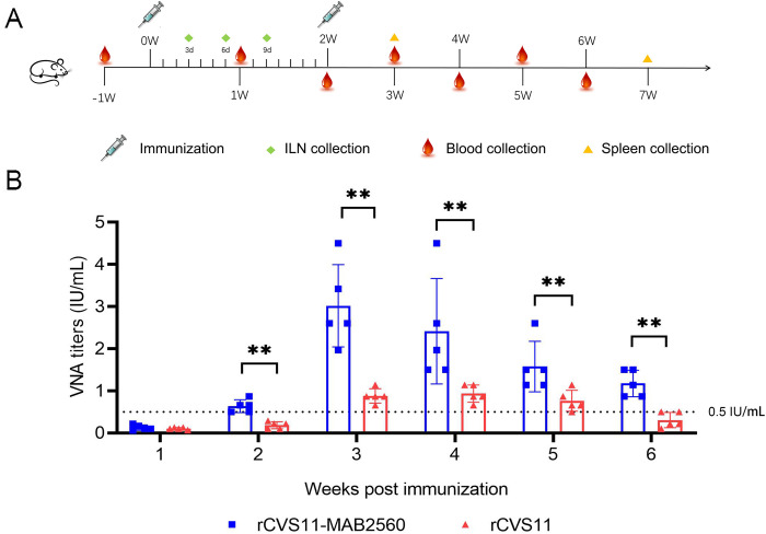
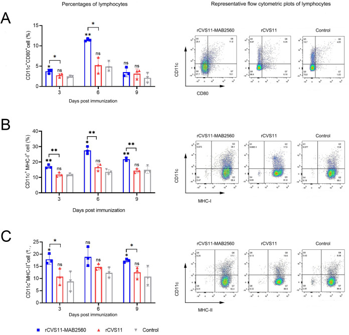
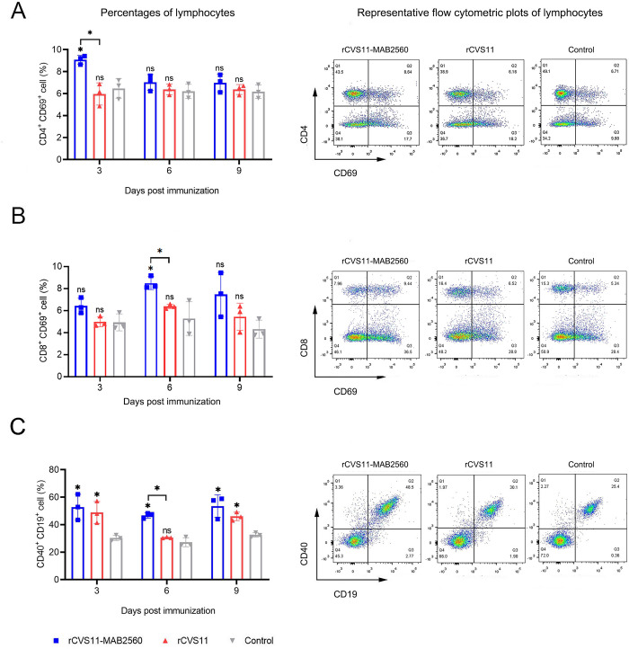
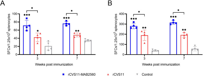
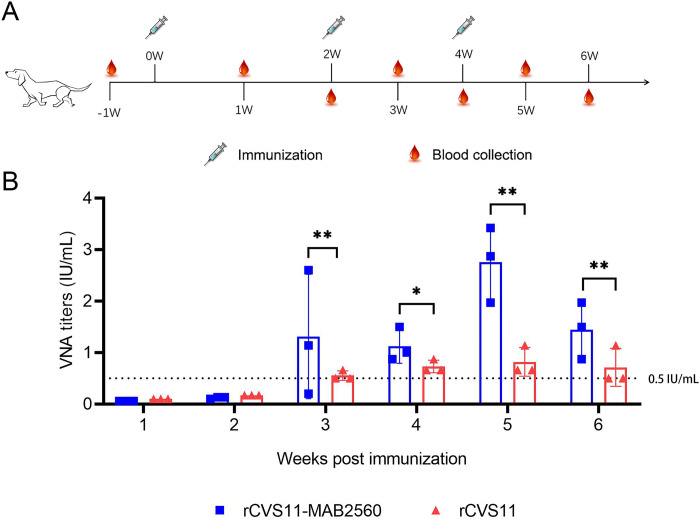
Methodology/principal findings: In this study, we constructed a recombinant virus, rCVS11-MAB2560, based on the reverse genetic system of the RABV CVS11 strain. The MAB2560 protein (a DC-targeting molecular) was chimeric expressed on the surface of the viral particles to help target and activate the DCs when this virus was used as inactivated vaccine. Our results demonstrated that inactivated rCVS11-MAB2560 was able to promote the recruitment and/or proliferation of DC cells, T cells and B cells in mice, and induce good immune memory after two immunizations. Moreover, the inactivated recombinant virus rCVS11-MAB2560 could produce higher levels of virus-neutralizing antibodies (VNAs) in both mice and dogs more quickly than rCVS11 post immunization.

Conclusions/significance: In summary, the recombinant virus rCVS11-MAB2560 chimeric-expressing the molecular adjuvant MAB2560 can stimulate high levels of humoral and cellular immune responses in vivo and can be used as an effective inactivated rabies vaccine candidate.


Conflict of interest statement

The authors have declared that no competing interests exist.

Figures

Fig 1
Fig 1. Construction and characterization of rCVS11-MAB2560.
(A) Construction diagram of the recombinant plasmid pCDNA3.0-CVS11-SP-MAB2560-TMCD. The MAB2560 gene was flanked by the signal peptide and the TMCD region from RABV CVS11 G protein, and was inserted into the CVS11 genome between G and L using the BisW I and Sac II restriction sites. (B) NA cells were infected with either the recombinant virus rCVS11-MAB2560 or rCVS11 at 0.1 MOI. After 48h, the cells were incubated with rabbit polyclonal MAB2560 antibody (1:500) as the primary antibody, with FITC-labeled goat anti-rabbit antibody as the secondary antibody (1:500). (C) NA cells were infected with rCVS11-MAB2560 or rCVS11 at 0.4 MOI. After 48h, the cells were incubated with antibodies against MAB2560 (1:200) and G protein (1:500). The nuclei were stained with DAPI. (D) Purified recombinant viruses rCVS11-MAB2560 and rCVS11 were analyzed using electron microscopy (EM) at a magnification of 40000x. (E) The purified viruses were quantified and equal amounts were analyzed with WB, with mouse anti-RABV G mAb (1:500) and rabbit polyclonal MAB2560 antibody (1:200) as the primary antibodies, and HRP-labeled goat anti-mouse IgG (1:10000) and HRP-labeled goat anti-rabbit IgG (1:20000) as the secondary antibodies.
Fig 2
Fig 2. Growth Characteristics of rCVS11-MAB2560.
(A and B) Multi-step and one-step growth curves of the recombinant viruses. The recombinant rCVS11-MAB2560 and rCVS11 viruses were separately inoculated into either NA or BSR cells at either MOI = 1 or MOI = 0.1. The cell cultures were collected on the 1st, 2nd, 3rd, and 4th days post-infection, and the viral titer was determined using the FAVN method. (C) Neurotropism of the recombinant virus rCVS11-MAB2560. The viral titers of the recombinant viruses rCVS11-MAB2560 or rCVS11 in NA cells compared to that in BSR cells. All of these experiments in A-C were repeated three times independently and the data were presented as the means ± SD for each group. ns, not significant; *, P<0.05; **, P<0.01. (D) Continual passage of the recombinant viruses, and determination of the viral titers.
Fig 3
Fig 3. rCVS11-MAB2560 induced stronger VNA in mice than rCVS11.
(A) Immunization scheme in mice. Six- to 8- week-old BALB/c mice were randomly divided into three groups (n = 20/group). The mice were immunized intramuscularly with 100μL of inactivated recombinant viruses, or with 100μL of PBS mixed with Gel02 adjuvant. The mice received a total of two immunizations separated by a 2-week interval. ILNs were collected on the 3rd, 6th, and 9th days post-immunization (dpi), mouse spleens were collected in the 3rd and 7th weeks following the initial immunization, and mouse blood was collected in the 1st, 2nd, 3rd, 4th, 5th, and 6th weeks following initial immunization. All the clipart used in this figure are quoted from OPENCLIPART (https://openclipart.org/). (B) RABV specific VNAs in mice sera at weeks 1, 2, 3, 4, 5, and 6 post-immunization were measured using a FAVN test. The black dotted line represents the standard 0.5 international unit (IU)/mL level which is recommend as protective neutralizing antibody level by WHO. The data were presented as the means ± SD for each group. **, P<0.01.
Fig 4
Fig 4. Activation of DCs by rCVS11-MAB2560 in vitro.
Bone marrow cells were harvested from healthy BALB/c mice, and DCs were separated and stimulated with the inactivated recombinant viruses rCVS11-MAB2560 and rCVS11. LPS was used as a positive control, and RPMI 1640 (mock) was used as a negative control. Single cell suspensions (106 cells/mL) were stained with antibodies against DC activation markers, and were then analyzed with flow cytometry. (A) The representative flow cytometric plots and percentages of CD11c+ CD80+ activated DCs (Top) and CD80+ MFI (Bottom) in BMDC. (B) The representative flow cytometric plots and percentages of CD11c+ MHC-I+ activated DCs (Top) and MHC-I+ MFI (Bottom) in BMDC. (C) The representative flow cytometric plots and percentages of CD11c+ MHC-II+ activated DCs (Top) and MHC-II+ MFI (Bottom) in BMDC. The data were presented as the means ± SD for each group. ns, not significant; *, P <0.05; **, P<0.01; ***, P<0.001.
Fig 5
Fig 5. Recruitment of DCs by rCVS11-MAB2560 in vivo.
On the 3rd, 6th and 9th dpi, ILNs were collected from the vaccinated BALB/c mice and single cell suspensions (106 cells/mL) were prepared. The single cell suspensions were stained with antibodies against DC activation markers and analyzed with flow cytometry. (A) The representative flow cytometric plots (6th dpi) and percentages of CD11c+ and CD80+ activated DCs in the ILNs of immunized mice. (B) The representative flow cytometric plots (6th dpi) and percentages of CD11c+ and MHC-I+ activated DCs in the ILNs of immunized mice. (C) The representative flow cytometric plots (3rd dpi) and percentages of CD11c+ and MHC-II+ activated DCs in the ILNs of immunized mice. The data were presented as the means ± SD for each group. ns, not significant; *, P <0.05; **, P<0.01; ***, P<0.001.
Fig 6
Fig 6. Recruitment of T and B cells by rCVS11-MAB2560.
On the 3rd, 6th and 9th dpi, ILNs were collected from the vaccinated BALB/c mice and single cell suspensions (106 cells/mL) were prepared. The suspensions were stained with antibodies against T and B cell activation markers and were analyzed using flow cytometry. (A) The representative flow cytometric plots (3rd dpi) and percentages of CD4+ and CD69+ recruited and/or activated CD4+ T cells in the ILNs of immunized mice. (B) The representative flow cytometric plots (6th dpi) and percentages of CD8+ and CD69+ recruited and/or activated CD8+ T cells in the ILNs of immunized mice. (C) The representative flow cytometric plots (6th dpi) and percentages of CD40+ and CD19+ recruited and/or activated B cells in the ILNs of immunized mice. The data were presented as the means ± SD for each group. ns, not significant; *, P <0.05.
Fig 7
Fig 7. Induction of Th1 and Th2 cytokine production by rCVS11-MAB2560.
Mouse spleens were collected at the 3rd and 7th week following initial immunization, and were prepared into splenocyte suspensions. RABV-specific IFN-γ (A) or IL-4 (B) SFCs were quantified using ELISpot assays. The data were presented as the means ± SD for each group. *, P <0.05; **, P<0.01; ***, P<0.001.
Fig 8
Fig 8. rCVS11-MAB2560 induced good immune memory after immunization in mice.
At the 3rd and 7th weeks following initial immunization, spleens were collected from the vaccinated BALB/c mice. Single cell suspensions of splenocyte cells (106 cells/mL) were stained with antibodies against TCM, T and B cell activation markers, and were analyzed with flow cytometry. (A) Flow gate strategy of CD4+ TCM (7th week post immunization). (B) The representative flow cytometric plots (7th week post immunization) and percentages of CD4+, CD44+ and CD62L+ recruited and/or activated CD4+ T cells in the spleens of immunized mice. (C) The representative flow cytometric plots (7th week post immunization) and percentages of CD4+, CD69+ recruited and/or activated CD4+ T cells in the spleens of immunized mice. (D) The representative flow cytometric plots (3rd week post immunization) and percentages of CD19+ and CD69+ recruited and/or activated B cells in the spleens of immunized mice. The data were presented as the means ± SD for each group. ns, not significant; *, P <0.05; **, P<0.01.
Fig 9
Fig 9. rCVS11-MAB2560 induces stronger VNA in dogs than rCVS11.
(A) Immunization scheme in dogs. Beagle dogs (17- to 21- months-old) were randomly divided into two groups (n = 3/group). The dogs were immunized intramuscularly with 1mL of inactivated rRABVs (108 TCID50/dog), or with 1mL of PBS mixed with Gel02 adjuvant. The dogs received three immunizations in total, at 2-week intervals. Dog blood was collected the 1st, 2nd, 3rd, 4th, 5th, and 6th week following the initial immunization. All the clipart used in this figure are quoted from OPENCLIPART (https://openclipart.org/). (B) RABV-specific VNAs in dog sera at weeks 1, 2, 3, 4, 5, and 6 post-immunization were measured using a FAVN test. The black dotted line represents the standard 0.5 international unit (IU)/mL level which is recommend as protective neutralizing antibody level by WHO. The data were presented as the means ± SD for each group. *, P <0.05; **, P<0.01.

Similar articles

Cited by

References

    1. Jackson AC. Research advances in rabies. Preface. Advances in virus research. 2011;79:xvii. Epub 2011/05/24. doi: 10.1016/b978-0-12-387040-7.00022–6 . - DOI - PubMed
    1. Gonzalez-Roldan JF, Undurraga EA, Meltzer MI, Atkins C, Vargas-Pino F, Gutierrez-Cedillo V, et al.. Cost-effectiveness of the national dog rabies prevention and control program in Mexico, 1990–2015. PLoS neglected tropical diseases. 2021;15(3):e0009130. Epub 2021/03/05. doi: 10.1371/journal.pntd.0009130 ; PubMed Central PMCID: PMC7963054. - DOI - PMC - PubMed
    1. Yamada A, Makita K, Kadowaki H, Ito N, Sugiyama M, Kwan NCL, et al.. A Comparative Review of Prevention of Rabies Incursion between Japan and Other Rabies-Free Countries or Regions. Japanese journal of infectious diseases. 2019;72(4):203–10. Epub 2018/12/26. doi: 10.7883/yoken.JJID.2018.431 . - DOI - PubMed
    1. Fernandes MES, Carnieli P Jr., Gregório ANF, Kawai JGC, Oliveira RN, Almeida LL, et al.. Phylogenetic analysis of rabies viruses isolated from cattle in southern Brazil. Virus genes. 2020;56(2):209–16. Epub 2020/01/20. doi: 10.1007/s11262-020-01730-y ; PubMed Central PMCID: PMC7223090. - DOI - PMC - PubMed
    1. Benavides JA, Velasco-Villa A, Godino LC, Satheshkumar PS, Nino R, Rojas-Paniagua E, et al.. Abortive vampire bat rabies infections in Peruvian peridomestic livestock. PLoS neglected tropical diseases. 2020;14(6):e0008194. Epub 2020/07/01. doi: 10.1371/journal.pntd.0008194 ; PubMed Central PMCID: PMC7351222. - DOI - PMC - PubMed

Publication types