. 2023 May 2;120(18):e2218085120.
doi: 10.1073/pnas.2218085120. Epub 2023 Apr 24.

In diverse conditions, intrinsic chromatin condensates have liquid-like material properties

Affiliations

In diverse conditions, intrinsic chromatin condensates have liquid-like material properties

Bryan A Gibson et al. Proc Natl Acad Sci U S A. .

Abstract

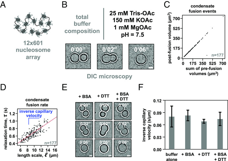
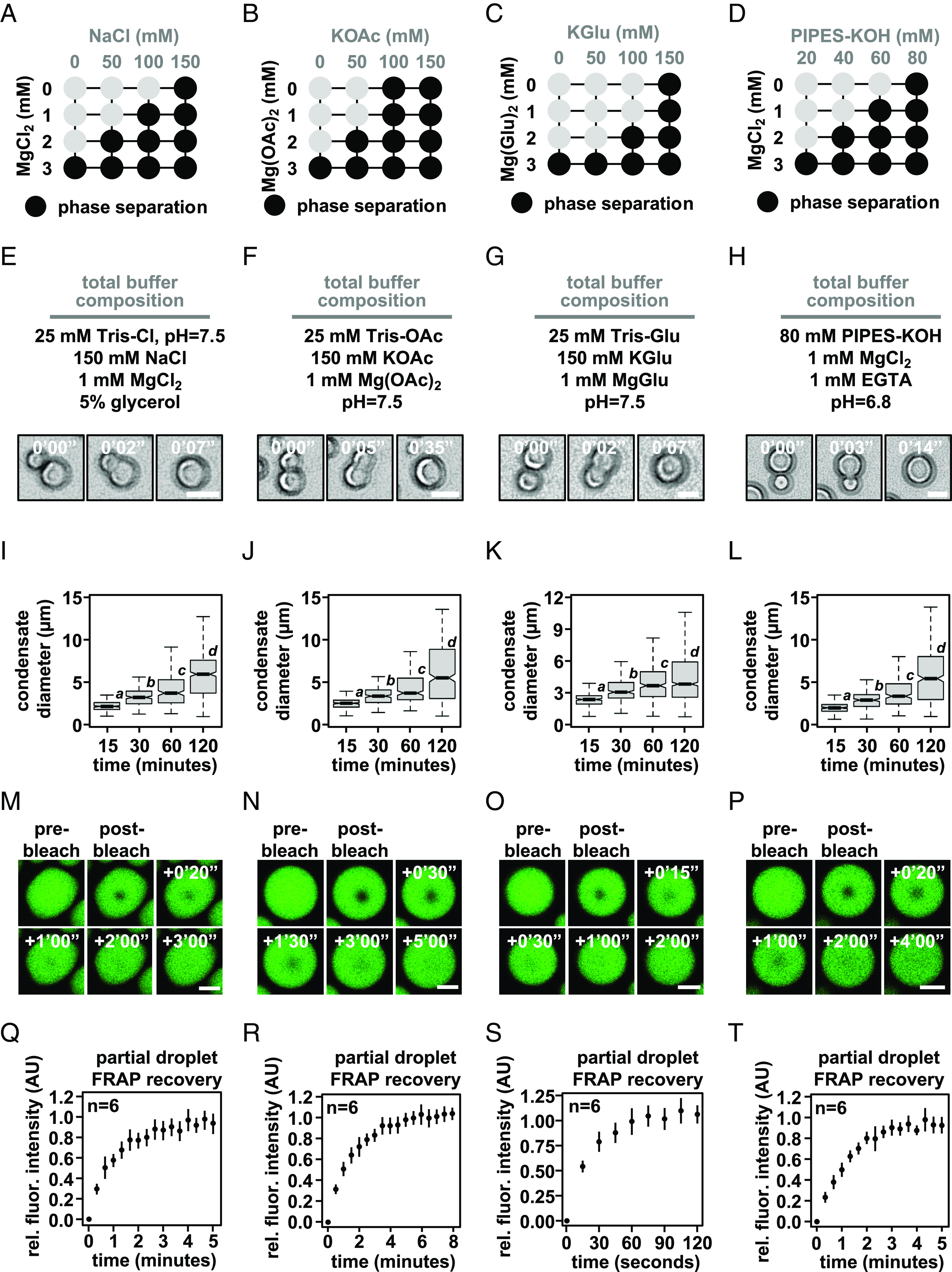
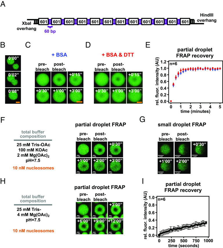
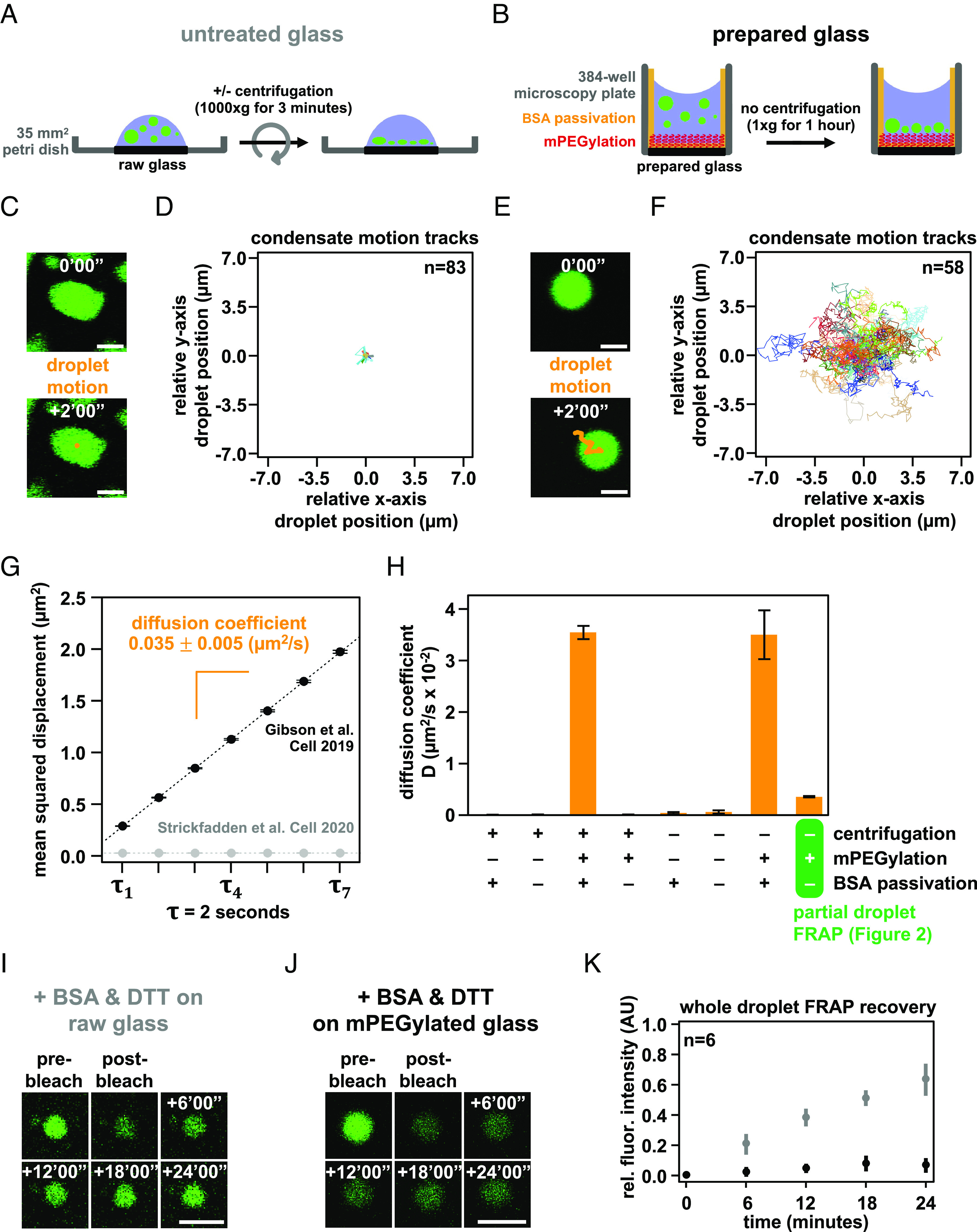
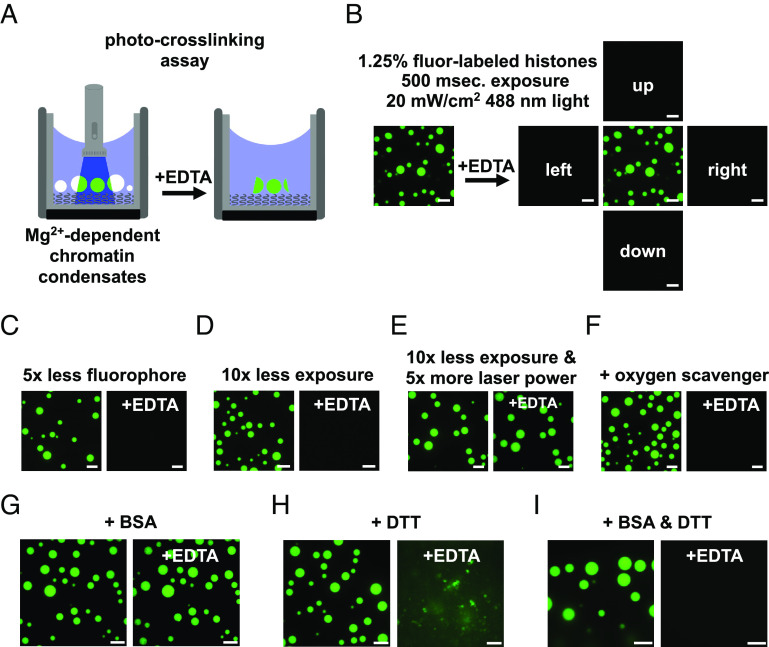
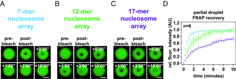
Nuclear DNA in eukaryotes is wrapped around histone proteins to form nucleosomes on a chromatin fiber. Dynamic folding of the chromatin fiber into loops and variations in the degree of chromatin compaction regulate essential processes such as transcription, recombination, and mitotic chromosome segregation. Our understanding of the physical properties that allow chromatin to be dynamically remodeled even in highly compacted states is limited. Previously, we reported that chromatin has an intrinsic capacity to phase separate and form dynamic liquid-like condensates, which can be regulated by cellular factors [B. A. Gibson et al., Cell 179, 470-484.e421 (2019)]. Recent contradictory reports claim that a specific set of solution conditions is required for fluidity in condensates that would otherwise be solid [J. C. Hansen, K. Maeshima, M. J. Hendzel, Epigenetics Chromatin 14, 50 (2021); H. Strickfaden et al., Cell 183, 1772-1784.e1713 (2020)]. We sought to resolve these discrepancies, as our ability to translate with confidence these biophysical observations to cells requires their precise characterization. Moreover, whether chromatin assemblies are dynamic or static affects how processes such as transcription, loop extrusion, and remodeling will engage them inside cells. Here, we show in diverse conditions and without specific buffering components that chromatin fragments form phase separated fluids in vitro. We also explore how sample preparation and imaging affect the experimental observation of chromatin condensate dynamics. Last, we describe how liquid-like in vitro behaviors can translate to the locally dynamic but globally constrained chromatin movement observed in cells.

Keywords: biomolecular condensate; chromatin; phase separation.


Conflict of interest statement

The authors declare no competing interest.

Figures

Fig. 1.
Fig. 1.
Intrinsic chromatin condensates are fluid without BSA and DTT. (A) Graphical depiction of the dodecameric nucleosomal arrays used for experimentation. (B) Differential interference contrast microscopy images of a fusion event between intrinsic chromatin condensates in the indicated buffer. (C) Dot plot representation of the inferred total volume of condensates before and after fusion. (D) Relaxation time versus length scale (sum of prefusion diameters) for 177 individual instances of condensate fusion in the buffer composition indicated in Fig. 1B. Inverse capillary velocity, the characteristic ratio of surface tension, η , and viscoscity, γ , is derived from the linear fit (red line) of the plots’ slope. (E) Differential interference contrast microscopy images of intrinsic chromatin condensate fusion in the buffer indicated in Fig. 1B supplemented with BSA (0.1 mg/mL, Left), DTT (5 mM, Middle), or BSA and DTT (0.1 mg/mL and 5 mM, respectively, Right). (F) Bar chart of inverse capillary velocities ( ± SD of 2 biological replicates) of intrinsic chromatin condensates in the buffer indicated in Fig. 1B, buffer with BSA, or buffer with BSA and DTT. For each condition, the fusion events per replicate are: buffer (177 and 68), +BSA (147 and 81), +DTT (183 and 68), +BSA+DTT (184 and 93). Scale bars, in white, are 4 μm.
Fig. 2.
Fig. 2.
Intrinsic chromatin condensates are fluid in diverse buffers. Phase diagrams for intrinsic chromatin condensate formation in (A) Tris-chloride, (B) Tris-acetate, (C) Tris-glutamate, and (D) PIPES-KOH buffers. Dark circles indicate the presence of condensates, and representative images are in SI Appendix, Fig. S2. With materials produced and experiments performed in the Narlikar lab, bright-field light microscopy images of intrinsic chromatin condensate fusion in (E) Tris-chloride, (F) Tris-acetate, (G) Tris-glutamate, and (H) PIPES-KOH buffers. Boxplots of intrinsic chromatin condensate diameters following induction of phase separation in (I) Tris-chloride, (J) Tris-acetate, (K) Tris-glutamate, or (L) PIPES-KOH-based buffers. Bars marked with different letters are significantly different from one another (Student’s t test, P < 1 × 10⁻⁷). Fluorescence microscopy images of partial-droplet FRAP of intrinsic chromatin condensates, in green, composed of nucleosomal arrays labeled with AlexaFluor 488 in (M) Tris-chloride, (N) Tris-acetate, (O) Tris-glutamate, or (P) PIPES-KOH-based buffers. Quantification of partial-droplet FRAP of intrinsic chromatin condensates in (Q) Tris-chloride, (R) Tris-acetate, (S) Tris-glutamate, or (T) PIPES-KOH-based buffers. Fluorescence signal is normalized to pre-bleach droplet intensity and error bars are SD of six technical replicates. Scale bars, in white, are 4 μm.
Fig. 3.
Fig. 3.
Intrinsic chromatin condensates are fluid in most conditions, but not in superphysiologic magnesium alone. (A) Graphical depiction of a long linker-length 12 × 601 DNA template (27). (B) Confocal fluorescence microscopy of intrinsic chromatin condensates composed of AlexaFluor 488-labeled long linker-length nucleosomal arrays, in green, undergoing fusion. Confocal fluorescence microscopy of partial-droplet FRAP of intrinsic chromatin condensates composed of AlexaFluor 488-labeled long linker-length nucleosomal arrays, in green, formed in the presence of (C) 0.1 mg/mL BSA or (D) 0.1 mg/mL BSA and 5 mM DTT. (E) Quantification of partial-droplet FRAP of intrinsic chromatin condensates with BSA or BSA and DTT, in blue and red, respectively. Fluorescence signal is normalized to pre-bleach droplet intensity and error bars are SD of six technical replicates. (F) Partial-droplet FRAP or (G) half-droplet FRAP of large or small intrinsic chromatin condensates, respectively, formed at 10 nM nucleosome concentration in minimal phase separation buffer. (H) Partial-droplet FRAP and (I) quantitation of fluorescence recovery for intrinsic chromatin condensates induced to form at 10 nM nucleosome concentration with 4 mM magnesium acetate. Scale bars, in orange and white, are 4 and 1 μm, respectively.
Fig. 4.
Fig. 4.
Condensate movement and dynamics is affected by microscopy glass preparation. Graphical depiction of techniques used to prepare intrinsic chromatin condensates for fluorescence microscopy imaging: (A) Intrinsic chromatin condensates can be spun onto raw glass using a centrifuge (27). (B) Alternatively, intrinsic chromatin condensates can be added to a 384-well microscopy plate and brought by gravity to rest on mPEGylated and BSA-passivated glass (11). Movement of a single or many intrinsic chromatin condensates, following their preparation for fluorescence microscopy imaging on untreated glass (C and D) and prepared glass (E and F). (C and E) The movement of an individual condensate across 2 min in 10 s intervals is overlaid in orange on fluorescence microscopy images of AlexaFluor 488-labeled intrinsic chromatin condensates, in green. (D and F) The relative movement of many condensates determined across 2 min in 500 ms intervals. (G) Plot of mean squared displacement ( ± SE) over lag time, τ , for intrinsic chromatin condensates between 4 and 8 μm in diameter following centrifugation onto untreated glass (gray dots) or settling by gravity onto prepared glass (black dots). The diffusion coefficient, indicated in orange ± SE, of intrinsic chromatin condensates can be calculated from the slope of the linear fit (dashed line) of the plotted data. For droplets centrifuged onto untreated glass, three replicates with 11,222, 8,114, and 14,092 trajectories extracted from 171, 147, and 238 droplets were used for analysis, respectively. For droplets settled onto passivated glass, three replicates with 6,563, 7,900, and 8,179 trajectories extracted from 106, 100, and 101 droplets were used for analysis, respectively. (H) Bar chart of the diffusion coefficients of intrinsic chromatin condensates following their preparation for microscopy with and without centrifugation, mPEGylation of the microscopy glass, and BSA passivation of the microscopy well. Error bars are SD of four technical replicates. Confocal fluorescence microscopy of whole-droplet FRAP of intrinsic chromatin condensates composed of AlexaFluor 488-labeled long linker-length nucleosomal arrays, in green, settled onto (I) untreated or (J) mPEGylated glass. (K) Quantification of whole-droplet FRAP recovery of intrinsic chromatin condensates on raw or mPEGylated glass, in gray and black, respectively. Fluorescence signal is normalized to pre-bleach droplet intensity and error bars are SD of six technical replicates. Panels CH used nucleosome arrays with a 25 base pair internucleosome linker length. Panels IK used nucleosome arrays with a 60 base pair internucleosome linker length. Scale bars, in white, are 4 μm.
Fig. 5.
Fig. 5.
DTT and BSA mitigate photocrosslinking during fluorescence microscopy. (A) Diagram depicting an assay to detect photocrosslinking of intrinsic chromatin condensates. (Left) Magnesium-dependent intrinsic chromatin condensates are exposed to fluorescent light prior to the addition of superstoichiometric quantities of EDTA. (Right) Photocrosslinked condensates fail to dissipate following chelation of magnesium. (B) Confocal fluorescence microscopy images of intrinsic chromatin condensates composed of nucleosomal arrays where 1 in 80 histone molecules are labeled with AlexaFluor 488. Images are following exposure to fluorescent light and both before (Left) and after (Right) the addition of EDTA. Confocal fluorescence microscopy images of intrinsic chromatin condensates imaged, as in Fig. 5B, with (C) less fluorophore, (D) less exposure, (E) more laser power with less exposure, or (F) the inclusion of oxygen scavenging components. Confocal fluorescence microscopy images of intrinsic chromatin condensates formed in the presence of (G) BSA, (H) DTT, or (I) BSA and DTT and imaged as described in Fig. 5B. Fluorescent microscopy images before and after the addition of EDTA were processed separately. All experiments were performed using nucleosome arrays with 25 base pair internucleosome repeat length. Scale bars, in white, are 10 μm.
Fig. 6.
Fig. 6.
Length-dependent effects on chromatin condensate dynamics. Confocal fluorescence microscopy images of partial-droplet FRAP of intrinsic chromatin condensates, in green, composed of AlexaFluor 488-labeled arrays that are (A) 7, (B) 12, or (C) 17 nucleosomes in length. (D) Quantification of partial-droplet FRAP of intrinsic chromatin condensates composed of 7, 12, or 17 nucleosome-long arrays in blue, green, and purple, respectively. Fluorescence signal is normalized to pre-bleach droplet intensity and error bars are SD of six technical replicates. Scale bars, in white, are 4 μm.

Similar articles

Cited by

References

    1. Olins D. E., Olins A. L., Chromatin history: Our view from the bridge. Nat. Rev. Mol. Cell Biol. 4, 809–814 (2003). - PubMed
    1. Misteli T., The self-organizing genome: Principles of genome architecture and function. Cell 183, 28–45 (2020). - PMC - PubMed
    1. Mirny L., Dekker J., Mechanisms of chromosome folding and nuclear organization: Their interplay and open questions. Cold Spring Harb. Perspect. Biol. 14, a040147 (2022). - PMC - PubMed
    1. Batty P., Gerlich D. W., Mitotic chromosome mechanics: How cells segregate their genome. Trends Cell Biol. 29, 717–726 (2019). - PubMed
    1. Rowley M. J., Corces V. G., Organizational principles of 3D genome architecture. Nat. Rev. Genet. 19, 789–800 (2018). - PMC - PubMed

Publication types