. 2023 Sep;12(23):e2300305.
doi: 10.1002/adhm.202300305. Epub 2023 May 14.

Gold-Enhanced Brachytherapy by a Nanoparticle-Releasing Hydrogel and 3D-Printed Subcutaneous Radioactive Implant Approach

Affiliations

Gold-Enhanced Brachytherapy by a Nanoparticle-Releasing Hydrogel and 3D-Printed Subcutaneous Radioactive Implant Approach

Mariia Kiseleva et al. Adv Healthc Mater. 2023 Sep.

Abstract

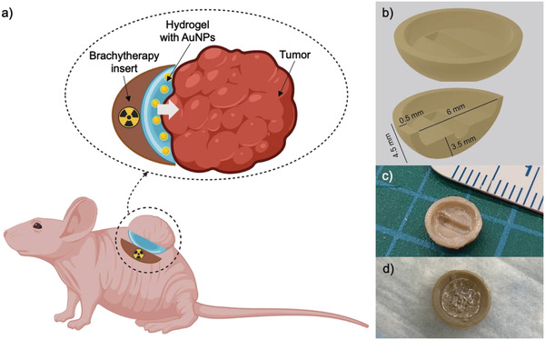
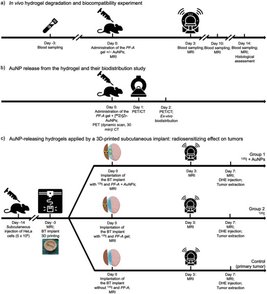
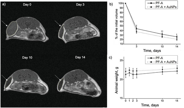
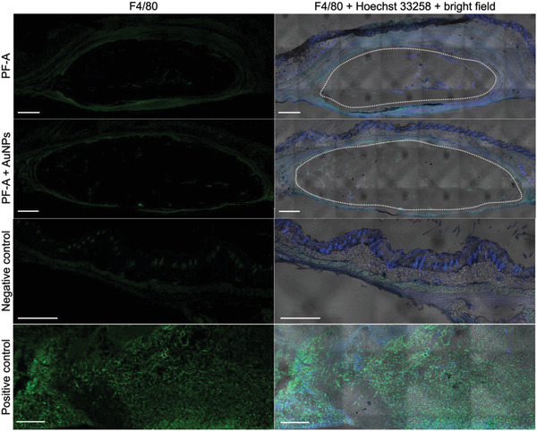
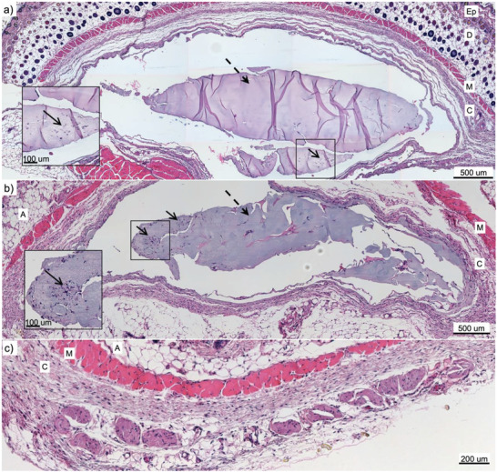
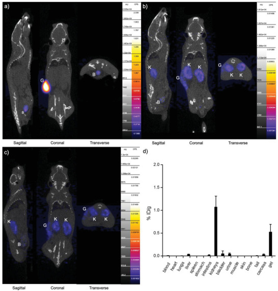
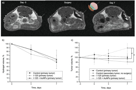
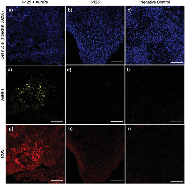
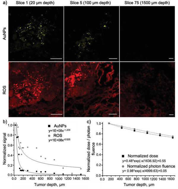
Brachytherapy (BT) is a widely used clinical procedure for localized cervical cancer treatment. In addition, gold nanoparticles (AuNPs) have been demonstrated as powerful radiosensitizers in BT procedures. Prior to irradiation by a BT device, their delivery to tumors can enhance the radiation effect by generating low-energy photons and electrons, leading to reactive oxygen species (ROS) production, lethal to cells. No efficient delivery system has been proposed until now for AuNP topical delivery to localized cervical cancer in the context of BT. This article reports an original approach developed to accelerate the preclinical studies of AuNP-enhanced BT procedures. First, an AuNP-containing hydrogel (Pluronic F127, alginate) is developed and tested in mice for degradation, AuNP release, and biocompatibility. Then, custom-made 3D-printed radioactive BT inserts covered with a AuNP-containing hydrogel cushion are designed and administered by surgery in mice (HeLa xenografts), which allows for measuring AuNP penetration in tumors (≈100 µm), co-registered with the presence of ROS produced through the interactions of radiation and AuNPs. Biocompatible AuNPs-releasing hydrogels could be used in the treatment of cervical cancer prior to BT, with impact on the total amount of radiation needed per BT treatment, which will result in benefits to the preservation of healthy tissues surrounding cancer.

Keywords: 3D printing; brachytherapy; gold nanoparticles; hydrogels; localized vaginal delivery; radiosensitizers; reactive oxygen species.


Conflict of interest statement

The authors declare no conflict of interest.

Figures

Figure 1
Figure 1
a) Schematic representation of the cupola‐shaped 3D‐printed radioactive BT inserts covered with a cushion of AuNP‐containing hydrogel, specifically designed to facilitate preclinical BT studies (image created with BioRender.com). b) A 3D drawing of the implant with typical dimensions. c) Photograph of the 3D‐printed and post‐processed BT insert accommodating a radioactive 125I seed prior to application of the AuNP‐containing radiation‐enhancing hydrogel layer. d) Photograph of the 3D‐printed and post‐processed BT insert with a hydrogel layer used for administration in the control group (without AuNPs).
Figure 2
Figure 2
Design of the in vivo experiments: a) timeline of the in vivo hydrogel degradation (by MRI) and biocompatibility experiment; b) timeline of the [89Zr]Zr‐AuNPs hydrogel release and biodistribution study (by PET); and c) timeline of the study to demonstrate the radiosensitizing effect on xenograft tumors, of AuNP‐releasing hydrogels applied by a 3D‐printed subcutaneous implant.
Figure 3
Figure 3
Visualization of BT inserts, tumors, and hydrogel cushions with MRI. a) Representative MRI images of mice from the 125I + AuNPs group, 3 days before, right after, and 7 days post‐surgery (the tumors are pointed with white arrows). b) Hydrogel volume (n = 5) and c) tumor volume (n = 5) quantified with MRI. The I‐125 + AuNPs group received a radioactive BT insert lined with a lens‐shaped USPION‐containing PF‐A + AuNPs hydrogel cushion, while the I‐125 group received a radioactive BT insert lined with a lens‐shaped USPION‐containing PF‐A hydrogel cushion (without AuNPs). The control (primary tumor) group was also exposed to the surgery and had a non‐radioactive BT insert lined with a lens‐shaped USPION‐containing PF‐A hydrogel cushion (without AuNPs). The secondary tumors developed on the other flank of the same mice were not exposed to the surgery but were used as an additional control (secondary tumor, no surgery). The data were analyzed using the one‐way ANOVA test (p < 0.05) followed by the Post Hoc Tukey HSD test and are presented as mean ± SD, p < 0.05 (*).
Figure 4
Figure 4
Results of the in vivo degradation study of AuNP‐containing hydrogels (PF‐A + AuNPs and PF‐A formulations). a) MRI scans of the mouse injected with PF‐A + AuNPs: white arrows indicate the hydrogel lumps. b) In vivo degradation profile of the two formulations (PF‐A + AuNPs and PF‐A), n = 6, paired Student's t‐test, p < 0.05 (*) and < 0.005 (**). c) No change was noted in the body weight of mice over a 14‐day course, n = 6. The data are presented as mean ± SD.
Figure 5
Figure 5
Histological assessment of the skin tissues surrounding the hydrogel implants (local inflammation response) 14 days after administration. Results are presented for both PF‐A and PF‐A + AuNPs groups. Cell nuclei are stained in blue (Hoechst 33258); macrophages are stained in green (F4/80 marker). The skin of CD‐1‐infected mice was used as a positive control, whereas the skin from the opposite flank of the experimental mice was used as a negative control. White ovals mark the cavities left by the fragile hydrogel volumes. The right side is a superposition of blue and green fluorescent signals on bright field optical microscopy [note: mosaics were reconstituted with several bright field images, which left geometrical patterns in the process (image artifact)]. The thickness of each slice was 20 µm, and the scale bar is 500 µm.
Figure 6
Figure 6
Histological assessment by H&E staining of local inflammation response in the skin tissues surrounding hydrogel implants (after 14 days). a) PF‐A group; b) PF‐A + AuNPs group; c) negative control skin. Cell nuclei appear in blue, while cytoplasm, elastin, and collagen are in pink. The gels are pointed with dashed arrows, whereas the slight presence of inflammatory cells at the surface of hydrogel volumes is pointed to with solid arrows. Ep: epidermis; D: dermis; A: adipose tissue; M: muscle; C: conjunctive tissue. The thickness of the slice is 5 µm.
Figure 7
Figure 7
AuNP release study by PET, from s.c. injected hydrogels at a) 30 min post‐injection (p.i.); b) day 1 p.i., c) day 2 p.i. B – bladder, G – gel, K – kidneys. d) Ex vivo biodistribution study at 2 days p.i.
Figure 8
Figure 8
Immunofluorescence analysis of the tumor samples from the groups I‐125 + AuNPs (a, d, g), I‐125 (b, e, h), and negative control (c, f, i) on Day 7 post‐surgery, the slice thickness is 20 µm, the scale bar is 500 µm.
Figure 9
Figure 9
Detection of AuNPs and ROS (fluorescence) at different depths of the tumor mass after treating it with BT in the presence of AuNP‐containing hydrogel (i.e., 125I + AuNPs group). a) Representative immunofluorescence images of several slices of the sample showing AuNPs and ROS signals (slice thickness 20 µm; scale bar 500 µm). The images at 20 µm (Slice 1) were reused from the Figure 8, panels d,g; b) Distribution of AuNPs and ROS signals across the depth of tumors (fitted to the power trendline); c) Distribution of the absorbed dose and photon fluence across the depth of tumors from the 125I seed (extracted from the Monte Carlo simulations, normalized, and fitted to the exponential trendline).

References

    1. Singh D., Vignat J., Lorenzoni V., Eslahi M., Ginsburg O., Lauby‐Secretan B., Arbyn M., Basu P., Bray F., Vaccarella S., The Lancet Global Health 2022, 11, 197. - PMC - PubMed
    1. Federico C., Sun J., Muz B., Alhallak K., Cosper P. F., Muhammad N., Jeske A., Hinger A., Markovina S., Grigsby P., Schwarz J. K., Azab A. K., Int. J. Radiat. Oncol., Biol., Phys. 2021, 109, 1483. - PMC - PubMed
    1. Abraham J., Gulley J. L., The Bethesda Handbook of Clinical Oncology, Lippincott Williams & Wilkins, Philadelphia, United States: 2022.
    1. Chao X. P., Song X. C., Wu H. W., You Y., Wu M., Li L., Front. Oncol. 2021, 11, 8. - PMC - PubMed
    1. de la Puente P., Azab A. K., J. Controlled Release 2014, 192, 19. - PubMed

Publication types