. 2023 Apr 24;14(1):2354.
doi: 10.1038/s41467-023-37984-x.

A small secreted protein NICOL regulates lumicrine-mediated sperm maturation and male fertility

Affiliations

A small secreted protein NICOL regulates lumicrine-mediated sperm maturation and male fertility

Daiji Kiyozumi et al. Nat Commun. .

Abstract

The mammalian spermatozoa produced in the testis require functional maturation in the epididymis for their full competence. Epididymal sperm maturation is regulated by lumicrine signalling pathways in which testis-derived secreted signals relocate to the epididymis lumen and promote functional differentiation. However, the detailed mechanisms of lumicrine regulation are unclear. Herein, we demonstrate that a small secreted protein, NELL2-interacting cofactor for lumicrine signalling (NICOL), plays a crucial role in lumicrine signalling in mice. NICOL is expressed in male reproductive organs, including the testis, and forms a complex with the testis-secreted protein NELL2, which is transported transluminally from the testis to the epididymis. Males lacking Nicol are sterile due to impaired NELL2-mediated lumicrine signalling, leading to defective epididymal differentiation and deficient sperm maturation but can be restored by NICOL expression in testicular germ cells. Our results demonstrate how lumicrine signalling regulates epididymal function for successful sperm maturation and male fertility.


Conflict of interest statement

The authors declare no competing interests.

Figures

Fig. 1
Fig. 1. Nicol encodes a secreted protein that is indispensable for male fertility.
a AlphaFold-predicted 3D structure of mouse NICOL. The signal peptide is depicted as faded. b RT-PCR analyses of Nicol expression in adult mouse organs, with Actb as an internal control. The initial to the fifth proximal segments of IS-caput region of epididymis was also separately analysed. Images are representative ones obtained from three independent biological replicates. c Litter sizes of WT, Nicol+/−, and Nicol−/− male (filled columns) and Nicol+/− and Nicol−/− female mice (unfilled columns). Average and standard error (S.E.) shown. d Litter sizes of Nicol−/− and Nicol−/−/CAG-Nicol males. Average and S.E. shown. ek Appearance of testis (e, f), testicular weight (g), HE staining of testis sections (h,i), and epididymal sperm morphologies (j, k) in Nicol+/− (e, h, j) and Nicol−/− (f, i, k) mice. Stages of spermatogenesis indicated by Roman numerals (h, i). Images are representative ones obtained from three independent biological replicates (e, f, hk). For bar plots, values are shown as mean ± S.E.M. P value = 0.5538 was determined by a two-tailed unpaired Students’ t-test (g). Scale bars, 2 mm (e, f), 100 µm (h, i), 20 µm (j, k).
Fig. 2
Fig. 2. Defective sperm maturation blocks sperm migration into the oviduct resulting in male sterility in Nicol−/− mice.
a Schematic representation of ejaculated sperm migrating from the uterus into the oviduct towards oocytes. bg Migration of red fluorescence-illuminated sperm from Nicol+/− (bd) and Nicol−/− (eg) mice ejaculated into reproductive tract in WT females. Bars, 1 mm. h, i Representative images of sperm–ZP binding assay using Nicol+/− (h) and Nicol−/− (i) sperm obtained from five independent biological replicates. Bars, 100 µm. j Sperm–ZP binding assay using sperm from Nicol+/− (green columns) and Nicol−/− (pink columns) mice. Average and S.E. shown. km In vitro fertilization capacity of sperm from Nicol+/− and Nicol−/− mice in assays with cumulus-free (k), cumulus-intact (l), and ZP-free (m) oocytes. n Development of fertilized eggs into blastocysts. o, p Representative images of blastocysts derived from WT oocytes inseminated with spermatozoa of WT (o) or Nicol−/− (p) males obtained from four independent biological replicates. Bars, 100 µm. For bar plots, values are shown as mean ± S.E.M. P value = 0.00001 (j), 0.0015 (k), 0.0075 (l), 0.9565 (m), and 0.3052 (n) were determined by a two-tailed unpaired Students’ t-test. *P < 0.05, **P < 0.01, ***P < 0.001.
Fig. 3
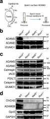
Fig. 3. Defective epididymal sperm maturation in Nicol−/− male mice.
a A schematic representation of ADAM3 processing during sperm transit through epididymal IS. b Expression of proteins associated with sperm ability to migrate into the oviduct and ZP binding of cauda epididymal sperm from Nicol+/− and Nicol−/− mice. c Expression of proteins associated with sperm ability to migrate into the oviduct and ZP binding in testicular germ cells from Nicol+/− and Nicol−/− mice. d Expression of OVCH2 and ADAM28 in WT, Nicol−/−, Nell2−/−, and Ros1−/− IS-caput epididymis. Gpx5 and GAPDH shown as internal controls. Images are representative ones obtained from three independent biological replicates (bd).
Fig. 4
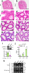
Fig. 4. Defective epididymal IS epithelium differentiation in Nicol−/− mice is a phenocopy of other lumicrine signalling-deficient mice.
ah HE staining of IS sections of epididymis from WT (a, c), Nicol−/− (b, d), Nell2−/− (e), Ros1−/− (f), efferent duct-ligated (EDL) (g), and W/Wv (h) mice. Bars, 500 μm (a, b), 100 μm (ch). Images are representative ones obtained from three (ad, g, h) or five (e, f) independent biological replicates. i Immunoblot detection of phosphorylated and total ERK1/2 in Nicol+/− (green columns) and Nicol−/− (pink columns) IS-caput epididymis. Average ERK phosphorylation levels and S.E. shown. j RNA expression of ERK downstream transcription factor genes Etv1, Etv4, and Etv5 in Nicol+/− and Nicol−/− IS-caput epididymis. Average and S.E. shown. RPKM, reads per kilobase per million. k RT-PCR analyses of Nell2, Nicol, Ros1, and Ptpn6 expression in Nicol−/− testis and caput epididymis. Actb shown as internal control. Images are representative ones obtained from three independent biological replicates. For bar plots, values are shown as mean ± S.E.M. P value = 0.0001 (i) and 0.006, 0.0004, and 0.0005 for Etv1, Etv4, and Etv5, respectively (j), were determined by a two-tailed unpaired Students’ t-test. *P < 0.05, **P < 0.01, ***P < 0.001.
Fig. 5
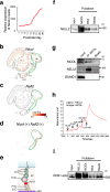
Fig. 5. NICOL forms a complex with NELL2.
a RNA expression profile of Nicol in developing testis. b–d t-distributed stochastic neighbour embedding plots of adult mouse testis single-cell RNA-seq data representing Nicol (b) and Nell2 (c) expression. Nicol expression in Nell2-expressing cells is also shown (d). Nicol is broadly expressed with highest expression in leptotene, pachytene, and diplotene spermatocyte populations, which also show the highest Nell2 expression. SGC spermatogonial stem cells, P Preleptotene spermatocytes, LZPD leptotene, zygotene, pachytene, and diplotene spermatocytes, St spermatids, SC Sertoli cells, EC endothelial cells, MC myoid cells, LC lymphatic cells. e Schematic representation of seminiferous tubule histology. SGC spermatogonial stem cell, PrSc preleptotene spermatocyte, PaSc pachytene spermatocyte, St spermatid, SC Sertoli cell, BTB blood–testis barrier, BM basement membrane. f In vitro NELL2 pulldown with NICOL or BSA. Images are representative ones obtained from three independent biological replicates. g Co-immunoprecipitation analyses of NICOL with NELL2 from Clgn-Nicol and Clgn-Nell2 testis lysate. Izumo sperm-egg fusion protein 1 (IZUMO1) shown as an internal negative control. Images are representative ones obtained from three independent biological replicates. h Kinetics of NELL2–NICOL interaction analysed by surface plasmon resonance technology. Binding of NICOL at indicated concentrations onto immobilized NELL2. i In vitro ROS1 ectodomain pulldown with NELL2, NICOL, or BSA. The image is representative one obtained from three independent biological replicates.
Fig. 6
Fig. 6. Testicular germ cell-specific expression of Nicol rescued epididymal differentiation, sperm maturation, and male infertility in Nicol−/− mice.
a Schematic representation of transgene Clgn-Nicol. b RT-PCR analyses of Clgn-Nicol transgene in brain (Br), testis (Te), and caput epididymis (Cap) in Nicol−/−/Clgn-Nicol mice. Actb shown as internal control. Images are representative ones obtained from three independent biological replicates. c, d HE staining of Nicol−/− (c) and Nicol−/−/Clgn-Nicol (d) IS. Bars, 100 µm. e Immunoblot analyses of phosphorylated ERK, ETV5, OVCH2, and ADAM28 in WT, Nicol−/−, and Nicol−/−/Clgn-Nicol IS-caput epididymis. Gpx5 and glyceraldehyde 3-phosphate dehydrogenase (GAPDH) shown as internal controls. Images are representative ones obtained from three independent biological replicates. f Immunoblot analyses of mature ADAM3 expression in WT, Nicol−/− and Nicol−/−/Clgn-Nicol cauda epididymal sperm. g Litter sizes of WT, Nicol−/−, and Nicol−/−/Clgn-Nicol males. Images are representative ones obtained from three independent biological replicates (e, f). For bar plots, values are shown as mean ± S.E.M.
Fig. 7
Fig. 7. Possible working model of NICOL in lumicrine signalling.
a The luminal fluid inside testicular seminiferous tubules flows and reaches epididymal IS via efferent duct. b The NELL2 and NICOL proteins secreted into seminiferous luminal fluid form a complex and reach the epididymal IS by the luminal flow. Spermatocytes seems to play a critical role in lumicrine factor secretion as they express both Nell2 and Nicol at the highest level in testicular cells. A contribution to the luminal NICOL protein by testicular and epididymal cells other than germ cells is not excluded, although such a non-germ cell contribution is dispensable. See also Fig. 4e for the schematic representation of a seminiferous tubule. c On IS cell surface NELL2-NICOL complex binds to ROS1 leading to its multimerization and subsequent self-activation, which eventually induces IS differentiation and sperm maturation.

Similar articles

Cited by

References

    1. Greep RO, Fevold HL. The spermatogenic and secretory function of the gonads of hypophysectomized adult rats treated with pituitary FSH and LH. Endocrinology. 1937;21:611–618. doi: 10.1210/endo-21-5-611. - DOI
    1. Russell L. Movement of spermatocytes from the basal to the adluminal compartment of the rat testis. Am. J. Anat. 1977;148:313–328. doi: 10.1002/aja.1001480303. - DOI - PubMed
    1. Challice CE. Electron microscope studies of spermiogenesis in some rodents. J. R. Microsc. Soc. 1953;73:115–127. doi: 10.1111/j.1365-2818.1953.tb01978.x. - DOI - PubMed
    1. Bedford JM. Effects of duct ligation on the fertilizing ability of spermatozoa from different regions of the rabbit epididymis. J. Exp. Zool. 1967;166:271–281. doi: 10.1002/jez.1401660210. - DOI - PubMed
    1. Orgebin-Crist MC. Sperm maturation in rabbit epididymis. Nature. 1967;216:816–818. doi: 10.1038/216816a0. - DOI - PubMed

Publication types