. 2023 Apr 24;14(4):288.
doi: 10.1038/s41419-023-05797-x.

CHST2-mediated sulfation of MECA79 antigens is critical for breast cancer cell migration and metastasis

Affiliations

CHST2-mediated sulfation of MECA79 antigens is critical for breast cancer cell migration and metastasis

Dan Zhang et al. Cell Death Dis. .

Abstract

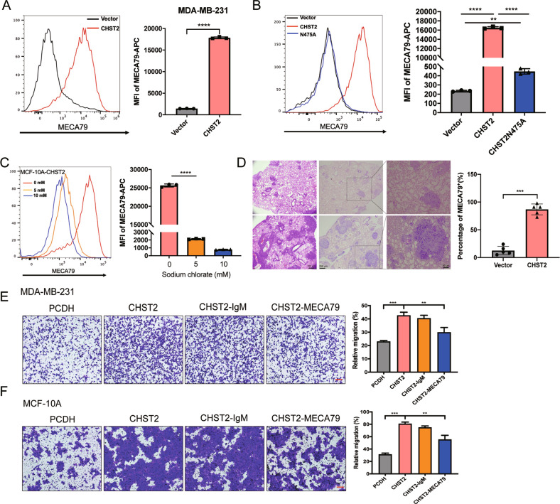
Snail is a denoted transcriptional repressor that plays key roles in epithelial-mesenchymal transition (EMT) and metastasis. Lately, a plethora of genes can be induced by stable expression of Snail in multiple cell lines. However, the biological roles of these upregulated genes are largely elusive. Here, we report identification of a gene encoding the key GlcNAc sulfation enzyme CHST2 is induced by Snail in multiple breast cancer cells. Biologically, CHST2 depletion results in inhibition of breast cancer cell migration and metastasis, while overexpression of CHST2 promotes cell migration and lung metastasis in nude mice. In addition, the expression level of MECA79 antigen is elevated and blocking the cell surface MECA79 antigen with specific antibodies can override cell migration mediated by CHST2 upregulation. Moreover, the sulfation inhibitor sodium chlorate effectively inhibits the cell migration induced by CHST2. Collectively, these data provide novel insights into the biology of Snail/CHST2/MECA79 axis in breast cancer progression and metastasis as well as potential therapeutic strategy for the diagnosis and treatment of breast cancer metastasis.


Conflict of interest statement

The authors declare no competing interests.

Figures

Fig. 1
Fig. 1. Snail induces the transcription and expression of CHST2 in breast cancer cells.
A The heatmap showed the representatives of differentially expressed genes regulated by Snail in MCF-10A cells. B The known target genes regulated by Snail are validated by qRT-PCR. Data were shown as mean ± SD, ****P < 0.0001, ***P < 0.001, **P < 0.01, *P < 0.05, compared with vector group. Overexpression of Snail increased CHST2 mRNA levels in MCF-10A cells (C), MDA-MB-231 cells (D) and MCF-7 cells (E). Data were shown as mean ± SD, ***P < 0.001, **P < 0.01, compared with vector group (left panels). Western blots showed the protein expression levels of CHST2 in breast cancer cells with stably expressing Snail (right panels). F Scatter plots showed the correlation of Snail expression with CHST2 expression in 1095 breast tumor specimens from TCGA dataset. The r value was calculated via Pearson’s ranking correlation coefficient analysis. G Human CHST2-Luc promoter reporter activity was measured after co-transfection with Snail expression plasmids in 293T cells. Human CHST2 gene promoter (−638 to +426) was subcloned into a pGL3 basic luciferase vector to create a CHST2-Luc reporter construct (top panel). The results were expressed as normalized β-galactosidase activity. Error bars show standard deviations (middle panel) from three replicate experiments, **P < 0.01, *P < 0.05. H ChIP analysis of Snail binding to CHST2 promoter region in MDA-MB-231 cells. The ChIP assays were performed in MDA-MB-231 cells with specific monoclonal antibody against Snail and the relative enrichment of Snail on the CHST2 promoter region was determined by qPCR, the results are expressed as normalized input. Error bars show standard deviations. ***P < 0.001, **P < 0.01, *P < 0.05, ns means no significance. All the data are representative of at least three independent experiments and presented as the means ± SD.
Fig. 2
Fig. 2. CHST2 is required for breast cancer cell migration and metastasis.
Protein levels of CHST2 were quantified by western blots in MDA-MB-231 cells with CHST2 knockdown (A) or CHST2 overexpression (D). Transwell assays show the migration and invasion abilities of MDA-MB-231-shCHST2 cells (B) or MDA-MB-231-CHST2 cells (E). Left panel: Representative images of migrated cells, right panel: Statistical analysis of the average migrated cells per field. Data were shown as mean ± SD from three independent experiments. ***P < 0.001, **P < 0.01, compared with vector groups, 100×, scale bar, 100 μm. C Tail-vein injection of MDA-MB-231 cells with knockdown of CHST2 for lung metastasis analysis. Left panel: Intensities of lung metastasis in mice at the 5th week were analyzed by bioluminescent imaging (BLI). Right panel: Quantification of lung photon flux at the first, 2nd, 4th, and 5th week. P value was determined using two-way ANOVA, *P < 0.05, compared with vector group (n = 7 and n = 5, respectively). F Overexpression of CHST2 in MDA-MB-231 cells promoted lung metastasis in vivo. Left panel: Intensities of lung metastasis in mice at the 6th week were analyzed by BLI. Right panel: Quantification of lung photon flux at the 0, 2nd, 4th, and 6th week. P value was measured by two-way ANOVA, *P < 0.05, compared with vector group (n = 7 and n = 6, respectively). G Overexpression of CHST2 increased the number of metastatic pulmonary surface nodules. Left panel: Shown are representative images of metastatic nodules. Right panel: The average number of metastatic tumor nodules shows a significant difference between the vector and CHST2 overexpression group (n = 7 and n = 6, respectively). Data were shown as mean ± SD, ***P < 0.001, compared with vector group.
Fig. 3
Fig. 3. CHST2 promotes cell migration in an enzymatic-dependent manner.
A The wound-healing assay showed migration capabilities of CHST2-overexpressed MCF-10A cells in the presence of 0, 5, 10 mM sodium chlorate for 48 h. Left panel: Representative images were shown. Right panel: The quantification of the percentage of wound closure. Data were shown as mean ± SD from four independent experiments, ***P < 0.001, *P < 0.05, Students t test. B The diagram of CHST2, the site of N475 which can be mono-glycosylated is critical for enzyme activity. C Western blots (WB) showed the protein level of MCF-10A-Vector, WT CHST2 and mutant CHST2-N475A cells in total cell lysates (TCL). D Transwell assays were performed in stable cell lines. Six fields chosen randomly were counted for statistical analysis. Data were shown as mean ± SD from three independent experiments. ***P < 0.001, *P < 0.05, Students t test. E Wound-healing assay showed the migration abilities of MCF-10A-Vector, CHST2 or mutant CHST2-N475A cells, left panel: Representative images were shown. Right panel: The quantification of the percentage of wound closure, Data were shown as mean ± SD. of four independent experiments, ***P < 0.001, **P < 0.01, *P < 0.05.
Fig. 4
Fig. 4. CHST2 is involved in MECA79 antigen synthesis to mediate breast cancer metastasis.
A FACS analysis of MDA-MB-231-Vector (black) and MDA-MB-231-CHST2 (red) cells stained by MECA79 mAb, left panel: Representative histograms were shown. Right panel: MFI of MECA79 antigen synthesis by breast cancer cells was shown compared with vector group, MFI, mean fluorescence intensity, the MFI of MECA79 less the MFI of isotype control equals the Delta MFI. Data were shown as mean ± SD, ****P < 0.0001, Student t test. B Flow cytometry analysis of the MECA79 surface expression in MCF-10A-Vector (black), CHST2 (red) or mutant CHST2-N475A (blue) cells, left panel: Representative histograms were shown. Right panel: Relative MFI of MECA79 antigen synthesis by MCF-10A cells was shown compared with the vector group. Data were shown as mean ± SD from three independent experiments. ****P < 0.0001, **P < 0.01, Student t test. C Flow cytometry analysis of the MECA79 surface expression in MCF-10A-CHST2 cells with treatment of 0, 5, 10 mM sodium chlorate for 48 h, left panel: Representative histograms were shown. Right panel: MFI of MECA79 antigen synthesis by MCF-10A-CHST2 cells was shown. Data were shown as mean ± SD from three independent experiments. ****P < 0.0001, Student t test. D H&E staining and Immunohistochemical staining of MECA79 antigen of the lung sections isolated from lung metastatic mice models. E, F Transwell assays in MDA-MB-231-Vector or MCF-10A-Vector and MDA-MB-231-CHST2 or MCF-10A -CHST2 cells with MECA79 or isotype antibody treated (left panel), Six fields chosen randomly were counted for statistical analysis (right panel) and data were shown as mean ± SD, ***P < 0.001, **P < 0.01. Student t test.
Fig. 5
Fig. 5. CHST2 is an essential target gene for Snail-induced cell migration.
Western blots showed the protein levels of Snail, CHST2. Knocking-down of CHST2 in Snail overexpression MDA-MB-231 cells (A) and Snail knockdown with CHST2 rescued cells (C). Transwell assays in MDA-MB-231-pLU-Snail cells (B) and MDA-MB-231-shSnail cells (D) with CHST2 knockdown and CHST2 rescue respectively. Three independent experiments were performed, six fields chosen randomly were counted for statistical analysis (right panels) and data were shown as mean ± SD, ***P < 0.001, **P < 0.01, *P < 0.05. Students’ t test.
Fig. 6
Fig. 6. Snail-CHST2 axis mediated breast cancer cell migration by enhanced MECA79 antigen synthesis.
A FACS analysis of MDA-MB-231-pLU-Vector (black) and MDA-MB-231-pLU-Snail (red) cells stained by MECA79 mAb, left panel: Representative histograms were shown. Right panel: MFI of MECA79 antigen synthesis by breast cancer cells was shown compared with the vector group. Data were shown as mean ± SD from three independent experiments. *P < 0.05, Student t test. B MECA79 surface expression in MDA-MB-7-pLU-Vector (black) cells and MCF-7-pLU-Snail cells with CHST2 knockdown was assessed by flow cytometry, left panel: Representative histograms were shown. Right panel: MFI of MECA79 antigen synthesis by cancer cells was shown compared with Snail group. Data were shown as mean ± SD, three independent experiments were performed. **P < 0.01, *P < 0.05, Student t test. C, D Transwell assays in MDA-MB-231-Vector or MCF-7-Vector and MDA-MB-231-Snail cells or MCF-7-Snail cells with MECA79 or isotype antibody treated (left panels), six fields chosen randomly were counted for statistical analysis (right panels) and data were shown as mean ± SD of three independent experiments, **P < 0.01, *P < 0.05. Student t test. E Schematic model for the role of Snail-CHST2-MECA79 sulfation axis in breast cancer cell migration and metastasis. Snail activated transcription of CHST2 via binding to promoter sequences, and simultaneously induced CHST2 expression which increased sulfation level in MECA79 antigen synthesis to enhance migration and metastasis of breast cancer cells. Furthermore, blocking cell surface MECA79 antigen inhibited cancer cell migration.

Similar articles

Cited by

References

    1. Friedl P, Wolf K. Tumour-cell invasion and migration: diversity and escape mechanisms. Nat Rev Cancer. 2003;3:362–74. doi: 10.1038/nrc1075. - DOI - PubMed
    1. Valastyan S, Weinberg RA. Tumor metastasis: molecular insights and evolving paradigms. Cell. 2011;147:275–92. doi: 10.1016/j.cell.2011.09.024. - DOI - PMC - PubMed
    1. Zou Y, Ye F, Kong Y, Hu X, Deng X, Xie J, et al. The single-cell landscape of intratumoral heterogeneity and the immunosuppressive microenvironment in liver and brain metastases of breast cancer. Adv Sci. 2023;10:e2203699. doi: 10.1002/advs.202203699. - DOI - PMC - PubMed
    1. Mittal V. Epithelial mesenchymal transition in tumor metastasis. Annu Rev Pathol. 2018;13:395–412. doi: 10.1146/annurev-pathol-020117-043854. - DOI - PubMed
    1. Liu P, Wang Z, Ou X, Wu P, Zhang Y, Wu S, et al. The FUS/circEZH2/KLF5/ feedback loop contributes to CXCR4-induced liver metastasis of breast cancer by enhancing epithelial-mesenchymal transition. Mol Cancer. 2022;21:198. doi: 10.1186/s12943-022-01653-2. - DOI - PMC - PubMed

Publication types

Substances