. 2023 Apr 24;13(1):6641.
doi: 10.1038/s41598-023-32459-x.

Structural insights into thrombolytic activity of destabilase from medicinal leech

Affiliations

Structural insights into thrombolytic activity of destabilase from medicinal leech

Egor Marin et al. Sci Rep. .

Abstract

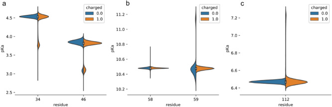
Destabilase from the medical leech Hirudo medicinalis belongs to the family of i-type lysozymes. It has two different enzymatic activities: microbial cell walls destruction (muramidase activity), and dissolution of the stabilized fibrin (isopeptidase activity). Both activities are known to be inhibited by sodium chloride at near physiological concentrations, but the structural basis remains unknown. Here we present two crystal structures of destabilase, including a 1.1 Å-resolution structure in complex with sodium ion. Our structures reveal the location of sodium ion between Glu34/Asp46 residues, which were previously recognized as a glycosidase active site. While sodium coordination with these amino acids may explain inhibition of the muramidase activity, its influence on previously suggested Ser49/Lys58 isopeptidase activity dyad is unclear. We revise the Ser49/Lys58 hypothesis and compare sequences of i-type lysozymes with confirmed destabilase activity. We suggest that the general base for the isopeptidase activity is His112 rather than Lys58. pKa calculations of these amino acids, assessed through the 1 μs molecular dynamics simulation, confirm the hypothesis. Our findings highlight the ambiguity of destabilase catalytic residues identification and build foundations for further research of structure-activity relationship of isopeptidase activity as well as structure-based protein design for potential anticoagulant drug development.


Conflict of interest statement

The authors declare no competing interests.

Figures

Figure 1
Figure 1
Phylogenetic analysis of Pfam “destabilase” protein family, annotated with information about muramidase and isopeptidase activities and source organisms. An outgroup protein (c-type lysozyme from hen egg Gallus gallus, colored with orange) is added as a distant relative of i-type lysozymes, indicating the root of the phylogenetic tree. A color of the first half of the organism taxonomy name corresponds to the muramidase activity and the second half—to the isopeptidase activity. Green and red colors mean presence and absence of the corresponded activity respectively, black means no information about the corresponding activity. Violet shows arthropods branch of the tree, yellow—the non-arthropods branch.
Figure 2
Figure 2
Multiple sequence alignment of destabilase homologs with confirmed muramidase and/or isopeptidase activities from Table 1. Color of the first half of the organism taxonomy name corresponds to the muramidase activity and the second half—to the isopeptidase activity. Green and red colors mean presence and absence of the confirmed corresponding activity, black means no information is available related to corresponding activity. Amino acids discussed in the manuscript are marked with light blue.
Figure 3
Figure 3
High salt destabilase structure and of its homologs. (a) Overall structure superposition of high salt destabilase chain A, TjL chain A (PDB ID 2DQA) and MlL chain C (PDB ID 4PJ2); lateral view on the inhibited enzymatic centers of high salt destabilase ((b,c) for chains A, B, correspondingly), TjL ((d,e) for chains A, B, correspondingly) and MlL ((f,g) for chains C, D, correspondingly). Residues potentially responsible for muramidase and isopeptidase activities or bonded to the inhibitor through the water molecules are shown as sticks. Inhibitors of muramidase activity are depicted as spheres, sticks or ribbon diagrams, respectively: (b,c) water coordinated Na+ ion; (d,e) NAG3; (f,g) Aeromonas hydrophila lysozyme inhibitor. Water and inhibitory molecules as well as hydrogen bonds are shown for alternative conformation A only in MlL and TjL structures and for alternative conformations with presence of sodium in destabilase structures. The variability of an inhibitor-bound TjL and MlL loop 44–51 confirms the importance of the loop flexibility for enzymatic activities of the proteins.
Figure 4
Figure 4
High and low salt destabilase structures. (a) Overall structure superposition of all destabilase structures; (b) front view of the catalytic cleft in the low salt destabilase structure; (c,d) front view of the catalytic cleft in the high salt destabilase structure in chains A and B, correspondingly; residues, potentially responsible for either muramidase or isopeptidase activity, are shown as sticks; Na+ ion and water molecules are shown as spheres. Water molecules and hydrogen bonds are shown only for alternative conformations with presence of sodium. Water coordinated Na+ ion occupies the cleft between catalytic dyad Glu34/Asp46 and fixates the flexible loop 44–51 in the closed conformation preventing a substrate from approaching the active site.
Figure 5
Figure 5
Structure-based pKa values, calculated over the 1 μs molecular dynamics simulations of the destabilase (high salt structure). (a) Glu34 and Asp46 residues, (b) Lys58 and Lys59 residues, (c) His112 residue. Blue and orange violin plots represent uncharged and charged His112 trajectories, respectively. Plot width is proportional to the total share of structures with the respective pKa value.

References

    1. Sig AK, Guney M, Uskudar Guclu A, Ozmen E. Medicinal leech therapy—An overall perspective. Integr. Med. Res. 2017;6:337. doi: 10.1016/j.imr.2017.08.001. - DOI - PMC - PubMed
    1. Kvist S, Manzano-Marín A, de Carle D, Trontelj P, Siddall ME. Draft genome of the European medicinal leech Hirudo medicinalis (Annelida, Clitellata, Hirudiniformes) with emphasis on anticoagulants. Sci. Rep. 2020;10:9885. doi: 10.1038/s41598-020-66749-5. - DOI - PMC - PubMed
    1. Babenko VV, et al. Draft genome sequences of Hirudo medicinalis and salivary transcriptome of three closely related medicinal leeches. BMC Genom. 2020;21:1. - PMC - PubMed
    1. Baskova IP, et al. Role of isopeptidolysis in the process of thrombolysis. Thromb. Res. 2018;165:18. doi: 10.1016/j.thromres.2018.03.007. - DOI - PubMed
    1. Baskova IP, Nikonov GI. Destabilase: An enzyme of medicinal leech salivary gland secretion hydrolyzes the isopeptide bonds in stabilized fibrin. Biokhimiya. 1985;50:424. - PubMed

Publication types