

Prevalence and hepatic histopathological findings of fascioliasis in sheep slaughtered in Jeddah, Saudi Arabia
- PMID: 37095133
- PMCID: PMC10126202
- DOI: 10.1038/s41598-023-33927-0
Prevalence and hepatic histopathological findings of fascioliasis in sheep slaughtered in Jeddah, Saudi Arabia
Abstract
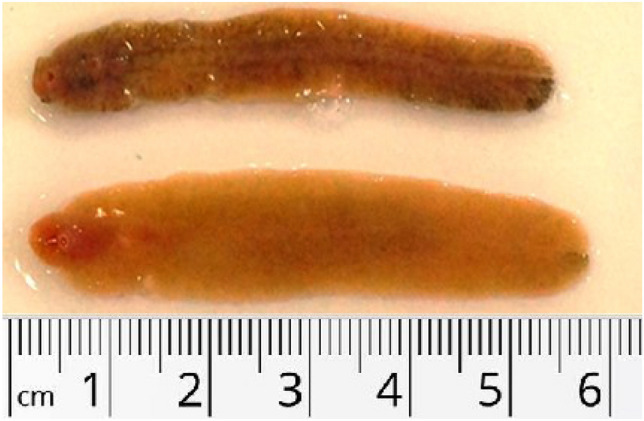
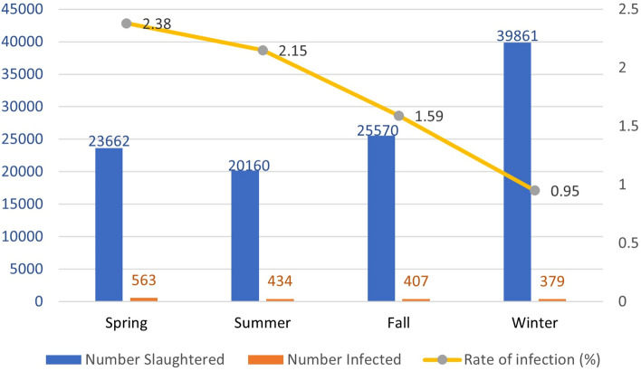
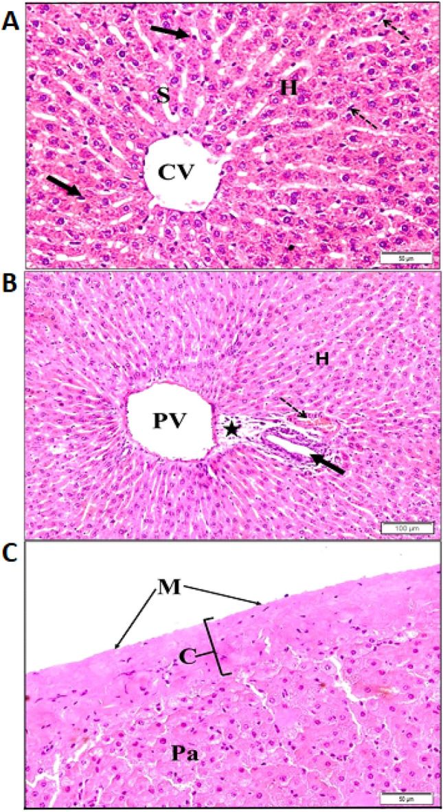
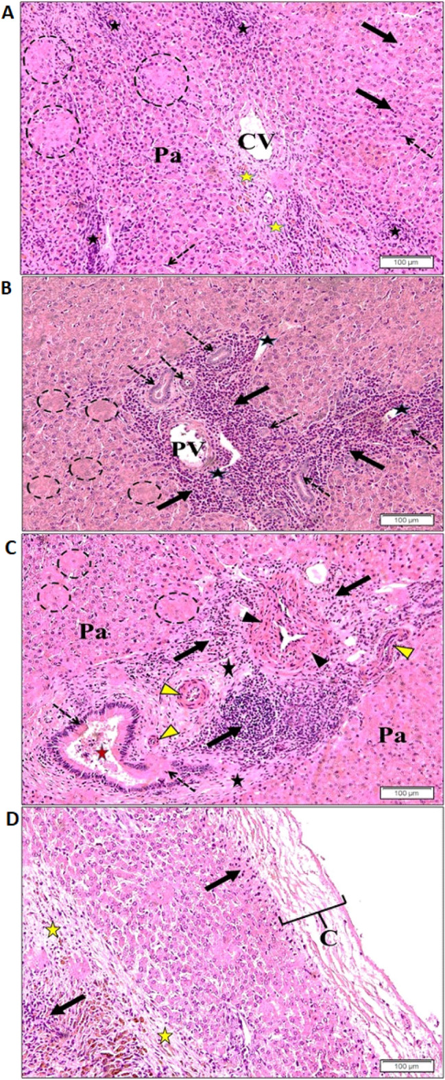
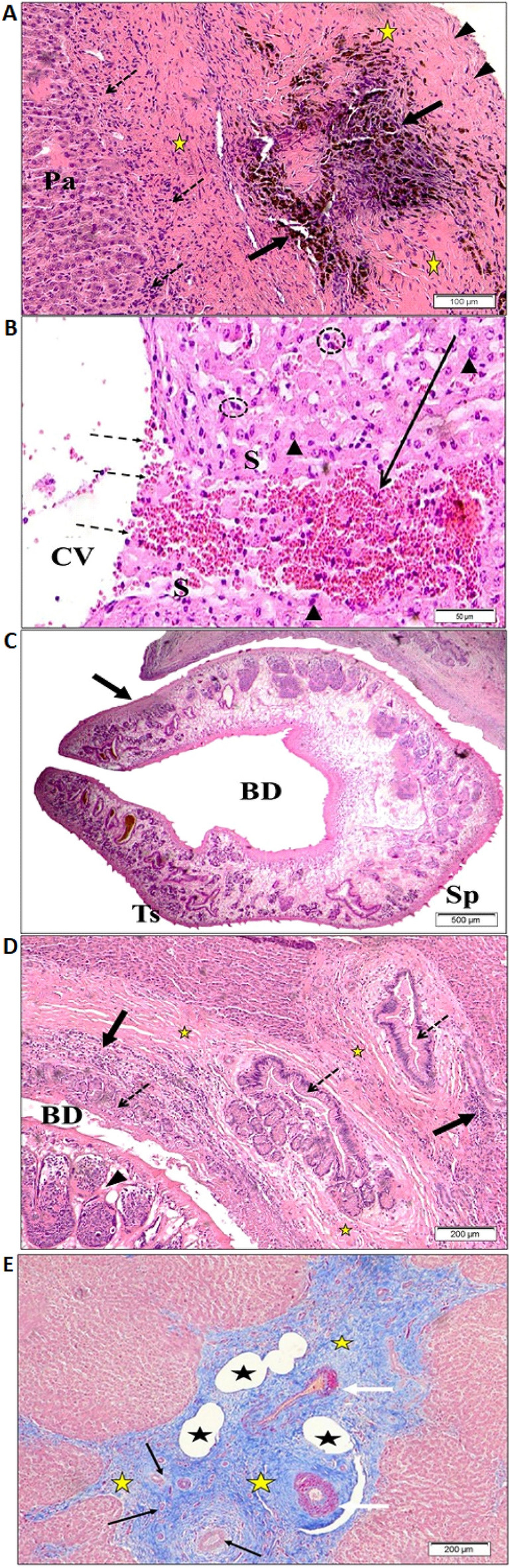
Hepatic fascioliasis is an important parasitic disease responsible for morbidity and mortality in many domestic ruminants, especially sheep, goats, and cattle, due to Fasciola (F.) hepatica and F. gigantica. This study aimed to determine the prevalence of fascioliasis in sheep slaughtered in Jeddah, Saudi Arabia, and to describe the morphological and histopathological changes in the liver. A total of 109,253 sheep slaughtered between July 2017 and July 2018 were screened to assess the prevalence of fascioliasis. The livers were grossly investigated for Fasciola infection and morphological changes. Tissue samples were collected for proper histopathological examinations. Livers of local and imported sheep represented infection rates of 0.67% and 2.12%, respectively, and the highest infection rate was in the spring season. Macroscopically, the affected liver showed hepatomegaly, thickened capsule and discoloration with necrosis, fibrosis, dilation of the bile duct, engorgement of the gallbladder and enlargement of the portal lymph nodes. Microscopic examination showed fibrotic thickening, calcification and hyperplasia of the bile ducts filled with debris, as well as massive hemorrhagic foci. Histopathological examinations of the infected liver showed a central vein region with disturbed parenchyma cells, focal lymphocytic infiltration, elongated endothelial cells, blood sinusoids that showed enlarged Kupffer cells, patches of lysed or necrotic hepatocytes, eosinophil infiltration, lymphocytes and proliferating fibroblast, thickening of hepatic artery and arteriolar walls. We concluded that fascioliasis among sheep slaughtered in Jeddah is not uncommon. The identified histopathological changes in the liver of infected sheep reflect tissue damage, which can lead to significant economic losses for the animals.
© 2023. The Author(s).
Conflict of interest statement
The authors declare no competing interests.
Figures

Similar articles
-
Prevalence of Fascioliasis in Slaughtered Cattle in Ağrı Province.Turkiye Parazitol Derg. 2023 Sep 18;47(3):156-159. doi: 10.4274/tpd.galenos.2023.39306. Turkiye Parazitol Derg. 2023. PMID: 37724364 English.
-
The prevalence and economic significance of Fasciola gigantica and Stilesia hepatica in slaughtered animals in the semi-arid coastal Kenya.Trop Anim Health Prod. 2006;38(6):475-83. doi: 10.1007/s11250-006-4394-4. Trop Anim Health Prod. 2006. PMID: 17243475
-
Molecular Confirmation of a Fasciola gigantica × Fasciola hepatica Hybrid in a Chadian Bovine.J Parasitol. 2020 Apr 1;106(2):316-322. doi: 10.1645/19-66. J Parasitol. 2020. PMID: 32330281
-
The Epidemiology and Control of Liver Flukes in Cattle and Sheep.Vet Clin North Am Food Anim Pract. 2020 Mar;36(1):109-123. doi: 10.1016/j.cvfa.2019.12.002. Vet Clin North Am Food Anim Pract. 2020. PMID: 32029178 Review.
-
Status of fasciolosis among domestic ruminants in Iran based on abattoir data: a systematic review and meta-analysis.Ann Parasitol. 2020;66(1):77–86. doi: 10.17420/ap6601.240. Ann Parasitol. 2020. PMID: 32198998
Cited by
-
Liver Histopathological and Immunohistochemical Evaluation from Fasciola hepatica Experimentally Infected and Reinfected Sheep.Animals (Basel). 2024 Jun 20;14(12):1833. doi: 10.3390/ani14121833. Animals (Basel). 2024. PMID: 38929451 Free PMC article.
References
-
- Zafar A, Khan M, Sindhu ZD, Abbas R, Masood S, Abbas Z, Mahmood M, Kashif M, Khan J, Hussain R, et al. Seroprevalence of Fasciola hepatica in small ruminants of District Chakwal, Punjab, Pakistan. J. Pak. Vet. 2019;39(1):96–101. doi: 10.29261/pakvetj/2018.024. - DOI
-
- Calvani NED, Ichikawa-Seki M, Bush RD, Khounsy S, Šlapeta J. Which species is in the faeces at a time of global livestock movements: Single nucleotide polymorphism genotyping assays for the differentiation of Fasciola spp. Int. J. Parasitol. 2020;50(2):91–101. doi: 10.1016/j.ijpara.2019.12.002. - DOI - PubMed
MeSH terms
LinkOut - more resources
Full Text Sources
