. 2023 Jun;30(6):1601-1614.
doi: 10.1038/s41418-023-01156-7. Epub 2023 Apr 24.

The biophysical property of the limbal niche maintains stemness through YAP

Affiliations

The biophysical property of the limbal niche maintains stemness through YAP

Swarnabh Bhattacharya et al. Cell Death Differ. 2023 Jun.

Abstract

The cell fate decisions of stem cells (SCs) largely depend on signals from their microenvironment (niche). However, very little is known about how biochemical niche cues control cell behavior in vivo. To address this question, we focused on the corneal epithelial SC model in which the SC niche, known as the limbus, is spatially segregated from the differentiation compartment. We report that the unique biomechanical property of the limbus supports the nuclear localization and function of Yes-associated protein (YAP), a putative mediator of the mechanotransduction pathway. Perturbation of tissue stiffness or YAP activity affects SC function as well as tissue integrity under homeostasis and significantly inhibited the regeneration of the SC population following SC depletion. In vitro experiments revealed that substrates with the rigidity of the corneal differentiation compartment inhibit nuclear YAP localization and induce differentiation, a mechanism that is mediated by the TGFβ-SMAD2/3 pathway. Taken together, these results indicate that SC sense biomechanical niche signals and that manipulation of mechano-sensory machinery or its downstream biochemical output may bear fruits in SC expansion for regenerative therapy.


Conflict of interest statement

The authors declare no competing interests.

Figures
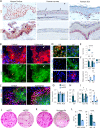
Fig. 1
Fig. 1. YAP is required to maintain LSC at undifferentiated state in vitro.
A Immunohistochemistry of YAP and K15 was performed on paraffin sections human limbus, cornea, and skin. B,C Primary human limbal epithelial and foreskin epidermal cells were co-cultured with NIH-3T3-J2 feeder cells, grown on a plastic plate for 4 days and the expression of the indicated markers was examined by immunostaining. The golden and white arrows indicate stem/progenitor or differentiated cell regions, respectively. DF Primary human LSCs were grown at low calcium (Day 0) or induced to differentiate in high calcium (Day 7) and the expression of the indicated markers was tested by immunostaining (D) that was further quantified for mean nuclear-to-cytoplasmic YAP intensity ratio (E) or by quantitative real-time PCR (qPCR) (F). GI Primary human limbal epithelial cells were transfected with esiRNA against YAP (esiYAP) or control esiRNA (esiCtl) and 48–72 h later, the expression of housekeeping genes (G), YAP (H) and the indicated markers (I) was examined by qPCR. JL Primary limbal epithelial cells were transfected with esiYAP or esiCtl or treated with Verteporfin or vehicle, and 48–72 h later, cells were subjected to a clonogenicity test (J, K) and the number of the colony was quantified (n = 3) (L). The qPCR data were normalized to the housekeeping gene GAPDH (mean ± standard error of mean, n = 4 biological replicates) as a fold increase compared to the control sample and the mean nuclear-to-cytoplasmic YAP intensity ratio from three independent experiments (E) is shown by the Tukey box-and-whisker plot followed by t-test with Welch’s correction. Statistical significance was assessed by t-test (*, p < 0.05; **, p < 0.01; ***, p < 0.001, ****, p < 0.0001). Immunostaining and Immunohistochemistry data are representative from 3 to 5 biological replicates. Nuclei were detected by DAPI counterstaining. Scale bars are 100 µm (A) and the rest are 50 µm.
Fig. 2
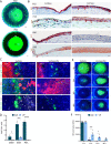
Fig. 2. YAP inhibitor perturbs LSC function in vivo.
A K15-GFP transgenic animals were sacrificed at P15 or P60 and the whole eye merged bright field and fluorescent images are shown. Green and white arrows indicate the limbus and cornea region, respectively. B Immunohistochemistry was performed on paraffin sections of the P15 or P60 wild-type mouse cornea using the indicated antibodies. Arrowheads indicate nuclear YAP or K15 expression in basal cells. C, D Daily sub-conjunctival injections (20 µl) of Verteporfin (20 µM) or vehicle (control) were performed for 4-days to adult K15-GFP transgenic mice. EdU was injected 6 h before eyes were enucleated and prepared for wholemount immunostaining. CD63 + outer quiescent LSCs, K15-GFP + inner active LSCs or K12 + corneal epithelial differentiated cells are shown, among them, EdU+ cells that are in mitosis and were quantified in (D) (n = 3 biological replicates). The basal layer was the focus point in the image acquisition. E,F The corneal epithelium of P60 mice was surgically removed using Algerbrush and animal were treated topically with Verteporfin (20 µM) or vehicle. At indicated time points, fluorescein dye staining was pictured (E) to follow epithelial healing and quantification is shown in (F) (n = 4 biological replicates). All data are represented as mean ± standard error of mean. Statistical significance was assessed by t-test (*, p < 0.05; ***, p < 0.001, ****, p < 0.0001). Immunohistochemistry and immunostaining data are representative from 3 to 5 biological replicates. Nuclei were detected by DAPI counterstaining. Scale bars are 500 µm (A) and the rest are 50 µm. Le Lens.
Fig. 3
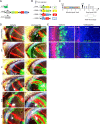
Fig. 3. YAP inhibitor perturbs dedifferentiation.
A Schematic illustration of triple transgenic animals (K15-GFP; Brainbow2.1; K14-CreERT2) used. B Examples of potential reorganization of the Brainbow cassette after tamoxifen induction. CE As illustrated in (C), 2–3 month old triple transgenic animals were injected with tamoxifen (Tam) to induce the random and irreversible expression of Confetti reporters (B). CE Four-months post Tam induction, fully developed Confetti + (RFP + ) limbal radial stripes were evident (see unwounded). Next, total limbal epithelial removal (LER, dotted white lines) performed by Algerbrush (see LER) and mice were sub-conjunctival injected with Verteporfin (VP, 20 µM) or control (Vehicle). For accurate clonal tracking over time, eyes with 2–3 RFP+ stripes were pictured in live animals (D). In vehicle treated mice, Confetti + (RFP+) corneal-committed cells repaired the denuded limbus by day 1 (D1) post LER and re-expressed K15-GFP by D10. In Verteporfin treated cornea, the recovery of K15-GFP was negligible. E On D15 post LER, eyes were enucleated and wholemount immunostaining for markers of the quiescent outer (GPHA2, CD63) LSCs or inner active (K15-GFP) LSCs are shown. Data represent 7 biological replicates. Nuclei were detected by DAPI counterstaining. Scale bars are 50 µm.
Fig. 4
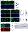
Fig. 4. Manipulation of niche rigidity impairs SC phenotype and corneal integrity.
A Atomic force microscopy measurement of the rigidity of the limbus (L) and the cornea (C) in wild type (WT) and Lox transgenic (LoxOE) mice at post-natal day 15 mice (P15) and P60 (the higher the Eapp the stiffer the tissue). Mean ± SD of the apparent Elastic modulus (in kPa). The plot shows the merge of three experiments performed on as many independent samples, using the same atomic force microscopy setup (deflection sensibility and cantilever spring constant). B Immunohistochemistry using anti-YAP antibody on paraffin sections of P60 cornea of the WT and LoxOE mice. Red arrows indicate nYAP and black arrows indicate cYAP in the limbus region. C Bright field binocular images of the eyes of the WT and LoxOE are shown. LoxOE corneas often display peripheral and/or central opacification and neovascularization (white arrows). D Wholemount immunostaining for conjunctiva (K4), cornea (K12) and immune cell (CD4) markers of WT and LoxOE cornea. Staining represents 3 biological replicates and statistical significance was assessed by t-test (*, p < 0.05; ***, p < 0.001, ****, p < 0.0001). Scale bars are 50 µm. Cj Conjunctiva, Peri Peripheral cornea.
Fig. 5
Fig. 5. Corneal rigidity induces differentiation and inhibits nuclear localization of YAP.
Primary human limbal epitheial cells were grown on a silicone substrate that mimics the rigidity of the human limbus (LR, 8 kPa) or cornea (CR, 20 kPa), coated with fibronectin and cultured for 4 days. Expression of stem/progenitor (K15, P63) or differentiation (K3, K12) genes was tested by quantitative real time PCR (qPCR) (A) or immunostaining with the indicated antibodies (B) and mean nuclear-to-cytoplasmic YAP intensity ratio was quantified (C). DF Primary limbal epithelial cells were grown on silicone substrate that mimics corneal rigidity, were cultured for 4 days with Blebbistatin (Bleb) or Vehicle (Veh), the expression of the indicated markers was tested by immunostaining (D), mean nuclear-to-cytoplasmic YAP intensity ratio was quantified (E) and the expression of the indicated genes was tested by qPCR (F). G Primary human limbal epithelial cells were seeded on pillars that mimic limbal or corneal rigidity overnight. Immunostaining of K15 and F-actin (phalloidin) is shown, and measurements of forces generated by K15-positive and K15-negative cells (undifferentiated and differentiated; see Methods) are shown in (G,H), n = 44 and 52 cells respectively from three independent experiments. The mean nuclear-to-cytoplasmic YAP intensity ratio in (C, E) is shown by Tukey box-and-whisker. The force generated by LSCs is shown in (G) by Tukey box-and-whisker plot followed by Mann–Whitney test. The qPCR data were normalized to the housekeeping gene and is presented (mean ± standard error of mean, n = 4 biological replicates) as fold increase compared to control sample and statistical analysis was performed by t-test (*, p < 0.05; **, p < 0.01; ***, p < 0.001, ****, p < 0.0001). Immunostaining data are representative from 3 biological replicates. Nuclei were detected by DAPI counterstaining. Scale bars are 5 µm (G) and the rest are 50 µm.
Fig. 6
Fig. 6. Stiffness-induced differentiation is mediated by SMAD2/3.
AH Primary human LSCs were grown on limbal or corneal rigidity and treated with Rho-Activator (Rho-Act) or Verteporfin (VP) for 4 days whereas treatment with TGFβ ligand or TGFβ pathway inhibitor (SB431542), or with control vehicle for 2 days. Cells were then fixed and immunostained with the indicated antibodies (A, C, E, G) and quantification of mean nuclear SMAD2/3 (nSMAD) (D), % of K12 + cells (F), or mean nuclear-to-cytoplasmic YAP intensity ratio and mean pLATS1/2 intensity is shown (H). I Immunohistochemistry of SMAD2/3 was performed on paraffin sections of the P60 mouse cornea. The regions of the limbus and corneal periphery are shown. Mean nuclear-to-cytoplasmic YAP intensity ratio, mean pLATS1/2 intensity, and mean nuclear SMAD2/3 intensity is shown by the Tukey box-and-whisker plot followed by Mann–Whitney test. % of K12 positive cells is shown by Tukey box-and-whisker plot followed by t-test with Welch’s correction (*, p < 0.05; **, p < 0.01; ***, p < 0.001, ****, p < 0.0001). Immunostaining, and immunohistochemistry, data are representative from 3 biological replicates. Nuclei were detected by DAPI counterstaining and scale bars are 50 µm.
Fig. 7
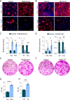
Fig. 7. Inhibition of actomyosin contractility or TGFβ pathway attenuates LSC differentiation on plastic.
AH Primary human LSCs were grown on plastic and treated with Blebbistatin (Bleb) or TGFβ pathway inhibitor (SB431542) or vehicle (control) for 4 days and the expression of the indicated markers was tested by immunostaining (A,B), quantitative real time PCR analysis (C,D) or cells were subjected to clonogenicity assay and colonies were visualized by Rhodamine stain (E,F) and the number of the colony was quantified (G,H) (n = 3). Real-time data was normalized to the housekeeping gene and is presented (mean ± standard error of mean, n = 4 biological replicates) as fold increase compared to control sample and statistical analysis was performed by t-test (*, p < 0.05; **, p < 0.01; ***, p < 0.001, ****, p < 0.0001). Immunostaining, data are representative from 3 biological replicates. Nuclei were detected by DAPI counterstaining. Scale bars are 50 µm.

References

    1. Spradling A, Drummond-Barbosa D, Kai T. Stem cells find their niche. Nature. 2001;414:98–104. doi: 10.1038/35102160. - DOI - PubMed
    1. Fuchs E, Tumbar T, Guasch G. Socializing with the Neighbors: Stem Cells and Their Niche. Cell. 2004;116:769–78. doi: 10.1016/S0092-8674(04)00255-7. - DOI - PubMed
    1. van Es JH, Sato T, van de Wetering M, Lyubimova A, Yee Nee AN, Gregorieff A, et al. Dll1+ secretory progenitor cells revert to stem cells upon crypt damage. Nat Cell Biol [Internet] 2012;14:1099–104. doi: 10.1038/ncb2581. - DOI - PMC - PubMed
    1. Rompolas P, Mesa KR, Greco V. Spatial organization within a niche as a determinant of stem-cell fate. Nat [Internet] 2013;502:513–8. doi: 10.1038/nature12602. - DOI - PMC - PubMed
    1. Nasser W, Amitai-Lange A, Soteriou D, Hanna R, Tiosano B, Fuchs Y, et al. Corneal-Committed Cells Restore the Stem Cell Pool and Tissue Boundary following Injury. Cell Rep. [Internet] 2018;22:323–31. doi: 10.1016/j.celrep.2017.12.040. - DOI - PubMed

Publication types

Substances