. 2023 Apr 24;14(1):2342.
doi: 10.1038/s41467-023-38115-2.

TMEM25 inhibits monomeric EGFR-mediated STAT3 activation in basal state to suppress triple-negative breast cancer progression

Affiliations

TMEM25 inhibits monomeric EGFR-mediated STAT3 activation in basal state to suppress triple-negative breast cancer progression

Jing Bi et al. Nat Commun. .

Abstract

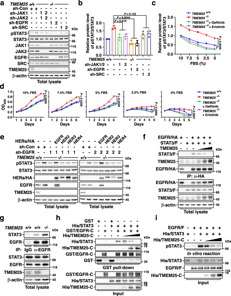
Triple-negative breast cancer (TNBC) is a subtype of breast cancer with poor outcome and lacks of approved targeted therapy. Overexpression of epidermal growth factor receptor (EGFR) is found in more than 50% TNBC and is suggested as a driving force in progression of TNBC; however, targeting EGFR using antibodies to prevent its dimerization and activation shows no significant benefits for TNBC patients. Here we report that EGFR monomer may activate signal transducer activator of transcription-3 (STAT3) in the absence of transmembrane protein TMEM25, whose expression is frequently decreased in human TNBC. Deficiency of TMEM25 allows EGFR monomer to phosphorylate STAT3 independent of ligand binding, and thus enhances basal STAT3 activation to promote TNBC progression in female mice. Moreover, supplying TMEM25 by adeno-associated virus strongly suppresses STAT3 activation and TNBC progression. Hence, our study reveals a role of monomeric-EGFR/STAT3 signaling pathway in TNBC progression and points out a potential targeted therapy for TNBC.


Conflict of interest statement

The authors declare no competing interests.

Figures

Fig. 1
Fig. 1. TMEM25 interacts with EGFR.
a, b TMEM25 interacts with EGFR endogenously. Cell lysates from MDA-MB-231 cells were subjected to anti-TMEM25 IP followed by immunoblotting to detect associated EGFR (a) or to anti-EGFR IP followed by immunoblotting to detect associated TMEM25 (b). c TMEM25 colocalizes with EGFR at plasma membrane. MDA-MB-231 cells transfected with C-terminal green fluorescent protein (GFP)-tagged TMEM25 (TMEM25/GFP) were subjected to immunofluorescence assay to detect localization of TMEM25/GFP (green) and endogenous EGFR (red). The nuclei were stained with DAPI (blue). The scale bars indicate 10 μm. d TMEM25 interacts with EGFR via both extracellular domain and cytosolic domain. HEK293T cells transfected with indicated combinations of full length (F), N-terminal extracellular/transmembrane domains (N), or cytosolic domain (C) of TMEM25/GFP and C-terminal Flag-tagged EGFR (EGFR/F) were subjected to anti-Flag IP followed by immunoblotting assay to detect associated TMEM25. e Direct interaction between cytosolic domains of TMEM25 and EGFR. Bacterially expressed and purified His-tagged TMEM25 cytosolic domain (His/TMEM25-C) and GST-tagged EGFR cytosolic domain (GST/EGFR-C) were subjected to GST pull-down assay. f TMEM25 interacts with EGFR but not HER2, HER3, or HER4. HEK293T cells transfected with indicated combinations of C-terminal hemagglutinin (HA)-tagged EGFR, HER2, HER3, or HER4 (HERs/HA) and C-terminal Flag-tagged TMEM25 (TMEM25/F) were subjected to anti-Flag IP followed by immunoblotting assay to detect associated HERs. The blotting experiments are representative of at least three biologically independent replicates (a, b, df). Source data are provided as a Source Data file.
Fig. 2
Fig. 2. TMEM25 inhibits TNBC progression.
a Effect of TMEM25 on growth of MDA-MB-231 cells. Cell viability of wild-type (TMEM25+/+), TMEM25 knockout (TMEM25−/−), and Flag-tagged TMEM25 overexpressing (TMEM25/F) MDA-MB-231 cells were determined using CCK-8 assay. Data were presented as mean ± SEM of three individual experiments. P values were determined by two-way ANOVA followed by Tukey test. b Effect of TMEM25 on colony formation of MDA-MB-231 cells. TMEM25+/+, TMEM25−/−, and TMEM25/F MDA-MB-231 cells were subjected to soft agar assay. Scale bar indicates 100 μm. At least three biological replicates were performed for each condition. ce TMEM25 suppresses xenograft tumor growth of MDA-MB-231 cells in nude mice. TMEM25+/+, TMEM25−/−, or TMEM25/F MDA-MB-231 cells were orthotopically injected into the mammary fat pad of nude mice to observe tumor formation. Tumor volumes were measured and plotted as mean ± SEM (n = 8 animals per group). P values were determined by two-way ANOVA followed by Tukey test (c). The tumors were obtained 13 days after tumor formation by sacrificing the mice (d), weighted and plotted as mean ± SEM (n = 8 animals per group). P values were determined by one-way ANOVA followed by Tukey test (e). fh TMEM25 suppresses spontaneous TNBC tumor growth in MMTV-PyMT mice. Tumor volumes in TMEM25+/+, TMEM25−/−, or TMEM25 transgenic (TMEM25wt/tg) MMTV-PyMT mice were measured at same ages and plotted as mean ± SEM of a pool of indicated number of mice per group. P values were determined by two-way ANOVA followed by Tukey test (f). Representative tumors were obtained from one TMEM25+/+, TMEM25−/−, or TMEM25wt/tg MMTV-PyMT mouse at age of 100 days (g). Tumors obtained from 100 days aged TMEM25+/+, TMEM25/− and TMEM25wt/tg MMTV-PyMT mice were weighted and plotted as mean ± SEM (n = 10 animals per group). P values were determined by one-way ANOVA followed by Tukey test (h). i Effect of TMEM25 on survival of MMTV-PyMT mice. Overall survival rates of TMEM25+/+, TMEM25−/−, or TMEM25wt/tg MMTV-PyMT mice in (f) were determined. P values were determined by log-rank (Mantel-Cox) test, 95% confidence interval of ratio. Source data are provided as a Source Data file.
Fig. 3
Fig. 3. TMEM25 negatively regulates STAT3 signaling.
a Knockout of TMEM25 induces phosphorylation of STAT3 in MEF cells. Primary MEF cells derived from TMEM25+/+ or TMEM25/− mice were serum-starved overnight and then treated with 100 ng/ml EGF for the indicated time before subjected to immunoblotting assay. b Overexpression of TMEM25 inhibits EGF treatment-induced phosphorylation of STAT3 in MEF cells. Primary MEF cells derived from TMEM25+/+ or TMEM25wt/tg mice were treated and analyzed as in (a). c Knockout of TMEM25 increases mRNA levels of representative STAT3 target genes. The mRNA levels of indicated genes in TMEM25+/+ or TMEM25−/− MDA-MB-231 cells were measured by real-time quantitative PCR using GAPDH as an internal control. Data are presented as mean ± SEM of three independent experiments. P values were determined by two-tailed unpaired Student’s t test. d, e TMEM25 inhibits STAT3 phosphorylation in the spontaneous breast tumors in MMTV-PyMT mice. Representative tumors from TMEM25+/+, TMEM25−/−, or TMEM25wt/tg MMTV-PyMT mice were subjected to immunoblotting assay (d) or IHC assay (e) as indicated. Scale bar indicates 100 μm. The blotting experiments are representative of at least three biologically independent replicates (a, b, d). Source data are provided as a Source Data file.
Fig. 4
Fig. 4. TMEM25 inhibits EGFR-mediated STAT3 phosphorylation.
a, b EGFR is required for TMEM25 knockout-induced STAT3 phosphorylation. Wild-type or TMEM25 −/− MDA-MB-231 cells transduced with lentivirus encoding control shRNA (sh-Con), shRNAs against JAK1 (sh-JAK1-1/2), JAK2 (sh-JAK2-1/2), EGFR (sh-EGFR-1/2), or SRC (sh-SRC-1/2) as indicated were serum starved overnight before subjected to immunoblotting assay (a). Relative pSTAT3/STAT3 ratios were determined and plotted as mean ± SEM (n = 3 individual experiments) (b). P values were determined by one-way ANOVA followed by Tukey test. c TMEM25 knockout enhances STAT3 phosphorylation via EGFR in different serum conditions. Wild-type or TMEM25 −/− MDA-MB-231 cells grown in indicated FBS concentrations were treated with/without EGFR inhibitor Gefitinib (20 μM, 24 h) or Erlotinib (20 μM, 4 h) before subjected to immunoblotting assay. Relative pSTAT3/STAT3 ratios were determined and plotted as mean ± SEM (n = 3 individual experiments). P values were determined by one-way ANOVA followed by Tukey test. d TMEM25 knockout-promoted cell proliferation and survival requires EGFR activity. Cell viabilities of wild-type and TMEM25 −/− MDA-MB-231 cells cultured in indicated concentrations of FBS with/without Gefitinib (20 μM) or Erlotinib (20 μM) were determined and plotted as mean ± SEM (n = 3 individual experiments). P values were determined by one-way ANOVA followed by Tukey test. e EGFR induces STAT3 phosphorylation in TMEM25 /− cells. TMEM25 −/− MDA-MB-231 cells with indicated combinations of sh-Con or sh-EGFR-1/2 and EGFR/HA, HER2/HA, HER3/HA, or HER4/HA were subjected to immunoblotting assay. f TMEM25 blocks EGFR and STAT3 interaction. HEK293T cells transfected with indicated combinations of TMEM25, EGFR/HA, or STAT3/F were subjected to Co-IP assay. g TMEM25 knockout enhances EGFR and STAT3 interaction. TMEM25 +/+ or TMEM25 −/ MDA-MB-231 cells were subjected to Co-IP assay. h TMEM25 inhibits STAT3 and EGFR interaction in vitro. Bacterially produced His-tagged STAT3 (His/STAT3), His-tagged TMEM25 cytosolic domain (His/TMEM25-C), and GST-tagged EGFR cytosolic domain (GST/EGFR-C) were subjected to GST pull-down assay. i TMEM25 blocks EGFR-mediated STAT3 phosphorylation in vitro. Affinity-purified EGFR/F from HEK293T and bacterially produced His/STAT3 and His/TMEM25-C were subjected to in vitro kinase assay. The blotting experiments are representative of at least three biologically independent replicates (a, ej). Source data are provided as a Source Data file.
Fig. 5
Fig. 5. Monomeric EGFR is able to phosphorylate STAT3 in the absence of TMEM25.
a EGFR-V948R mutant does not form dimer. HEK293T cells transfected with indicated combinations of EGFR/HA, EGFR/F, EGFR-V948R/HA, or EGFR-V948R/F were subjected to Co-IP assay. b EGFR-V948R is able to phosphorylate STAT3. Lentivirus encoding Flag-tagged wild-type (WT) EGFR, EGFR-V948R, or EGFR-D837N were transduced into TMEM25 and EGFR double knockout (TMEM25 /−EGFR/−) MDA-MB-231 cells as indicated. The cells were serum starved overnight and then subjected to immunoblotting assay. c EGFR-V948R binds STAT3. HEK293T cells transfected with indicated combinations of STAT3/F and EGFR/HA (WT or V948R) were subjected to Co-IP assay. d EGFR-V948R can directly phosphorylate STAT3 in vitro. Exogenously expressed EGFR/F (WT, V948R, or D837N) in EGFR−/− MDA-MB-231 cells were serum starved overnight and then affinity purified for subjecting to in vitro kinase assay with bacterially produced STAT3. e Phosphorylation of Y1068 or Y1086 is not essential for EGFR-mediated phosphorylation of STAT3 in the absence of TMEM25. Lentivirus encoding Flag-tagged wild-type EGFR, EGFR-Y1068F, EGFR-Y1086F, or EGFR-Y1068,1086F (2YF) were transduced into TMEM25−/−EGFR−/ MDA-MB-231 cells as indicated. The cells were serum starved overnight and then subjected to immunoblotting assay. f EGFR-Y1068F, EGFR-Y1086F, and EGFR-2YF can phosphorylate STAT3 in vitro. Exogenously expressed EGFR/F (WT, Y1068F, Y1086F, or 2YF) in EGFR−/− MDA-MB-231 cells were serum starved overnight and then affinity purified for subjecting to in vitro kinase assay with bacterially produced STAT3. The blotting experiments are representative of at least three biologically independent replicates (af). Source data are provided as a Source Data file.
Fig. 6
Fig. 6. TMEM25 expression and mutations in clinical samples and potential therapeutic application of TMEM25 in TNBC treatment.
a TMEM25 protein levels are frequently decreased and negatively correlated with phospho-STAT3 levels in TNBC. TMEM25 and phospho-STAT3 levels in 28 human TNBC samples were determined and their correlation coefficient r value and p value were determined by Spearman’s rank correlation analysis and two-tailed unpaired t test. bd Wild-type but not R326W or L338F TMEM25 suppresses tumor growth in MMTV-PyMT mice. MMTV-PyMT mice were orthotopically injected into mammary fat pad once at 8 weeks old with control AAV, or AAV encoding TMEM25 (WT, R326W, or L338F). Tumor volumes were measured and plotted as mean ± SEM (n = 8 animals per group). P values were determined by two-way ANOVA followed by Tukey test (b). Representative tumors obtained from one mouse injected with control AAV or AAV encoding indicated TMEM25 were presented (c). All tumors were weighted and plotted as mean ± SEM (n = 8 animals per group). P values were determined by one-way ANOVA followed by Tukey test (d). e Supply of wild-type but not R326W or L338F TMEM25 prolongs survival time of MMTV-PyMT mice. Overall survival rates of MMTV-PyMT mice injected with control AAV or AAV encoding indicated TMEM25 were determined (n = 8 animals per group). P values were determined by log-rank (Mantel-Cox) test, 95% confidence interval of ratio. fh AAV-delivered TMEM25 suppresses tumor growth in patient-derived xenograft (PDX) models. NCG mice (6–8 weeks old) were intratumorously injected with control AAV, or AAV encoding TMEM25 every other day. Tumor volumes were measured and plotted as mean ± SEM (n = 6 animals per group). P values were determined by two-way ANOVA followed by Tukey test (f). Tumors obtained from NCG mice 22 days after transplantation and injected with control AAV or AAV encoding TMEM25 were presented (g). All tumors were weighted and plotted as mean ± SEM (n = 6 animals per group). P values were determined by two-tailed unpaired Student’s t test (h). i A schematic model of TMEM25/EGFR/STAT3 signaling. Source data are provided as a Source Data file.

References

    1. Burgess AW. EGFR family: structure physiology signalling and therapeutic targets. Growth Factors. 2008;26:263–274. doi: 10.1080/08977190802312844. - DOI - PubMed
    1. Zhang X, Gureasko J, Shen K, Cole PA, Kuriyan J. An allosteric mechanism for activation of the kinase domain of epidermal growth factor receptor. Cell. 2006;125:1137–1149. doi: 10.1016/j.cell.2006.05.013. - DOI - PubMed
    1. Jura N, et al. Mechanism for activation of the EGF receptor catalytic domain by the juxtamembrane segment. Cell. 2009;137:1293–1307. doi: 10.1016/j.cell.2009.04.025. - DOI - PMC - PubMed
    1. Ferguson KM. Structure-based view of epidermal growth factor receptor regulation. Annu. Rev. Biophys. 2008;37:353–373. doi: 10.1146/annurev.biophys.37.032807.125829. - DOI - PMC - PubMed
    1. Normanno N, et al. Epidermal growth factor receptor (EGFR) signaling in cancer. Gene. 2006;366:2–16. doi: 10.1016/j.gene.2005.10.018. - DOI - PubMed

Publication types