. 2023 Jun;24(6):941-954.
doi: 10.1038/s41590-023-01499-w. Epub 2023 Apr 24.

High-dimensional analysis of 16 SARS-CoV-2 vaccine combinations reveals lymphocyte signatures correlating with immunogenicity

Collaborators, Affiliations

High-dimensional analysis of 16 SARS-CoV-2 vaccine combinations reveals lymphocyte signatures correlating with immunogenicity

Nicolás Gonzalo Nuñez et al. Nat Immunol. 2023 Jun.

Abstract

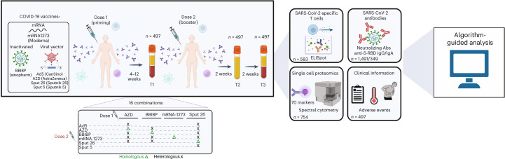
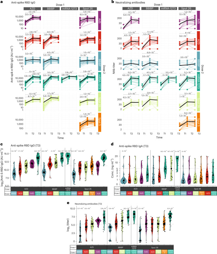
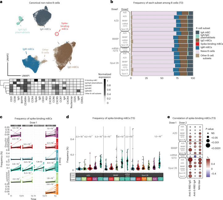
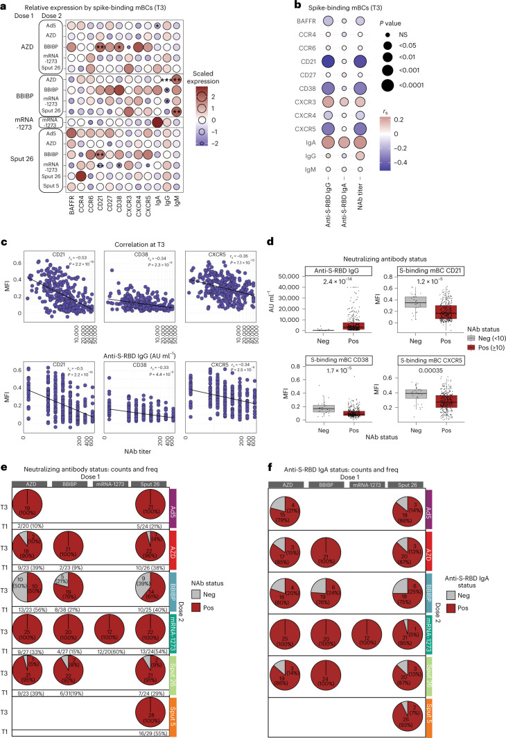
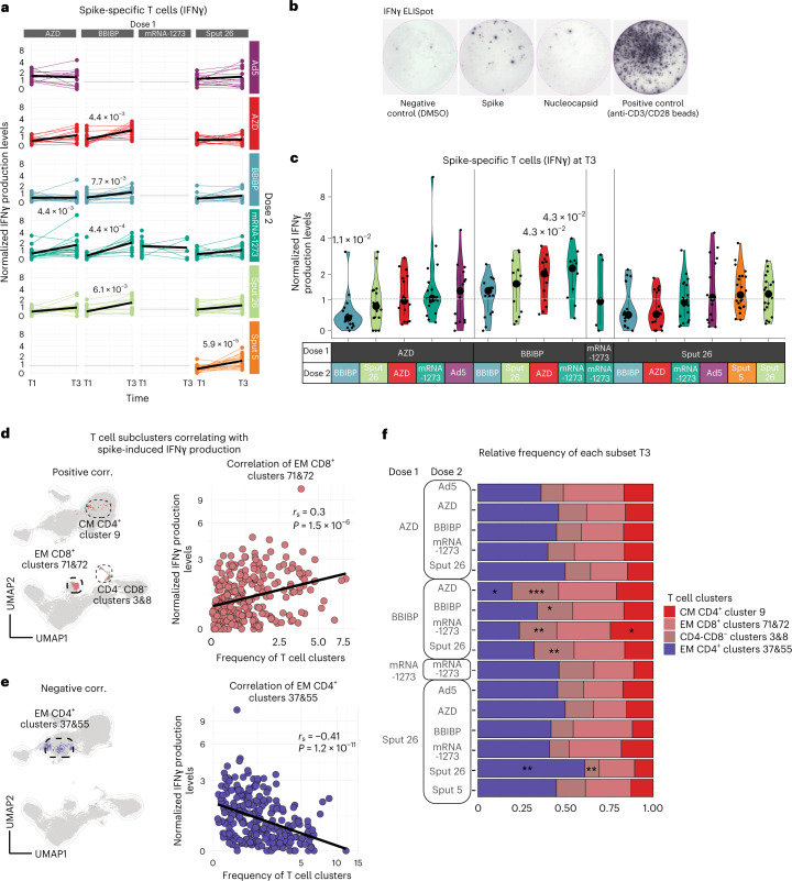
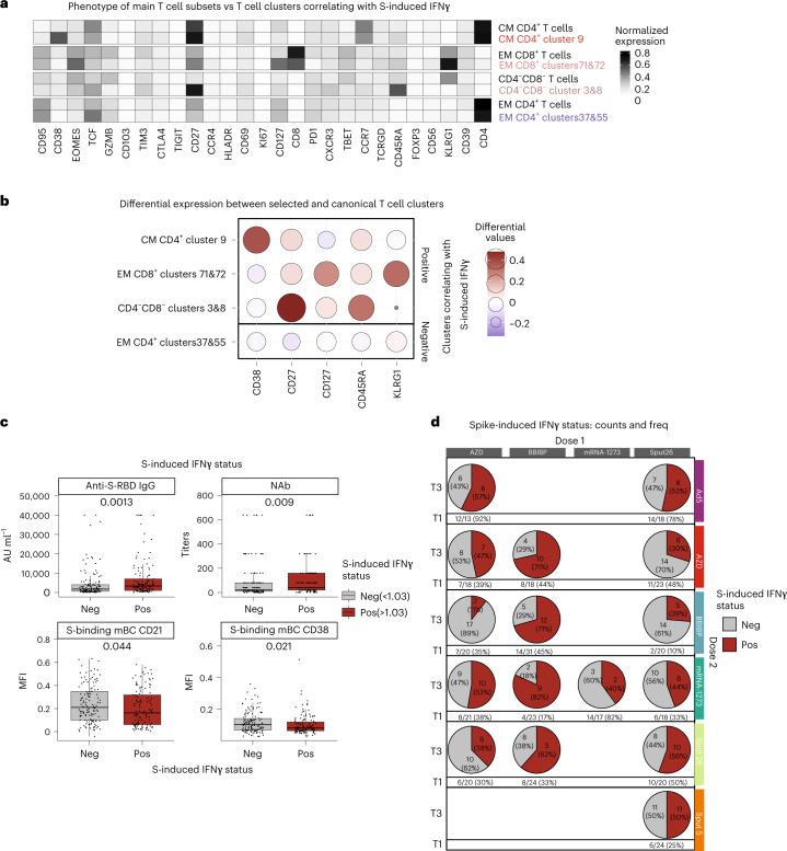
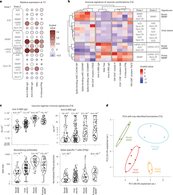
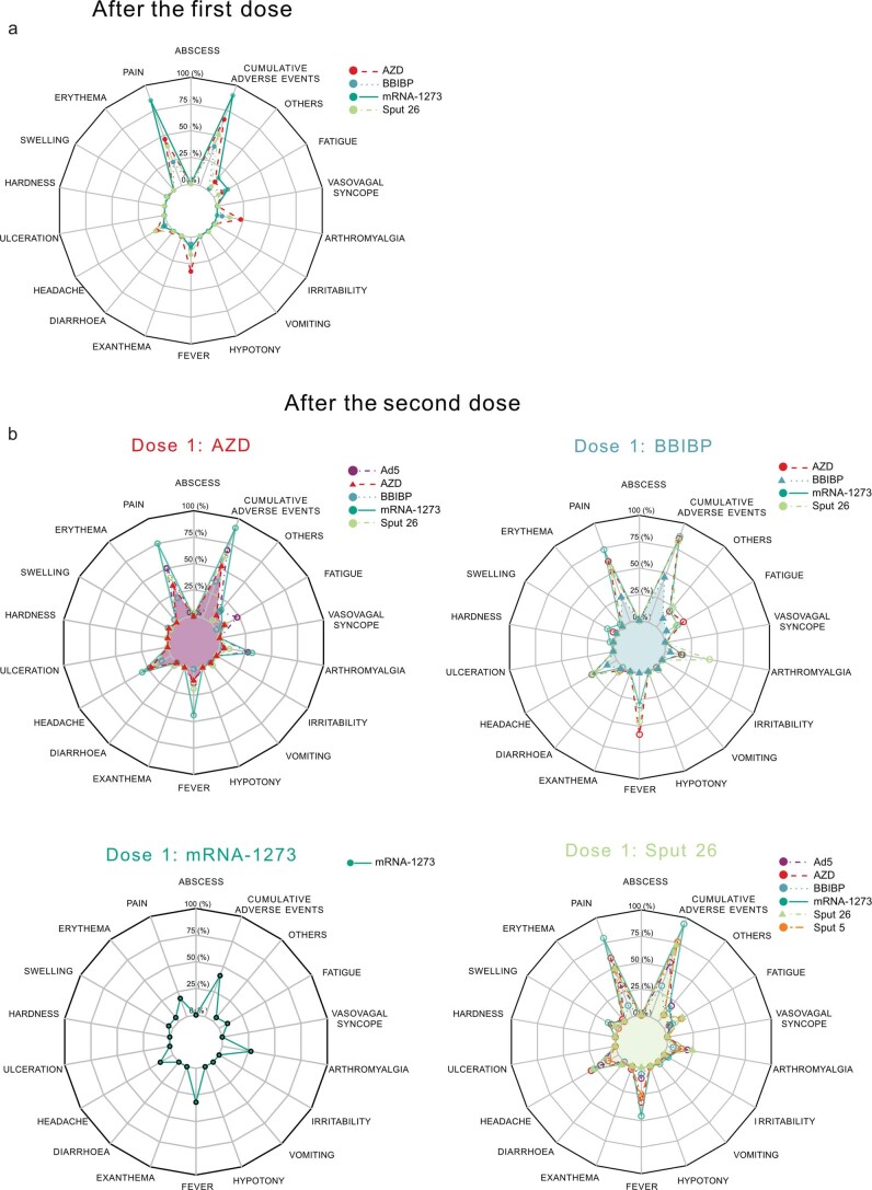
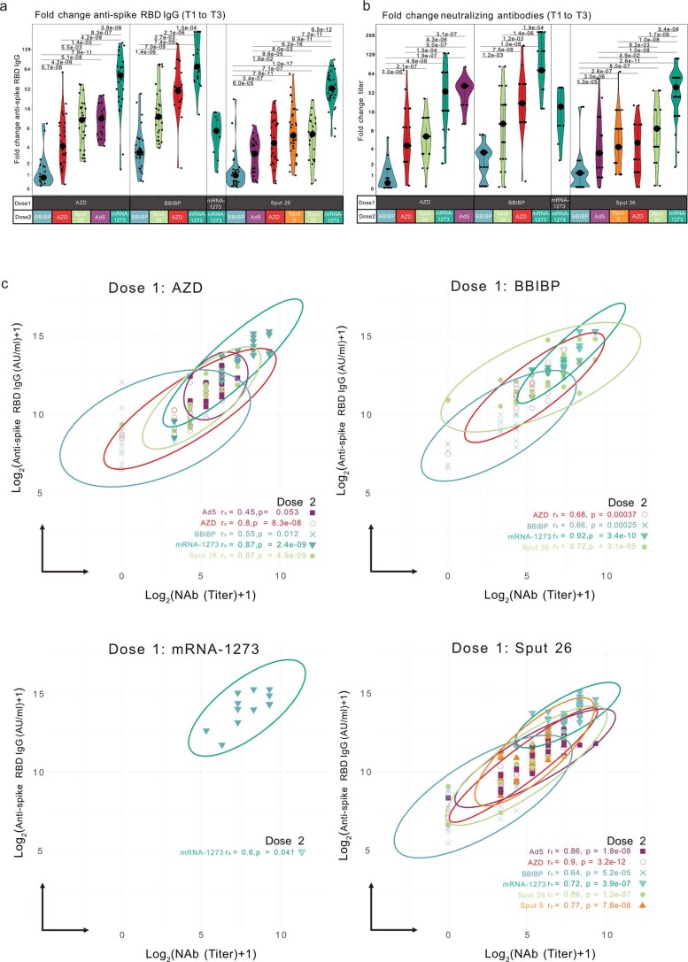
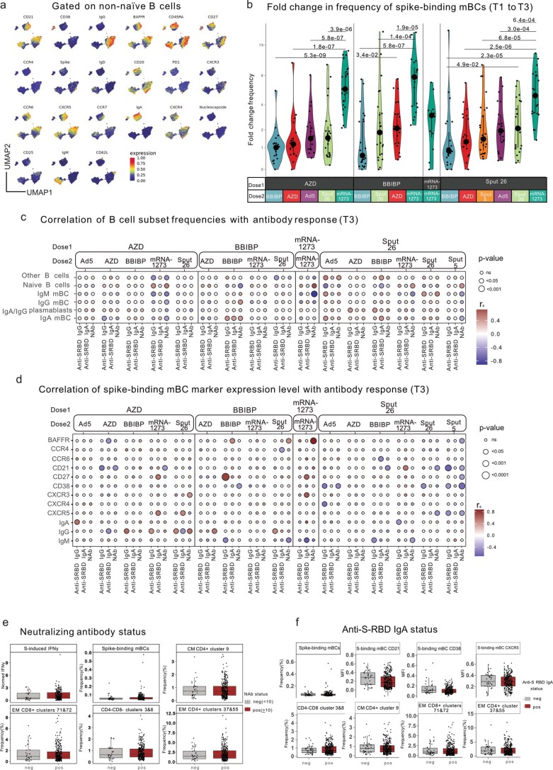
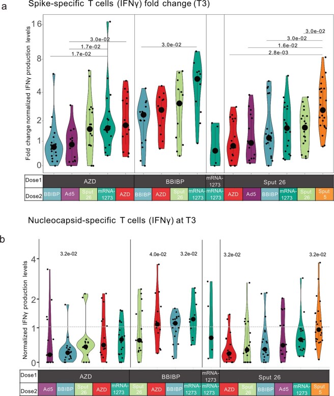
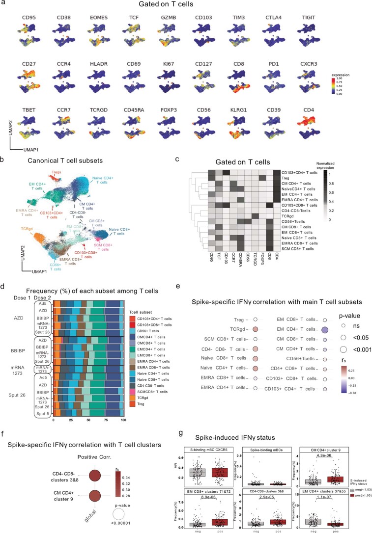
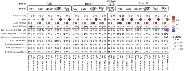
The range of vaccines developed against severe acute respiratory syndrome coronavirus 2 (SARS‑CoV‑2) provides a unique opportunity to study immunization across different platforms. In a single-center cohort, we analyzed the humoral and cellular immune compartments following five coronavirus disease 2019 (COVID-19) vaccines spanning three technologies (adenoviral, mRNA and inactivated virus) administered in 16 combinations. For adenoviral and inactivated-virus vaccines, heterologous combinations were generally more immunogenic compared to homologous regimens. The mRNA vaccine as the second dose resulted in the strongest antibody response and induced the highest frequency of spike-binding memory B cells irrespective of the priming vaccine. Priming with the inactivated-virus vaccine increased the SARS-CoV-2-specific T cell response, whereas boosting did not. Distinct immune signatures were elicited by the different vaccine combinations, demonstrating that the immune response is shaped by the type of vaccines applied and the order in which they are delivered. These data provide a framework for improving future vaccine strategies against pathogens and cancer.

Trial registration: ClinicalTrials.gov NCT04988048.


Conflict of interest statement

All authors declare no competing interests.

Figures

Fig. 1
Fig. 1. Schematic of vaccination regimens and data analysis pipeline.
Participants were vaccinated with one of sixteen COVID-19 vaccine combination regimens and donated blood at timepoints T1 (4–12 weeks after dose 1), T2 (2 weeks after dose 2) and T3 (4 weeks after dose 2). PBMCs collected at T1 and T3 were used for IFNγ ELISpot assays and high-dimensional spectral flow cytometry analysis. Sera and plasma collected at T1, T2 and T3 were analyzed for anti-S-RBD IgG levels and NAb titers, respectively. Anti-S-RBD IgA levels were measured at T3. Participants were monitored for adverse events. The data collected were analyzed using dimensionality reduction, FlowSOM-based clustering algorithms and statistical testing. IFNγ, interferon-gamma.
Fig. 2
Fig. 2. Antibody response in participants after vaccination with various regimens.
a,b, Longitudinal (a) anti-S-RBD IgG levels (n = 1354) and (b) NAb titers (n = 1355) measured at timepoints T1, T2 and T3. Black lines show the median. ce, Anti-S-RBD IgG levels (n = 420) (c), anti-S-RBD IgA levels (n = 349) (d) and NAb titers (n = 421) at T3 (e). Large black dots depict the median of each group, and the vertical line spans the interquartile range. The horizontal line shows the overall mean of all participants, and P values indicate differences between the respective group and the overall mean of all participants (ce). P values were calculated using the Mann–Whitney–Wilcoxon test and corrected for multiple hypothesis testing with the Benjamini–Hochberg method. Only statistically significant P values (P < 0.05) are shown. Source data
Fig. 3
Fig. 3. Characterization of spike-binding mBCs from participants receiving different vaccine regimens.
a, UMAP showing the FlowSOM-guided manual metaclustering of nonnaive B cells (IgD/IgM) for all vaccine groups (n = 799). The heatmap indicates the median intensity of normalized marker expression (range: 0–1) for the identified B cell subsets. b, Relative frequencies of B cell subsets from each vaccine regimen at T3 (n = 347). c, Longitudinal analysis of spike-binding mBC frequencies among total B cells at timepoints T1 and T3. Black lines show the median (n = 754). d, Frequencies of spike-binding mBCs among total B cells at T3. Large black dots show the median of each group, and the vertical line spans the interquartile range. The horizontal line indicates the overall mean of all participants. P values (shown above groups) indicate differences between the respective group and the overall mean of all participants (n = 347). e, Correlations of spike-binding mBC frequencies with anti-S-RBD IgG levels, anti-S-RBD IgA levels and NAb titers for each vaccine regimen at T3 (n = 347). P values were calculated using the Mann–Whitney–Wilcoxon test and corrected for multiple hypothesis testing with the Benjamini–Hochberg method (c and d). Only statistically significant P values (P < 0.05) are displayed. Color indicates Spearman’s rank correlation coefficient (rs), and the bubble size indicates the P value. Source data
Fig. 4
Fig. 4. Phenotypes of spike-binding mBCs from participants receiving different vaccine regimens.
a, Scaled expression of phenotypic markers by spike-binding mBCs at T3 for each group. P values indicate differences between the respective group and the overall mean of all participants for each marker (n = 347). *P < 0.05, **P < 0.01, ***P < 0.001. b, Correlations between phenotypic marker expression by spike-binding mBCs and antibody response (anti-S-RBD IgG levels, anti-S-RBD IgA levels and NAb titers) across all vaccine regimens at T3. Color indicates Spearman’s rank correlation coefficient (rs), and the bubble size indicates the P value (n = 347). c, Spearman’s rank correlations between CD21, CD38 and CXCR5 expression and anti-S-RBD antibodies and NAb titers (n = 339). d, Participants were classified as negative (<10) or positive (≥10) for NAb titers and mean antibody levels. Spike-binding mBC marker expression was compared between negative and positive participants (n = 347). Boxes bound the IQR divided by the median, and Tukey-style whiskers extend to a maximum of 1.5× IQR beyond the box. Dots are participant data points. P values were calculated using the Mann–Whitney–Wilcoxon test and corrected for multiple hypothesis testing with the Benjamini–Hochberg method (a and d). Only statistically significant P values (P < 0.05) are displayed. e, The count and percentage of participants at T1 and T3 classified as negative or positive responders, grouped by vaccine regimen (n = 407 at T1, n = 347 at T3). f, Counts and percentages of participants negative or positive for anti-S RBD IgA at T3 per vaccine regimen (n = 349). IQR, interquartile range. Source data
Fig. 5
Fig. 5. Spike-specific T cell responses to antigen re-encounter after delivery of various vaccine regimens.
a,b, Normalized IFNγ production after stimulation of PBMCs with SARS-CoV-2 spike peptide pool measured by ELISpot assay. Longitudinal IFNγ response at T1 and T3. Black lines show the median (n = 581) (a). Representative images of one IFNγ ELISpot assay (b). c, Normalized IFNγ responses at T3 after stimulation with SARS-CoV-2 spike peptide pool (n = 255). Large black dots show the median of each group, and the vertical line spans the IQR. The horizontal line indicates the positivity threshold. P values indicate differences between the respective group and the overall mean of all participants. P values were calculated using the Mann–Whitney–Wilcoxon test and the Benjamini–Hochberg method to control for multiple hypothesis testing; only statistically significant P values (P < 0.05) are shown (a and c). d,e, UMAP of the T cell compartment (CD3+) showing the clusters with frequencies (d) positively and (e) negatively correlated with SARS-CoV-2 spike peptide-induced IFNγ response. Scatter plots show the frequencies of the clusters with the (d) highest and (e) lowest Spearman’s rank correlation coefficients (rs) when compared to IFNγ production (n = 255). f, Relative frequency per vaccine combination of the T cell subclusters with the highest positive or negative correlations to the SARS-CoV-2 spike peptide-induced IFNγ levels (n = 347). P values indicate differences between the specific group and the overall mean frequency of all participants of each T cell cluster per vaccine combination and were calculated using the Mann–Whitney–Wilcoxon test and the Benjamini–Hochberg method to control for multiple hypothesis testing; *P < 0.05, **P < 0.01 and ***P < 0.001. Only statistically significant P values (P < 0.05) are shown. Source data
Fig. 6
Fig. 6. Phenotypes of the T cell subclusters with the highest positive and negative correlation to SARS-CoV-2 spike peptide-induced IFNγ production.
a, Heatmap showing the median intensity of normalized marker expression (range: 0–1) for canonical T cell subsets and identified T cell subclusters (n = 347). b, Differential marker expression by the specific T cell subclusters compared to the canonical T cell subsets that positively and negatively correlated with SARS-CoV-2 spike peptide-induced IFNγ responses, filtered for markers with at least 0.2 differential expression. Color and bubble size indicate the differential expression values compared to the canonical cell type (n = 347). c, Participants were classified as negative (<1.03, the detection threshold) or positive (>1.03) for IFNγ response and mean antibody levels and spike-binding mBC marker expression levels were compared between negative and positive participants. Boxes bound the IQR divided by the median, and Tukey-style whiskers extend to a maximum of 1.5× IQR beyond the box. Dots are participant data points (n = 255). d, The count and percentage of participants classified as negative or positive responders, grouped by vaccine regimen at T1 and T3 (n = 328 at T1, n = 255 at T3). Source data
Fig. 7
Fig. 7. Immune signatures of specific vaccine combinations.
a, Scaled and centered levels of neutralizing (NAb) and spike-specific (anti-S-RBD IgG) antibodies as well as SARS-CoV-2 spike peptide-induced T cell IFNγ production for each vaccine combination at T3 (n = 347). b, Scaled and centered values per column of the top humoral and cellular immune features for each immune signature, displayed in a heatmap with k-means clustering applied to the rows and columns (n = 347). c, Anti-S-RBD IgG levels, anti-S-RBD IgA levels, NAb titers and SARS-CoV-2 spike peptide-induced IFNγ production from all participants grouped by immune signature (n = 347). Large black dots depict the median of the group, and the vertical line spans the IQR. P values indicate differences between the respective group and the overall mean of all participants and were calculated using the Mann–Whitney–Wilcoxon test and the Benjamini–Hochberg method to control for multiple hypothesis testing. Only significant P values (P < 0.05) are displayed. d, Principal component analysis of the top humoral and cellular immune features for each immune signature at T3 (n = 347). Source data
Extended Data Fig. 1
Extended Data Fig. 1. Local and systemic adverse events following vaccination.
Reported adverse events and percentage of participants experiencing them are reported per vaccination group a, after the first vaccine dose and b, after the second dose (n = 497). Shaded areas indicate the homologous vaccine combinations. Source data
Extended Data Fig. 2
Extended Data Fig. 2. Anti-S-RBD IgG levels and neutralizing antibody titers are correlated and respond differently to various vaccine combinations.
Fold change in a, anti-S-RBD IgG levels (n = 420) and b, neutralizing antibody titers (n = 421) at T3 compared to the mean per group at T1. Large black dots depict the median of the group, and the vertical line spans the interquartile range. P-values were calculated using the Mann-Whitney-Wilcoxon test and the Benjamini-Hochberg method to correct for multiple hypothesis testing. Only significant P values (P < 0.05) are displayed. c, Correlation between the anti-S-RBD IgG levels and neutralizing antibody titers for every group (n = 420). The log transformation was performed by adding 1 to the value before taking the log2. The Spearman’s rank correlation coefficients (rs) and P values (P) are indicated. Source data
Extended Data Fig. 3
Extended Data Fig. 3. Analysis of the B cell compartment after vaccination.
a, UMAP showing the distribution of markers expressed by non-naïve B cells (IgD-/IgM-) among all vaccine groups (n = 799). b, Fold change of spike-binding mBC frequencies at T3 compared to the mean per group at T1 (n = 347). Large black dots show the median, and the vertical line spans the interquartile range. P-values were calculated using the Mann-Whitney-Wilcoxon test between groups with the same dose 1 and the Benjamini-Hochberg method to correct for multiple hypothesis testing. Only significant P values (P < 0.05) are displayed. c, Correlation among B cell subsets and antibody responses (anti-S-RBD IgG levels, anti-S-RBD IgA levels and neutralizing antibody titers) (n = 347). d, Correlation among spike-binding mBC marker expression levels and antibody responses. Color indicates the Spearman’s rank correlation coefficient (rs), and the circle size indicates the P value (n = 347). e, Participants were classified as negative (< 10) or positive (≥ 10) for neutralizing antibodies (n = 347). f, Participants were positive (>0.32 ng/ml) or negative for anti-S RBD IgA (n = 345). (e,f) The means of each listed parameter were compared between negative and positive responders using the Mann-Whitney-Wilcoxon test and corrected for multiple hypothesis testing with the Benjamini-Hochberg method. Boxes bound the interquartile range (IQR) divided by the median, and Tukey-style whiskers extend to a maximum of 1.5 × IQR beyond the box. Dots are participant data points. Source data
Extended Data Fig. 4
Extended Data Fig. 4. Spike- and nucleocapsid-specific cellular IFNγ responses to antigen re-encounter after different prime-boost vaccine combinations.
a, Fold change (value at T3/group mean at T1) of the IFNγ response after stimulation of PBMCs with SARS-CoV-2 spike peptide pool, measured by ELISpot assay. Statistical tests were performed between groups with the same dose 1 (n = 255). b, Normalized IFNγ responses at T3 after stimulation of PBMCs with SARS-CoV-2 nucleocapsid peptide pool, measured by ELISpot assay (n = 254). The horizontal line indicates the cut-off threshold. P values indicate differences between the respective group and the overall mean of all participants. (a-b) P-values were calculated using the Mann-Whitney-Wilcoxon test and the Benjamini-Hochberg method to correct for multiple hypothesis testing. Only significant P values (P < 0.05) are displayed. Large black dots depict the median of the group, and the vertical line spans the interquartile range. Source data
Extended Data Fig. 5
Extended Data Fig. 5. Canonical T cell subsets and T cell subclusters related to the spike-specific IFNγ response.
a, UMAP showing the distribution of markers expressed by T cells among all vaccine groups (n = 799). b, UMAP showing the FlowSOM-guided manual metaclustering of T cells (CD3 + ) for all vaccine groups combined. c, Heatmap showing the median intensity of normalized marker expression (range 0-1) for each canonical T cell subset (n = 799). d, Frequencies of canonical T cell subsets relative to the total T cells for each vaccine regimen (n = 347). e, Spearman’s rank correlations (rs) of spike peptide-induced T cell response (IFNγ release) with the frequencies of (e) the canonical T cell subsets and f, T cell subclusters (n = 255). g, Participants were classified as negative (< 1.03, the cut-off threshold) or positive (> 1.03) for IFNγ response (n = 255). The means of each listed parameter were compared between negative and positive responders using the Mann-Whitney-Wilcoxon test and corrected for multiple hypothesis testing with the Benjamini-Hochberg method. Only significant P values (P < 0.05) are displayed. Source data
Extended Data Fig. 6
Extended Data Fig. 6. Immune parameters associated with positive and negative humoral and cellular responses.
Spearman’s rank correlations (rs) of the top humoral and cellular immune features with the neutralizing antibody (NAb) titers, anti-S-RBD IgG levels and SARS-CoV-2 spike peptide- induced T cell IFNγ response (n = 347). Source data
Extended Data Fig. 7
Extended Data Fig. 7. Data cleaning for high dimensional flow cytometry data analysis.
Representative flow cytometry gating strategy for data cleaning of a, B and b, T cell panels.

References

    1. Sadarangani M, Marchant A, Kollmann TR. Immunological mechanisms of vaccine-induced protection against COVID-19 in humans. Nat. Rev. Immunol. 2021;21:475–484. doi: 10.1038/s41577-021-00578-z. - DOI - PMC - PubMed
    1. Pozzetto B, et al. Immunogenicity and efficacy of heterologous ChAdOx1–BNT162b2 vaccination. Nature. 2021;600:701–706. doi: 10.1038/s41586-021-04120-y. - DOI - PubMed
    1. Barros-Martins J, et al. Immune responses against SARS-CoV-2 variants after heterologous and homologous ChAdOx1 nCoV-19/BNT162b2 vaccination. Nat. Med. 2021;27:1525–1529. doi: 10.1038/s41591-021-01449-9. - DOI - PMC - PubMed
    1. Pereson MJ, et al. Heterologous gam-covid-vac (sputnik V)/mRNA-1273 (Moderna) vaccination induces a stronger humoral response than homologous sputnik V in a real-world data analysis. Clin. Microbiol. Infect. 2022;1273:382–1388. - PMC - PubMed
    1. Hillus D, et al. Safety, reactogenicity, and immunogenicity of homologous and heterologous prime-boost immunisation with ChAdOx1 nCoV-19 and BNT162b2: a prospective cohort study. Lancet Respir. Med. 2021;9:1255–1265. doi: 10.1016/S2213-2600(21)00357-X. - DOI - PMC - PubMed

Publication types

Associated data