. 2023 Apr 24;24(1):212.
doi: 10.1186/s12864-023-09310-8.

Candidate variants in DNA replication and repair genes in early-onset renal cell carcinoma patients referred for germline testing

Affiliations

Candidate variants in DNA replication and repair genes in early-onset renal cell carcinoma patients referred for germline testing

Elena V Demidova et al. BMC Genomics. .

Erratum in

Abstract

Background: Early-onset renal cell carcinoma (eoRCC) is typically associated with pathogenic germline variants (PGVs) in RCC familial syndrome genes. However, most eoRCC patients lack PGVs in familial RCC genes and their genetic risk remains undefined.

Methods: Here, we analyzed biospecimens from 22 eoRCC patients that were seen at our institution for genetic counseling and tested negative for PGVs in RCC familial syndrome genes.

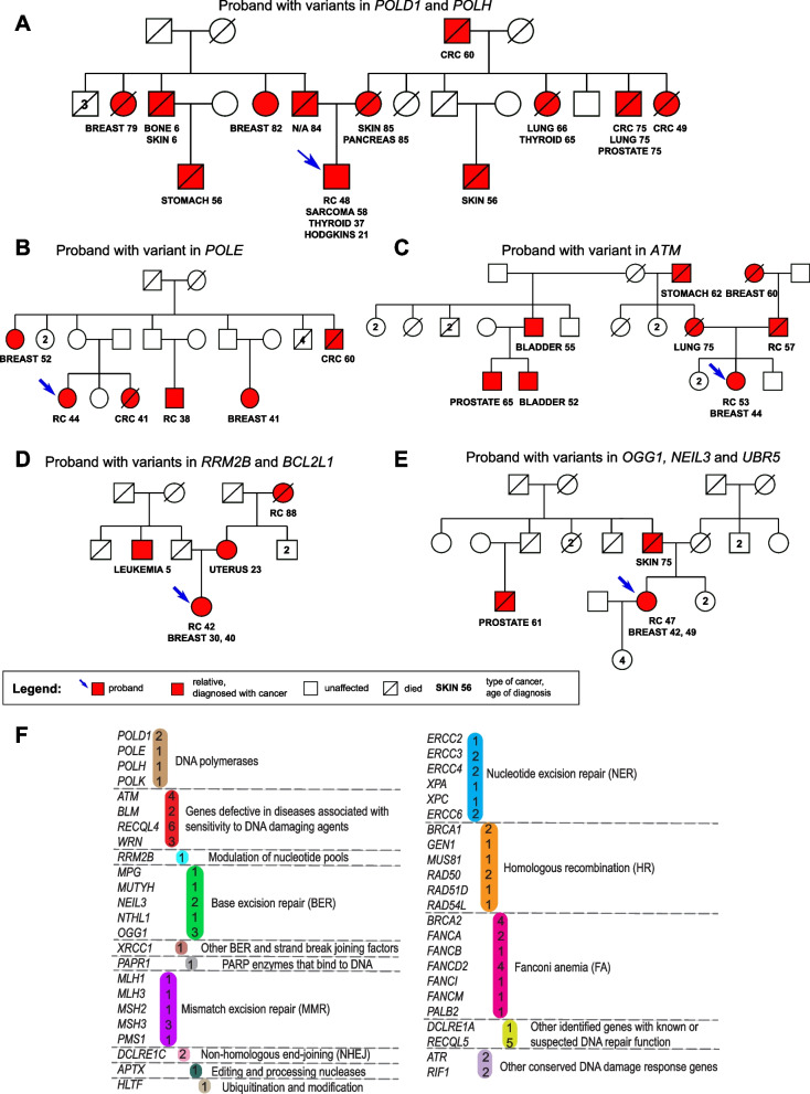
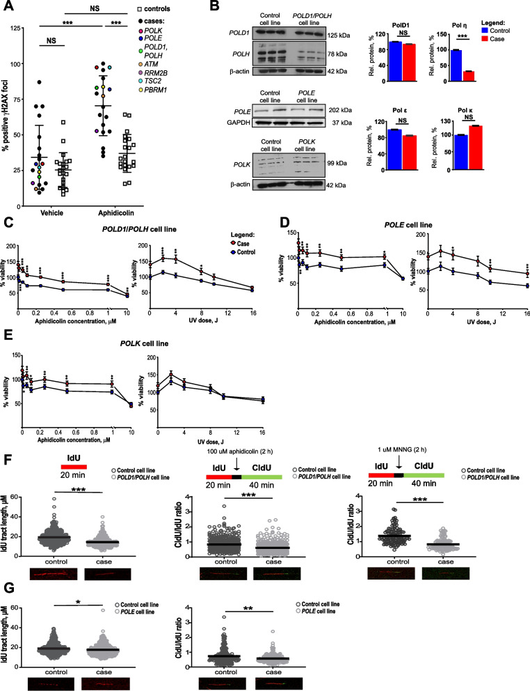
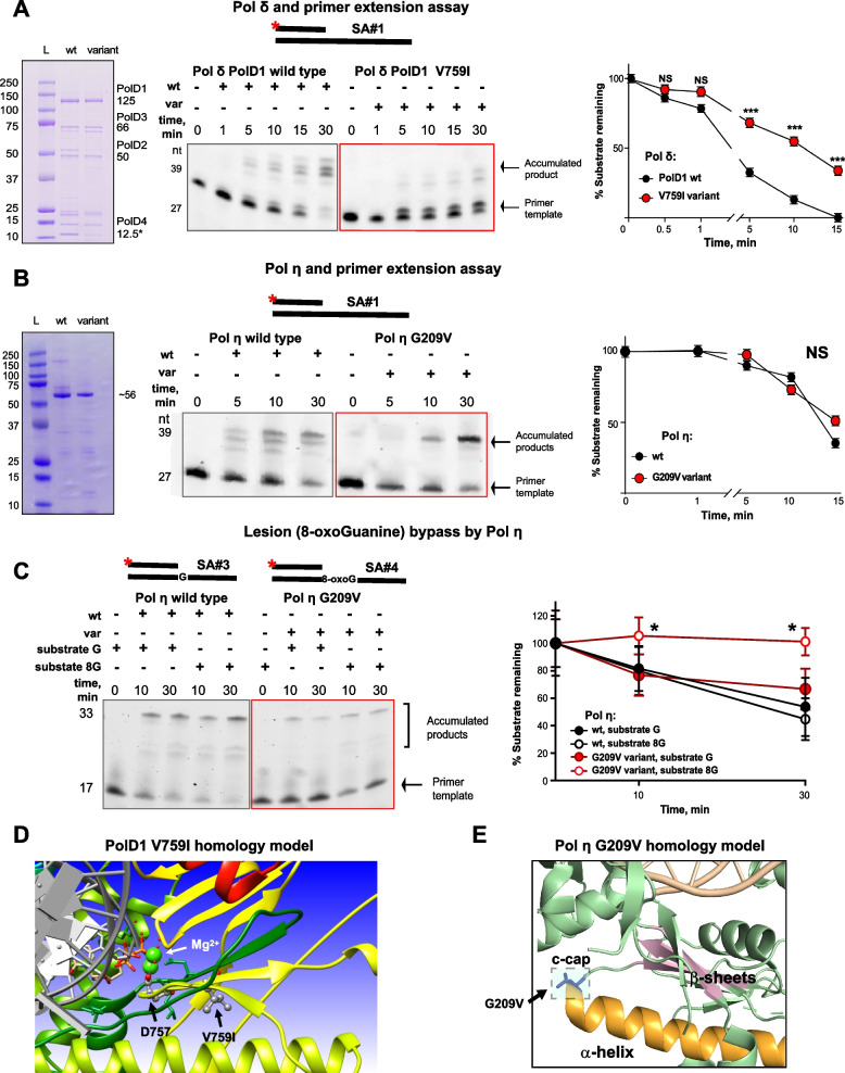
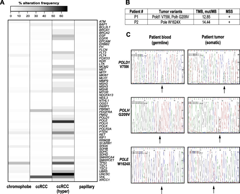
Results: Analysis of whole-exome sequencing (WES) data found enrichment of candidate pathogenic germline variants in DNA repair and replication genes, including multiple DNA polymerases. Induction of DNA damage in peripheral blood monocytes (PBMCs) significantly elevated numbers of [Formula: see text]H2AX foci, a marker of double-stranded breaks, in PBMCs from eoRCC patients versus PBMCs from matched cancer-free controls. Knockdown of candidate variant genes in Caki RCC cells increased [Formula: see text]H2AX foci. Immortalized patient-derived B cell lines bearing the candidate variants in DNA polymerase genes (POLD1, POLH, POLE, POLK) had DNA replication defects compared to control cells. Renal tumors carrying these DNA polymerase variants were microsatellite stable but had a high mutational burden. Direct biochemical analysis of the variant Pol δ and Pol η polymerases revealed defective enzymatic activities.

Conclusions: Together, these results suggest that constitutional defects in DNA repair underlie a subset of eoRCC cases. Screening patient lymphocytes to identify these defects may provide insight into mechanisms of carcinogenesis in a subset of genetically undefined eoRCCs. Evaluation of DNA repair defects may also provide insight into the cancer initiation mechanisms for subsets of eoRCCs and lay the foundation for targeting DNA repair vulnerabilities in eoRCC.

Keywords: DNA repair; DNA replication; Germline; Renal cancer.


Conflict of interest statement

All authors declare that there is no competing interest regarding this manuscript. M.J.H. performs collaborative research (with no funding) with the following: Myriad Genetics, Invitae Corporation, Ambry Genetics, Foundation Medicine, Inc. He also performs collaborative research (with no funding) and is part of a Precision Oncology Alliance funded by Caris Life Sciences (cover travel and meals at meetings). S.A. performs collaborative research (with no funding) with Caris Life Sciences, Foundation Medicine, Inc., Ambry Genetics, and Invitae Corporation. S.A.’s spouse is employed by Akoya Biosciences and has stocks in Akoya Biosciences, HTG Molecular Diagnostics, Abcam Plc., and Senzo Health. S.A., M.J.H., E.A.G., I.G.S. have patents and/or pending patents related to cancer diagnostics/treatment.

Figures

Fig. 1
Fig. 1
Select pedigrees from the eoRCC patient cohort and enrichment of predicted pathogenic variants in DNA repair genes in the cohort. A-E. Pedigrees of eoRCC patients with variants in: A— POLD1 and POLH; B— POLE; C— ATM; D— RRM2B and BCL2L1; E— OGG1, NEIL3 and UBR5. F. Summary of variants in genes and pathways, identified in the cohort. In color—number of variants identified for each gene. For detailed information, see Supplementary tables 1 and 2
Fig. 2
Fig. 2
Cell-based functional analysis revealed defects in DNA repair and DNA replication in lymphocytes from eoRCC patients. A. γH2AX foci immune fluorescence staining in primary PBMCs from eoRCC patients versus matched controls, at baseline or post treatment with aphidicolin (2 h). PBMCs from patients showed statistically significant elevation of γH2AX foci post treatment with aphidicolin. Data were normalized and are presented as percent of positive γH2AX foci. B. Representative Western blots showing expression of PolD1, Pol η, Pol ε and Pol κ in EBV-transformed cell lines carrying variants versus matched controls (without the variants). Data quantification was performed based on 3 independent biological repeats, technical repeats are presented on gels. C-E. Relative viability of EBV-transformed cell lines was assessed by CTB assay at baseline or after treatment with aphidicolin or UV. Data were normalized to CTB values for controls and are presented as percent cellular viability for POLD1/POLH (C), POLE (D), and POLK (E) cell lines. Data from 3 independent biological repeats are presented. F-G. Difference in DNA replication fork elongation/restoration in EBV-transformed cell lines (F— POLD1, POLH; G— POLE lines) at the baseline and post replications stress was assessed using DNA fiber assay. At baseline the EBV-transformed cells were labeled with IdU for 20 min, for fork restoration cells then were treated with 100 µM aphidicolin or 1 uM MNNG for 2 h, and then labeled with CldU for 40 min. For all conditions, post labeling, cells were lysed, and DNA fibers stretched onto glass-slides, fixed, denatured, blocked, and stained with corresponding antibodies. Fiber images were captured using the Nikon TS2R Inverted Microscope and analyzed in ImageJ software. Data for 3 independent repeats are presented as IdU tract length or CldU/IdU tract length ratio. For all graphs: *** for p < 0.001, ** for p < 0.01, * for p < 0.05 and NS for p > 0.05, unpaired, non-parametric t-test, Mann–Whitney criteria
Fig. 3
Fig. 3
Structural and biochemical assays revealed altered enzymatic activities of the PolD1 and Pol η variants. A. Pol δ complex and primer extension assay. On the left—representative gel image of purified wt and variant Pol δ protein complexes, containing 4 subunits: PolD1 (125 kDa), PolD2 (50 kDa), PolD3 (66 kDa) and PolD4 (12.5 kDa). Center and right – Pol δ complex primer extension assay with quantification. Representative gel image showing reactions performed with 20 nM Cy-3 labeled DNA-duplex template (SA#1), 20 nM of indicated proteins and 500 uM dNTPs. PolD1 V759I complex extended DNA-template less efficiently comparing to wt protein complex. Data for 3 independent repeats are presented. B. Pol η and primer extension assay. On the left—representative gel image of purified wt and variant Pol η catalytic cores (432 amino acids), molecular weight ~ 56 kDa. Center and right – Pol η primer extension assay with quantification. Representative gel image showing reactions performed with 20 nM Cy-3 labeled DNA-duplex template (SA#1), 20 nM of indicated proteins and 500 uM dNTPs. Data for 3 independent repeats is presented. C. Pol η lesion (8-oxoG) bypass assay with quantification. Representative gel image showing reactions performed with 20 nM Cy-3 labeled DNA-duplex template with 8-oxoG in the position, opposite to 3`-OH group (SA#4), and template of the same sequence without lesion (SA#3), 20 nM of indicated proteins and 500 uM dNTPs. Data for 3 independent repeats are presented. For A-C: *** for p < 0.001, ** for p < 0.01, * for p < 0.05 and NS for p > 0.05, unpaired, non-parametric t-test, Mann–Whitney criteria. All template sequences may be found in Supplementary Table 4. D-E. Homology modeling structures using yeast protein templates for human PolD1 V759I (D) and for human Pol η G209V (E)
Fig. 4
Fig. 4
Renal tumors carrying polymerase variants showed high TMB, MSS, and no LOH. A. Percent alteration frequency in 897 tumors from TCGA in different histological types of RCC: chromophobe (n = 66), ccRCC—clear cell renal cell carcinoma (n = 538), ccRCC (hyper)—hypermutated samples (n = 12), papillary (n = 293). B. TMB and MSS data are presented for Pt #1 (POLD1 V759I, POLH G209V) and Pt #2 (POLE W1624X). C. Tumor and normal Sanger sequencing for variants in Pt #1 (POLD1 V759I, POLH G209V) and Pt #2 (POLE W1624X) showing no LOH. Arrows show variants of interest on sequencing tracks
Fig. 5
Fig. 5
Structure mapping of the novel PolD1 and Pol ε variants from hypermutated ccRCCs in TCGA. A-E. DNA-bound PolD1 3D-model was refined from PDB:3IAY. The colored functional domains are exonuclease (light blue, residues 131–477) and polymerase (green, residues 550–978). A. Red spheres represent known cancer drivers. B. Blue spheres represent variants of uncertain significance in ccRCC. C-E. Fragments of PolD1 model showing variants: A810S and R823G (C), D893N, A810S, and R978C (D), P151S and E245K (E). F. DNA-bound N-terminal domain of Pol ε was refined from PDB: 4M8O. The colored functional domains are N-terminal subdomain (dark grey, residues 31–281), exonuclease (wheat, residues 282–527), polymerase (light pink, palm: 528–950; cyan, fingers: 769–833; lime, thumb: 951–1186). Red spheres represent known cancer drivers (structure above). Blue spheres represent variants of uncertain significance in ccRCC (structure below). G. A fragment of Pol ε model showing variant P696L. H. 3D-model of whole-length Pol ε (without DNA). Structure was refined as described in Methods based on [34]. The colored functional domains are N-terminal subdomain (dark grey, residues 31–281), exonuclease (wheat, residues 282–527), polymerase (light pink, palm: 528–950; cyan, fingers: 769–833; lime, thumb: 951–1186), C-terminal domain (light grey, residues 1308–2222). Blue spheres represent variants of uncertain significance in ccRCC. I. Fragments of Pol ε model showing variants S803L and F753

References

    1. Sung H, Siegel RL, Rosenberg PS, Jemal A. Emerging cancer trends among young adults in the USA: analysis of a population-based cancer registry. The Lancet Public Health. 2019;4(3):e137–e147. doi: 10.1016/S2468-2667(18)30267-6. - DOI - PubMed
    1. Rosner G, Gluck N, Carmi S, Bercovich D, Fliss-Issakov N, Ben-Yehoyada M, Aharon-Caspi S, Kellerman E, Strul H, Shibolet O, et al. POLD1 and POLE gene mutations in Jewish cohorts of early-onset colorectal cancer and of multiple colorectal adenomas. Dis Colon Rectum. 2018;61(9):1073–1079. doi: 10.1097/DCR.0000000000001150. - DOI - PubMed
    1. Carlo MI, Mukherjee S, Mandelker D, Vijai J, Kemel Y, Zhang L, Knezevic A, Patil S, Ceyhan-Birsoy O, Huang KC, et al. Prevalence of germline mutations in cancer susceptibility genes in patients with advanced renal cell carcinoma. JAMA Oncol. 2018;4(9):1228–1235. doi: 10.1001/jamaoncol.2018.1986. - DOI - PMC - PubMed
    1. Linehan WM, Bratslavsky G, Pinto PA, Schmidt LS, Neckers L, Bottaro DP, Srinivasan R. Molecular diagnosis and therapy of kidney cancer. Annu Rev Med. 2010;61:329–343. doi: 10.1146/annurev.med.042808.171650. - DOI - PMC - PubMed
    1. Linehan WM, Srinivasan R, Schmidt LS. The genetic basis of kidney cancer: a metabolic disease. Nat Rev Urol. 2010;7(5):277–285. doi: 10.1038/nrurol.2010.47. - DOI - PMC - PubMed