

Ischemic optic neuropathy as first presentation in patient with m.3243 A > G MELAS classic mutation
- PMID: 37095452
- PMCID: PMC10123965
- DOI: 10.1186/s12883-023-03198-3
Ischemic optic neuropathy as first presentation in patient with m.3243 A > G MELAS classic mutation
Abstract
Background: Mitochondrial encephalomyopathy, lactic acidosis, and stroke-like episodes (MELAS) syndrome is a systemic disorder in which multi-organ dysfunction may occur from mitochondrial metabolism failure. Maternally inherited mutations in the MT-TL1 gene are the most frequent causes for this disorder. Clinical manifestations may include stroke-like episodes, epilepsy, dementia, headache and myopathy. Among these, acute visual failure, usually in association with cortical blindness, can occur because of stroke-like episodes affecting the occipital cortex or the visual pathways. Vision loss due to optic neuropathy is otherwise considered a typical manifestation of other mitochondrial diseases such as Leber hereditary optic neuropathy (LHON).
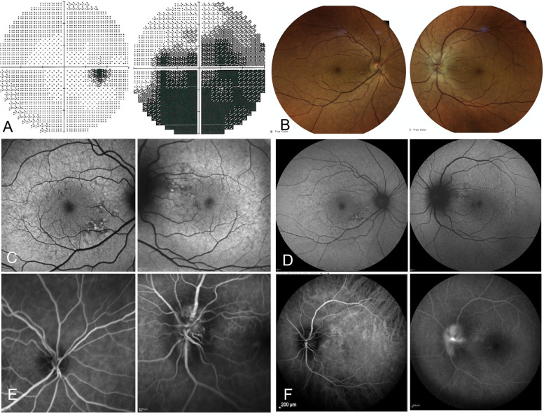
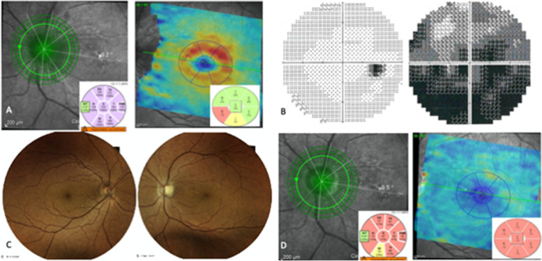
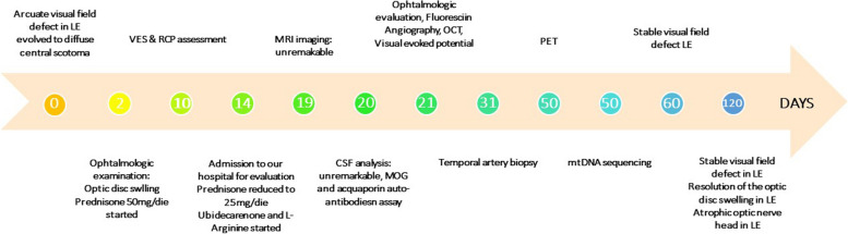
Case presentation: Here we describe a 55-year-old woman, sister of a previously described patient with MELAS harbouring the m.3243A > G (p.0, MT-TL1) mutation, with otherwise unremarkable medical history, that presented with subacute, painful visual impairment of one eye, accompanied by proximal muscular pain and headache. Over the next weeks, she developed severe and progressive vision loss limited to one eye. Ocular examination confirmed unilateral swelling of the optic nerve head; fluorescein angiography showed segmental perfusion delay in the optic disc and papillary leakage. Neuroimaging, blood and CSF examination and temporal artery biopsy ruled out neuroinflammatory disorders and giant cell arteritis (GCA). Mitochondrial sequencing analysis confirmed the m.3243A > G transition, and excluded the three most common LHON mutations, as well as the m.3376G > A LHON/MELAS overlap syndrome mutation. Based on the constellation of clinical symptoms and signs presented in our patient, including the muscular involvement, and the results of the investigations, the diagnosis of optic neuropathy as a stroke-like event affecting the optic disc was performed. L-arginine and ubidecarenone therapies were started with the aim to improve stroke-like episode symptoms and prevention. The visual defect remained stable with no further progression or outbreak of new symptoms.
Conclusions: Atypical clinical presentations must be always considered in mitochondrial disorders, even in well-described phenotypes and when mutational load in peripheral tissue is low. Mitotic segregation of mitochondrial DNA (mtDNA) does not allow to know the exact degree of heteroplasmy existent within different tissue, such as retina and optic nerve. Important therapeutic implications arise from a correct diagnosis of atypical presentation of mitochondrial disorders.
Keywords: MELAS; Mitochondrial disease; NAION; Optic neuropathy; Stroke-like episodes.
© 2023. The Author(s).
Conflict of interest statement
The authors declare no competing interests.
Figures
References
Publication types
MeSH terms
Substances
Grants and funding
LinkOut - more resources
Full Text Sources
Medical
