

Transcriptome profiling of a synergistic volumetric muscle loss repair strategy
- PMID: 37095469
- PMCID: PMC10124022
- DOI: 10.1186/s12891-023-06401-1
Transcriptome profiling of a synergistic volumetric muscle loss repair strategy
Abstract
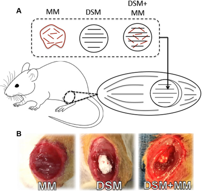
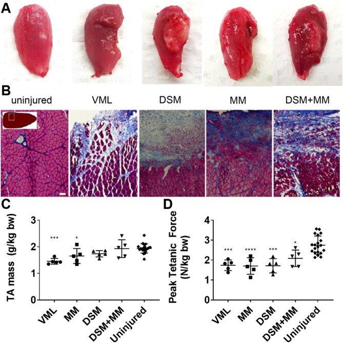
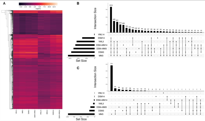
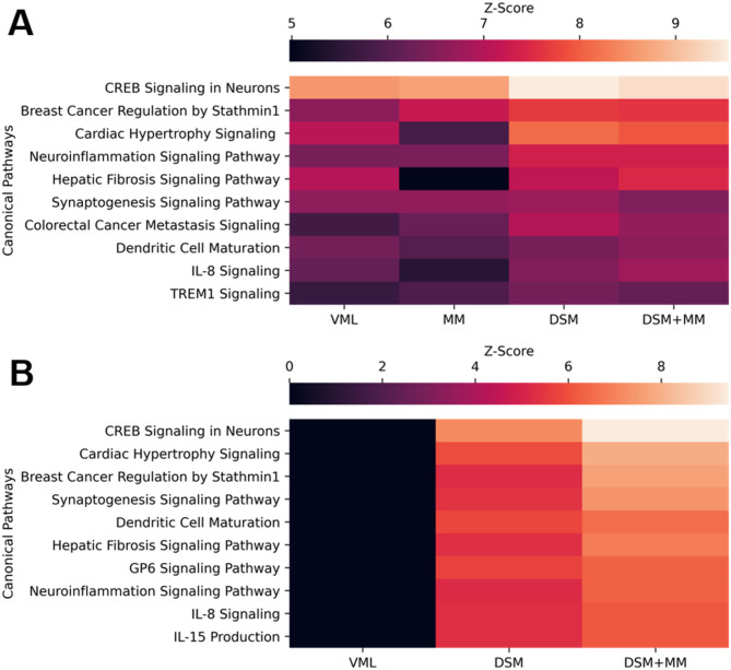
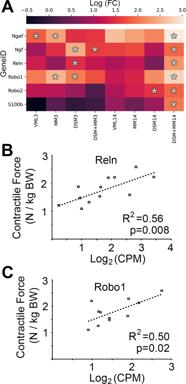
Volumetric muscle loss overwhelms skeletal muscle's ordinarily capable regenerative machinery, resulting in severe functional deficits that have defied clinical repair strategies. In this manuscript we pair the early in vivo functional response induced by differing volumetric muscle loss tissue engineering repair strategies that are broadly representative of those explored by the field (scaffold alone, cells alone, or scaffold + cells) to the transcriptomic response induced by each intervention. We demonstrate that an implant strategy comprising allogeneic decellularized skeletal muscle scaffolds seeded with autologous minced muscle cellular paste (scaffold + cells) mediates a pattern of increased expression for several genes known to play roles in axon guidance and peripheral neuroregeneration, as well as several other key genes related to inflammation, phagocytosis, and extracellular matrix regulation. The upregulation of several key genes in the presence of both implant components suggests a unique synergy between scaffolding and cells in the early period following intervention that is not seen when either scaffolds or cells are used in isolation; a finding that invites further exploration of the interactions that could have a positive impact on the treatment of volumetric muscle loss.
Keywords: Biomaterial implant; Muscle injury and repair; Pre-clinical animal model research; Transcriptomic profiling; Volumeric muscle loss.
© 2023. The Author(s).
Conflict of interest statement
The authors declare no competing interests.
Figures

Similar articles
-
Codelivery of Infusion Decellularized Skeletal Muscle with Minced Muscle Autografts Improved Recovery from Volumetric Muscle Loss Injury in a Rat Model.Tissue Eng Part A. 2016 Oct;22(19-20):1151-1163. doi: 10.1089/ten.TEA.2016.0134. Epub 2016 Sep 23. Tissue Eng Part A. 2016. PMID: 27570911 Free PMC article.
-
Perfusable adipose decellularized extracellular matrix biological scaffold co-recellularized with adipose-derived stem cells and L6 promotes functional skeletal muscle regeneration following volumetric muscle loss.Biomaterials. 2024 Jun;307:122529. doi: 10.1016/j.biomaterials.2024.122529. Epub 2024 Mar 11. Biomaterials. 2024. PMID: 38489911
-
Implantation of in vitro tissue engineered muscle repair constructs and bladder acellular matrices partially restore in vivo skeletal muscle function in a rat model of volumetric muscle loss injury.Tissue Eng Part A. 2014 Feb;20(3-4):705-15. doi: 10.1089/ten.TEA.2012.0761. Epub 2013 Dec 19. Tissue Eng Part A. 2014. PMID: 24066899 Free PMC article.
-
Mechanisms by which acellular biologic scaffolds promote functional skeletal muscle restoration.Biomaterials. 2016 Oct;103:128-136. doi: 10.1016/j.biomaterials.2016.06.047. Epub 2016 Jun 24. Biomaterials. 2016. PMID: 27376561 Review.
-
Naturally derived and synthetic scaffolds for skeletal muscle reconstruction.Adv Drug Deliv Rev. 2015 Apr;84:208-21. doi: 10.1016/j.addr.2014.08.011. Epub 2014 Aug 29. Adv Drug Deliv Rev. 2015. PMID: 25174309 Free PMC article. Review.
Cited by
-
In vivo evaluation of decellularized skeletal muscle matrices for skeletal muscle repair: A systematic review.Bioeng Transl Med. 2025 Mar 4;10(4):e70009. doi: 10.1002/btm2.70009. eCollection 2025 Jul. Bioeng Transl Med. 2025. PMID: 40708976 Free PMC article. Review.
-
Tissue Engineered 3D Constructs for Volumetric Muscle Loss.Ann Biomed Eng. 2024 Sep;52(9):2325-2347. doi: 10.1007/s10439-024-03541-w. Epub 2024 Jul 31. Ann Biomed Eng. 2024. PMID: 39085677 Free PMC article. Review.
-
Biomaterial-Based Regenerative Strategies for Volumetric Muscle Loss: Challenges and Solutions.Adv Wound Care (New Rochelle). 2025 Mar;14(3):159-175. doi: 10.1089/wound.2024.0079. Epub 2024 Jul 10. Adv Wound Care (New Rochelle). 2025. PMID: 38775429 Review.
References
-
- Allen RE, Sheehan SM, Taylor RG, Kendall TL, Rice GM. Hepatocyte growth factor activates quiescent skeletal muscle satellite cells in vitro. J Cell Physiol. 1995;165(2):307–12. - PubMed
-
- Sheehan SM, Tatsumi R, Temm-Grove CJ, Allen RE. HGF is an autocrine growth factor for skeletal muscle satellite cells in vitro. Muscle & Nerve: Official Journal of the American Association of Electrodiagnostic Medicine. 2000;23(2):239–45. - PubMed
-
- Tatsumi R, Anderson JE, Nevoret CJ, Halevy O, Allen RE. HGF/SF is present in normal adult skeletal muscle and is capable of activating satellite cells. Dev Biol. 1998;194(1):114–28. - PubMed
MeSH terms
Grants and funding
LinkOut - more resources
Full Text Sources
Molecular Biology Databases
