

In-Vivo fluorescent nanosensor implants based on hydrogel-encapsulation: investigating the inflammation and the foreign-body response
- PMID: 37095500
- PMCID: PMC10123989
- DOI: 10.1186/s12951-023-01873-8
In-Vivo fluorescent nanosensor implants based on hydrogel-encapsulation: investigating the inflammation and the foreign-body response
Abstract
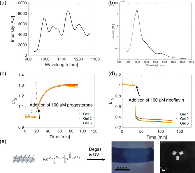
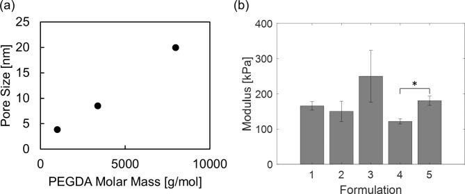
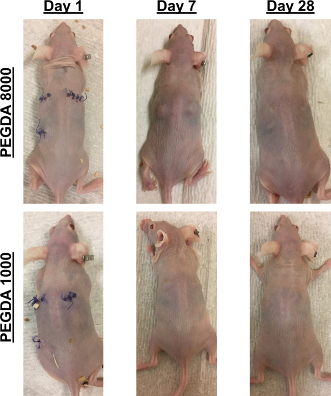
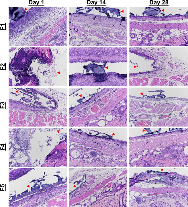
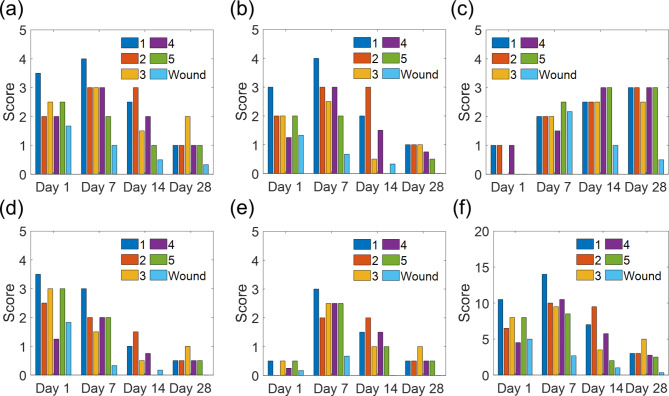
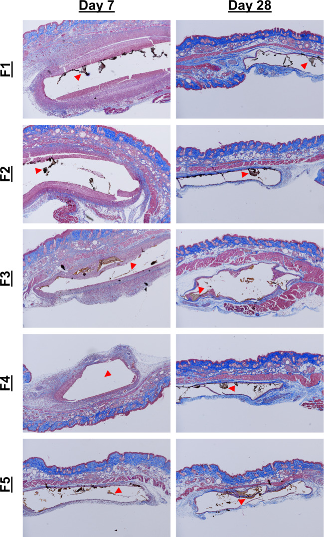
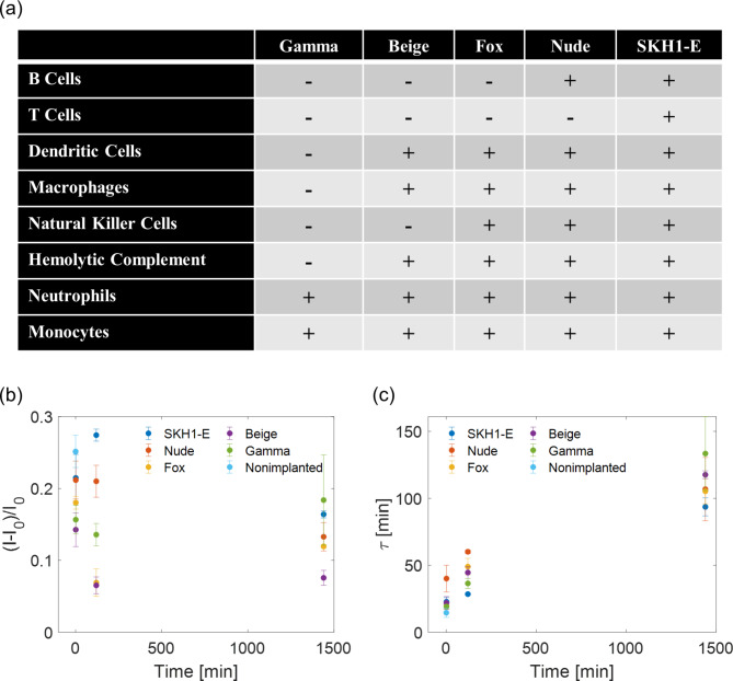
Nanotechnology-enabled sensors or nanosensors are emerging as promising new tools for various in-vivo life science applications such as biosensing, components of delivery systems, and probes for spatial bioimaging. However, as with a wide range of synthetic biomaterials, tissue responses have been observed depending on cell types and various nanocomponent properties. The tissue response is critical for determining the acute and long term health of the organism and the functional lifetime of the material in-vivo. While nanomaterial properties can contribute significantly to the tissue response, it may be possible to circumvent adverse reactions by formulation of the encapsulation vehicle. In this study, five formulations of poly (ethylene glycol) diacrylate (PEGDA) hydrogel-encapsulated fluorescent nanosensors were implanted into SKH-1E mice, and the inflammatory responses were tracked in order to determine the favorable design rules for hydrogel encapsulation and minimization of such responses. Hydrogels with higher crosslinking density were found to allow faster resolution of acute inflammation. Five different immunocompromised mice lines were utilized for comparison across different inflammatory cell populations and responses. Degradation products of the gels were also characterized. Finally, the importance of the tissue response in determining functional lifetime was demonstrated by measuring the time-dependent nanosensor deactivation following implantation into animal models.
Keywords: Carbon nanotube; Hydrogel; Implants; Inflammation; Nanosensor.
© 2023. The Author(s).
Figures
Similar articles
-
Implanted Nanosensors in Marine Organisms for Physiological Biologging: Design, Feasibility, and Species Variability.ACS Sens. 2019 Jan 25;4(1):32-43. doi: 10.1021/acssensors.8b00538. Epub 2018 Dec 11. ACS Sens. 2019. PMID: 30525471
-
Inflammation via myeloid differentiation primary response gene 88 signaling mediates the fibrotic response to implantable synthetic poly(ethylene glycol) hydrogels.Acta Biomater. 2019 Dec;100:105-117. doi: 10.1016/j.actbio.2019.09.043. Epub 2019 Sep 27. Acta Biomater. 2019. PMID: 31568879 Free PMC article.
-
In vivo bone and soft tissue response to injectable, biodegradable oligo(poly(ethylene glycol) fumarate) hydrogels.Biomaterials. 2003 Aug;24(19):3201-11. doi: 10.1016/s0142-9612(03)00168-6. Biomaterials. 2003. PMID: 12763447
-
Determination of the in vivo degradation mechanism of PEGDA hydrogels.J Biomed Mater Res A. 2014 Dec;102(12):4244-51. doi: 10.1002/jbm.a.35096. Epub 2014 Feb 13. J Biomed Mater Res A. 2014. PMID: 24464985 Free PMC article.
-
Synthesis of stiffness-tunable and cell-responsive Gelatin-poly(ethylene glycol) hydrogel for three-dimensional cell encapsulation.J Biomed Mater Res A. 2016 Oct;104(10):2401-11. doi: 10.1002/jbm.a.35779. Epub 2016 May 30. J Biomed Mater Res A. 2016. PMID: 27170015
Cited by
-
Tissue Reaction to Low-Density Polyacrylamide Gel as a Carrier for Microimplants in the Adipose Fin of Rainbow Trout.Gels. 2023 Aug 5;9(8):629. doi: 10.3390/gels9080629. Gels. 2023. PMID: 37623084 Free PMC article.
-
Single-Walled Carbon Nanotubes as Optical Transducers for Nanobiosensors In Vivo.ACS Nano. 2024 Dec 31;18(52):35164-35181. doi: 10.1021/acsnano.4c13076. Epub 2024 Dec 18. ACS Nano. 2024. PMID: 39696968 Free PMC article. Review.
-
Noninvasive Injectable Optical Nanosensor-Hydrogel Hybrids Detect Doxorubicin in Living Mice.Adv Opt Mater. 2024 Jun 17;12(17):2303324. doi: 10.1002/adom.202303324. Epub 2024 Apr 20. Adv Opt Mater. 2024. PMID: 39450264 Free PMC article.
-
In vivo biocompatibility assessment of 3D printed bioresorbable polymers for brain tissue regeneration. A feasibility study.Regen Ther. 2024 Oct 23;26:941-955. doi: 10.1016/j.reth.2024.10.004. eCollection 2024 Jun. Regen Ther. 2024. PMID: 39512739 Free PMC article.
References
-
- Rabbani M, Hoque ME, Mahbub Z, Bin. Nanosensors in biomedical and environmental applications: perspectives and prospects. Nanofabrication Smart Nanosensor Appl. 2020;163–86. 10.1016/B978-0-12-820702-4.00007-6.
-
- Koman VB et al. A wavelength-induced frequency filtering method for fluorescent nanosensors in vivo.Nat. Nanotechnol.17, (2022). - PubMed
-
- Barbosa AI, Rebelo R, Reis RL, Bhattacharya M, Correlo V. M. Current nanotechnology advances in diagnostic biosensors. Med Devices Sensors. 2021;4:e10156. doi: 10.1002/mds3.10156. - DOI
MeSH terms
Substances
Grants and funding
LinkOut - more resources
Full Text Sources
