

Application effect of an artificial intelligence-based fundus screening system: evaluation in a clinical setting and population screening
- PMID: 37095516
- PMCID: PMC10127070
- DOI: 10.1186/s12938-023-01097-9
Application effect of an artificial intelligence-based fundus screening system: evaluation in a clinical setting and population screening
Abstract
Background: To investigate the application effect of artificial intelligence (AI)-based fundus screening system in real-world clinical environment.
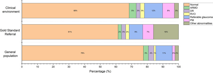
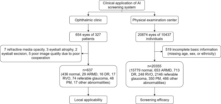
Methods: A total of 637 color fundus images were included in the analysis of the application of the AI-based fundus screening system in the clinical environment and 20,355 images were analyzed in the population screening.
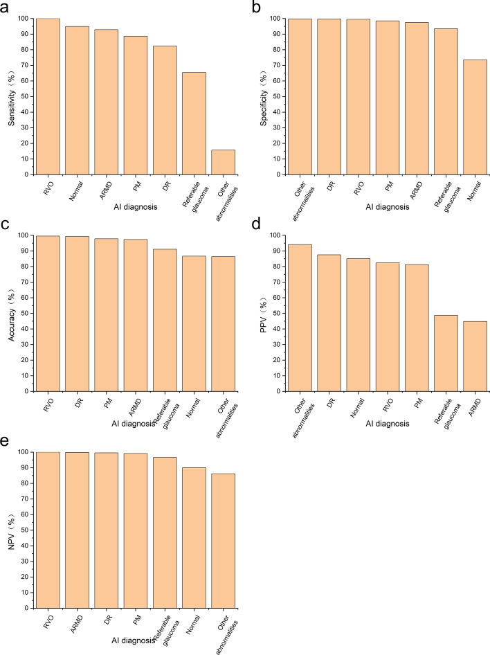
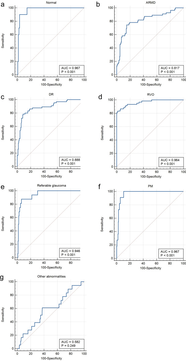
Results: The AI-based fundus screening system demonstrated superior diagnostic effectiveness for diabetic retinopathy (DR), retinal vein occlusion (RVO) and pathological myopia (PM) according to gold standard referral. The sensitivity, specificity, accuracy, positive predictive value (PPV) and negative predictive value (NPV) of three fundus abnormalities were greater (all > 80%) than those for age-related macular degeneration (ARMD), referable glaucoma and other abnormalities. The percentages of different diagnostic conditions were similar in both the clinical environment and the population screening.
Conclusions: In a real-world setting, our AI-based fundus screening system could detect 7 conditions, with better performance for DR, RVO and PM. Testing in the clinical environment and through population screening demonstrated the clinical utility of our AI-based fundus screening system in the early detection of ocular fundus abnormalities and the prevention of blindness.
Keywords: Artificial intelligence; Color fundus photography; Early screening; Ocular fundus abnormalities; Prevention of blindness.
© 2023. The Author(s).
Conflict of interest statement
None of the authors have any financial/conflicting interests to disclose.
Figures
Similar articles
-
Comparison of 21 artificial intelligence algorithms in automated diabetic retinopathy screening using handheld fundus camera.Ann Med. 2024 Dec;56(1):2352018. doi: 10.1080/07853890.2024.2352018. Epub 2024 May 13. Ann Med. 2024. PMID: 38738798 Free PMC article.
-
Application of artificial intelligence-based dual-modality analysis combining fundus photography and optical coherence tomography in diabetic retinopathy screening in a community hospital.Biomed Eng Online. 2022 Jul 20;21(1):47. doi: 10.1186/s12938-022-01018-2. Biomed Eng Online. 2022. PMID: 35859144 Free PMC article.
-
Development of an artificial intelligence system to classify pathology and clinical features on retinal fundus images.Clin Exp Ophthalmol. 2019 May;47(4):484-489. doi: 10.1111/ceo.13433. Epub 2018 Nov 15. Clin Exp Ophthalmol. 2019. PMID: 30370587
-
An overview of artificial intelligence in diabetic retinopathy and other ocular diseases.Front Public Health. 2022 Oct 28;10:971943. doi: 10.3389/fpubh.2022.971943. eCollection 2022. Front Public Health. 2022. PMID: 36388304 Free PMC article. Review.
-
Artificial Intelligence in the assessment of diabetic retinopathy from fundus photographs.Semin Ophthalmol. 2020 Nov 16;35(7-8):325-332. doi: 10.1080/08820538.2020.1855358. Epub 2021 Feb 4. Semin Ophthalmol. 2020. PMID: 33539253 Review.
Cited by
-
Differentiating Choroidal Melanomas and Nevi Using a Self-Supervised Deep Learning Model Applied to Clinical Fundoscopy Images.Ophthalmol Sci. 2024 Nov 8;5(2):100647. doi: 10.1016/j.xops.2024.100647. eCollection 2025 Mar-Apr. Ophthalmol Sci. 2024. PMID: 39802204 Free PMC article.
-
Classification of Color Fundus Photographs Using Fusion Extracted Features and Customized CNN Models.Healthcare (Basel). 2023 Aug 7;11(15):2228. doi: 10.3390/healthcare11152228. Healthcare (Basel). 2023. PMID: 37570467 Free PMC article.
-
Interpretation of Clinical Retinal Images Using an Artificial Intelligence Chatbot.Ophthalmol Sci. 2024 May 23;4(6):100556. doi: 10.1016/j.xops.2024.100556. eCollection 2024 Nov-Dec. Ophthalmol Sci. 2024. PMID: 39139542 Free PMC article.
-
Artificial intelligence in pathologic myopia: a review of clinical research studies.Front Med (Lausanne). 2025 Apr 23;12:1572750. doi: 10.3389/fmed.2025.1572750. eCollection 2025. Front Med (Lausanne). 2025. PMID: 40337273 Free PMC article.
-
A hybrid multi model artificial intelligence approach for glaucoma screening using fundus images.NPJ Digit Med. 2025 Feb 27;8(1):130. doi: 10.1038/s41746-025-01473-w. NPJ Digit Med. 2025. PMID: 40016437 Free PMC article.
References
MeSH terms
Grants and funding
- 2019D01C004/Natural Science Foundation of Xinjiang Uygur Autonomous Region
- 2019Q145/Xinjiang Uygur Autonomous Region Innovation Environment (Talents, Bases) Special Project (Special Talent Project-Tianshan Youth Project)
- KDYY202018/The Pearl River Scholar Tianshan Talent Cooperation's Expert Studio Innovation Team
- 202102020736/Science and Technology Program of Guangzhou
LinkOut - more resources
Full Text Sources
Medical
