. 2023 Apr 24;24(1):92.
doi: 10.1186/s13059-023-02925-w.

A desert lncRNA HIDEN regulates human endoderm differentiation via interacting with IMP1 and stabilizing FZD5 mRNA

Affiliations

A desert lncRNA HIDEN regulates human endoderm differentiation via interacting with IMP1 and stabilizing FZD5 mRNA

Pei Lu et al. Genome Biol. .

Abstract

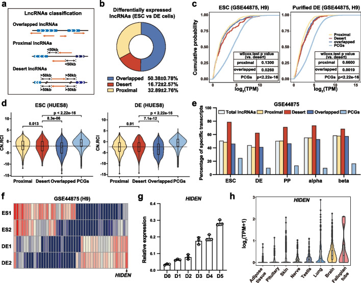
Background: Extensive studies have revealed the function and mechanism of lncRNAs in development and differentiation, but the majority have focused on those lncRNAs adjacent to protein-coding genes. In contrast, lncRNAs located in gene deserts are rarely explored. Here, we utilize multiple differentiation systems to dissect the role of a desert lncRNA, HIDEN (human IMP1-associated "desert" definitive endoderm lncRNA), in definitive endoderm differentiation from human pluripotent stem cells.

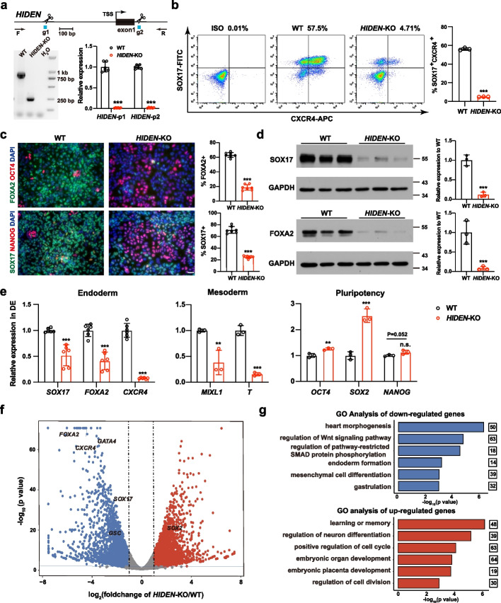
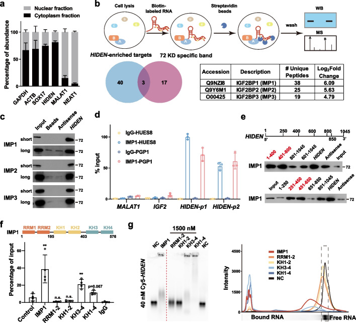
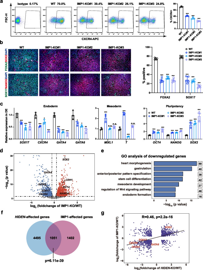
Results: We show that desert lncRNAs are highly expressed with cell-stage-specific patterns and conserved subcellular localization during stem cell differentiation. We then focus on the desert lncRNA HIDEN which is upregulated and plays a vital role during human endoderm differentiation. We find depletion of HIDEN by either shRNA or promoter deletion significantly impairs human endoderm differentiation. HIDEN functionally interacts with RNA-binding protein IMP1 (IGF2BP1), which is also required for endoderm differentiation. Loss of HIDEN or IMP1 results in reduced WNT activity, and WNT agonist rescues endoderm differentiation deficiency caused by the depletion of HIDEN or IMP1. Moreover, HIDEN depletion reduces the interaction between IMP1 protein and FZD5 mRNA and causes the destabilization of FZD5 mRNA, which is a WNT receptor and necessary for definitive endoderm differentiation.

Conclusions: These data suggest that desert lncRNA HIDEN facilitates the interaction between IMP1 and FZD5 mRNA, stabilizing FZD5 mRNA which activates WNT signaling and promotes human definitive endoderm differentiation.

Keywords: Desert lncRNA; Endoderm differentiation; FZD5; Human pluripotent stem cell; IMP1.


Conflict of interest statement

The authors declare that they have no competing interests.

Figures

Fig. 1
Fig. 1
Desert lncRNAs are highly expressed during human endoderm differentiation. a The classification of overlapped lncRNAs, proximal lncRNAs and desert lncRNAs. b Pie chart showing the average proportion of differentially expressed overlapped, proximal, and desert lncRNAs identified between human PSCs and DE cells (data from three PSC lines). c The expression level of lncRNAs and PCGs in H9 ESCs and DE cells. The curves were colored by the category of lncRNAs. The p value between desert lncRNAs and other subsets were listed in the chart. d The subcellular localization of lncRNAs and PCGs, calculated by “relative concentration index” (RCI) in HUES8 ESCs and DE cells. CN.RCI = log2(CE/NE) + log2(CE/NM) (CE: cytoplasmic elution component, NE: nuclear elution component, NM: nuclear insoluble component). e The cell expression specificity of lncRNAs and PCGs in ESCs, DE, pancreatic endocrine (PP), pancreatic alpha and beta cells, calculated by specificity score. f Heatmap of differentially expressed desert lncRNAs between ESCs and DE cells. Red indicates higher expression while blue indicates lower expression. The lncRNAs list was shown in Additional file 2: Table S1. g Time course expression of HIDEN during endoderm differentiation from human HUES8 ESCs was detected by RT-qPCR (n = 3). h The expression of HIDEN in 30 human tissues from GTEx database. The top eight tissues with high expression were shown
Fig. 2
Fig. 2
HIDEN is a desert lncRNA required for endoderm differentiation. a Generation of HIDEN knockout (KO) PSCs by CRISPR/Cas9. Top: sgRNAs used to delete the promoter of HIDEN and genomic PCR primers used to detect the promoter deletion. Bottom: The HIDEN RNA level was quantified using two sets of primers in HIDEN-KO DE cells compared to wildtype (n = 6). b Flow cytometric analysis of SOX17+CXCR4+ cells in wildtype and HIDEN-KO DE cells. The statistical results were shown on the right (n = 3). c Immunofluorescent staining of DE markers (SOX17, FOXA2) and pluripotency markers (OCT4, NANOG) in wildtype and HIDEN-KO DE cells. Quantitative results were shown on the right (n = 7). Scale bar, 50 μm. d The protein levels of DE markers (SOX17 and FOXA2) were determined in wildtype and HIDEN-KO DE cells (n = 3). GAPDH was used as internal control. e The RNA expression levels of representative endoderm genes (n = 6), mesoderm genes (n = 3) and pluripotency genes (n = 3) in wildtype and HIDEN-KO DE cells. f Scatterplot showing differentially expressed genes identified by RNA-seq of wildtype and HIDEN-KO DE cells (n = 3). Upregulated and downregulated genes upon HIDEN-KO were shown in red and blue, respectively. g GO analysis of the downregulated or upregulated genes in HIDEN-KO DE cells compared to wildtype
Fig. 3
Fig. 3
HIDEN physically interacted with IMP1. a Subcellular localization of HIDEN in DE cells determined by RT-qPCR following nucleo-cytoplasmic separation (n = 6). b The schematic diagram of RNA pulldown was shown on the top. Venn diagram indicates the overlapped hints identified by mass spectrometry of the specific band around 72 kDa (unique peptides ≥ 4, red circle) and whole extracts (unique peptides ≥ 10 in HIDEN-pulldown group and log2(fold-change (HIDEN/Antisense)) > 2.32, blue circle). The unique peptides and log2(fold-change) of IMP1/2/3 compared to antisense in mass spectrometry data of HIDEN-pulldown group were shown in the table. c Immunoblot for IMP1, IMP2, and IMP3 after RNA pulldown in DE cells. Beads and antisense were used as negative controls. Pictures captured for short and long exposure time were shown. d IMP1 RIP followed by RT-qPCR in DE cells of two PSC lines, PGP1 and HUES8 (n = 3). RNA levels were normalized to input. e Mapping the IMP1-binding region in HIDEN in 293 T cells. Top, diagrams of full-length HIDEN and the deletion fragments used in RNA pulldown. Bottom, immunoblot for IMP1 in protein samples pulled down by different HIDEN fragments. f Mapping the HIDEN-binding domain in IMP1 protein. Domain structure of IMP1 protein (top). FLAG-tagged IMP1 or IMP1 mutants and HIDEN were co-overexpressed in 293 T cells and FLAG RIP was performed to examine the enrichment of HIDEN (bottom). g Electrophoretic mobility shift assay (EMSA) results of in vitro binding assays. Cy5-labeled HIDEN RNA (40 nM) were transcribed in vitro and His-tagged IMP1 proteins (1500 nM) were purified from E. coli
Fig. 4
Fig. 4
IMP1 deficiency inhibits endoderm differentiation. a Flow cytometric analysis of CXCR4-positive cells in wildtype (WT) and IMP1-KO DE cells. The statistical results were shown on the right (n = 3). b Immunofluorescent staining of DE markers (SOX17, FOXA2) and pluripotency markers (NANOG, SOX2) in WT and IMP1-KO DE cells. Quantitative results were shown on the right (n = 5). Scale bar = 50 μm. c RNA levels of representative endoderm genes, mesoderm genes and pluripotency genes in WT and IMP1-KO DE cells determined by RT-qPCR (n = 3). d Scatterplot showing differentially expressed genes in RNA-seq of WT and IMP1-KO DE cells. Upregulated and downregulated genes upon IMP1 knockout were shown in red and blue, respectively. e GO analysis of the downregulated genes in DE cells upon IMP1 knockout. f The overlap of differentially expressed genes upon HIDEN-KO and IMP1-KO. g The correlation of HIDEN-KO and IMP1-KO affected genes
Fig. 5
Fig. 5
WNT signaling pathway acts as downstream of HIDEN/IMP1. a Venn diagram indicates the overlapped genes of differentially expressed PCGs upon HIDEN knockout in DE cells and IMP1-enriched targets identified by IMP1 RIP-seq in DE cells. b GO analysis of the overlapped genes from a. c,HIDEN knockout led to reduced active β-catenin and unaltered total β-catenin level compared to wildtype after two days’ DE differentiation, as shown by Western blot (c) and statistical results (d) (n = 6). e The protein level of β-catenin in nuclear fraction of WT or HIDEN-KO DE cells (n = 3). f The TCF-luciferase activity in 293 T when transfected with HIDEN, antisense control or empty vector (n = 3). Cells treated with 1 μM CHIR-99021 were used as positive controls. g-j Flow cytometric analysis of SOX17-positive cells (g), the presentative endoderm genes expression revealed by RT-qPCR (h) and immunostaining (i-j) in wildtype or HIDEN-KO cells after manipulating WNT signaling through small molecular inhibitors during DE differentiation (n = 3). WNT signaling activator CHIR-99021, with different concentrations (0.5 μM, 1 μM and 2 μM) at (g) and 1 μM at (h-j), and WNT signaling inhibitor WNT-C59 (1 μM) were used. NT indicated for non-treated group. i Scale bar = 50 μm. j Quantitative results of SOX17- and FOXA2-positive cells were shown (n = 6)
Fig. 6
Fig. 6
HIDEN-IMP1 stabilized FZD5 mRNA to contribute to DE differentiation. a Venn diagram indicated the overlapping of WNT-associated differentially expressed genes during DE differentiation (yellow circle), WNT-associated HIDEN-regulated genes (green circle), and IMP1-enriched WNT-associated genes (pink circle). The WNT-associated genes were listed in Additional file 8: Table S6 (from online WNT website: http://web.stanford.edu/group/nusselab/cgi-bin/wnt/). b The time course expression of FZD5 during DE differentiation, as shown by RT-qPCR (n = 3). c The relative expression of FZD5 in wildtype or HIDEN-KO DE cells was determined by RT-qPCR (n = 3). d RT-qPCR following IMP1 RIP was performed for determination of the indicated transcripts enrichment by IMP1 in wildtype or HIDEN-KO DE cells (n = 3). RNA levels were normalized to input. U1 and MALAT1 were considered as negative controls. e The visualization of IMP1 binding on FZD5 mRNA identified by IMP1 RIP-seq in wildtype or HIDEN-knockout DE cells. A detailed description was in Methods. f RNA stability assay of FZD5 in wildtype or HIDEN-KO DE cells treated with Actinomycin D (n = 3). g Flow cytometric analysis of SOX17+CXCR4+ cells in wildtype or FZD5-KO DE cells (n = 3). h RNA levels of representative endoderm genes, mesoderm genes and pluripotency genes in wildtype or FZD5-KO DE cells, determined by RT-qPCR (n = 3). i Immunofluorescent staining of DE markers (SOX17, FOXA2) and pluripotency markers (OCT4, SOX2) in wildtype or FZD5-KO DE cells. Quantitative results of SOX17- and FOXA2-positive cells were shown on the bottom (n = 5). Scale bar = 50 μm. j The functional model of HIDEN in human DE differentiation

Similar articles

Cited by

References

    1. Sigova AA, et al. Divergent transcription of long noncoding RNA/mRNA gene pairs in embryonic stem cells. Proc Natl Acad Sci U S A. 2013;110:2876–2881. doi: 10.1073/pnas.1221904110. - DOI - PMC - PubMed
    1. Engreitz JM, et al. Local regulation of gene expression by lncRNA promoters, transcription and splicing. Nature. 2016;539:452–455. doi: 10.1038/nature20149. - DOI - PMC - PubMed
    1. Lu P, Li M, Zhang D, Jiang W. Lnc-ing pluripotency maintenance and early differentiation in human pluripotent stem cells. FASEB J. 2021;35:e21438. doi: 10.1096/fj.202002278R. - DOI - PubMed
    1. Fatica A, Bozzoni I. Long non-coding RNAs: new players in cell differentiation and development. Nat Rev Genet. 2014;15:7–21. doi: 10.1038/nrg3606. - DOI - PubMed
    1. Flynn RA, Chang HY. Long noncoding RNAs in cell-fate programming and reprogramming. Cell Stem Cell. 2014;14:752–761. doi: 10.1016/j.stem.2014.05.014. - DOI - PMC - PubMed

Publication types

MeSH terms