

Acute obstructive pyelonephritis due to pyosalpinx: a case report
- PMID: 37095582
- PMCID: PMC10127100
- DOI: 10.1186/s13256-023-03900-6
Acute obstructive pyelonephritis due to pyosalpinx: a case report
Abstract
Background: A pyosalpinx is the acute inflammation of the fallopian tube, which fills up and swells with pus. It commonly results from inadequate or delayed treatment of pelvic inflammatory disease.
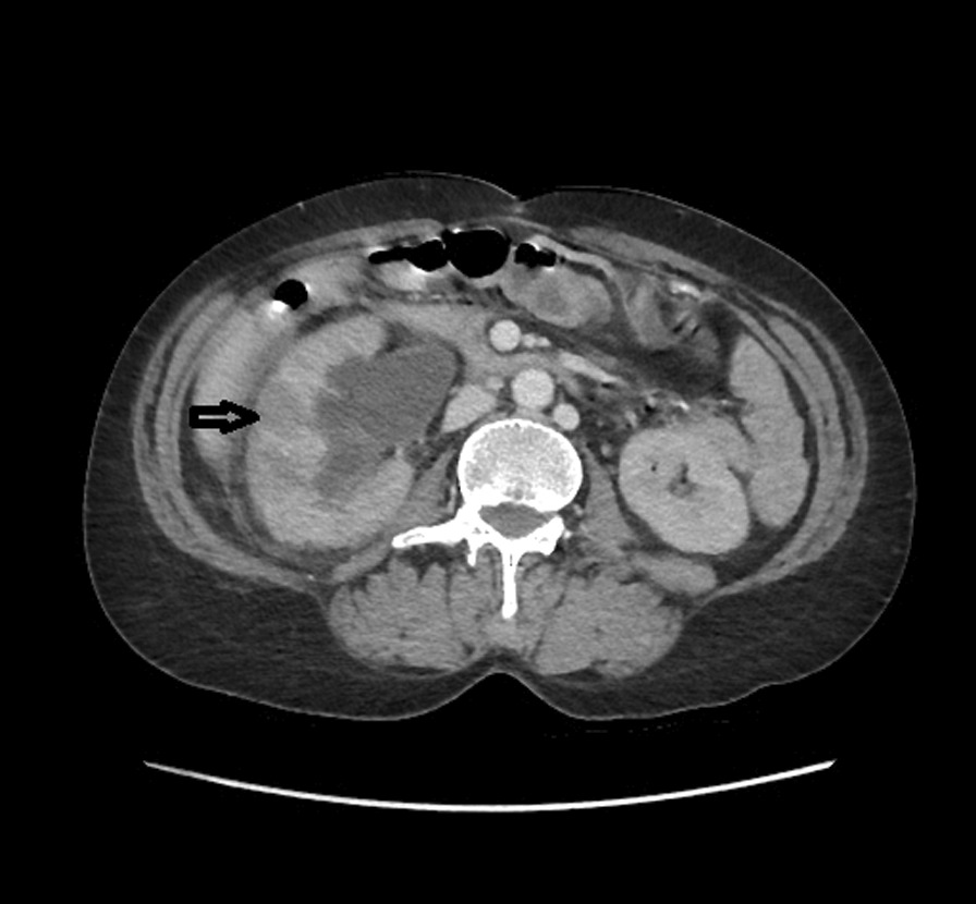
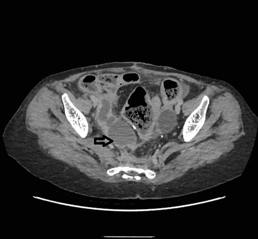
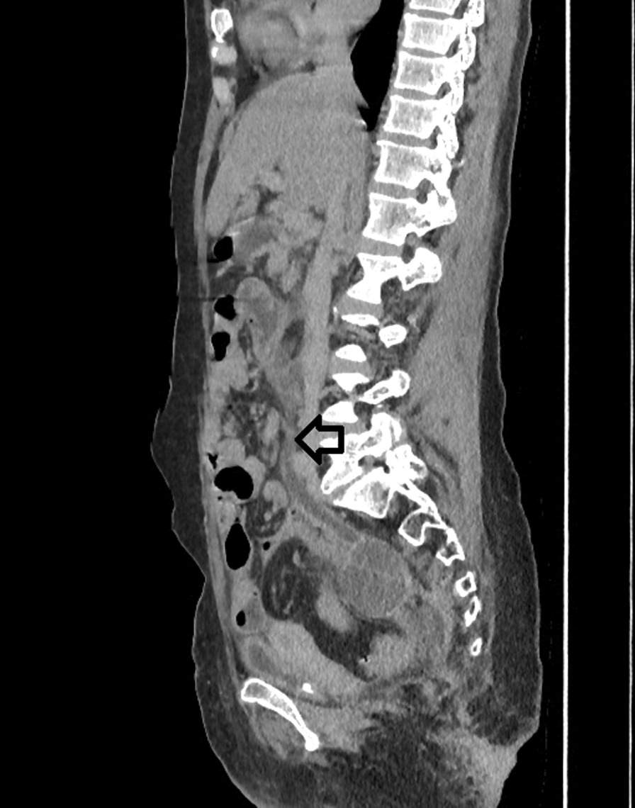
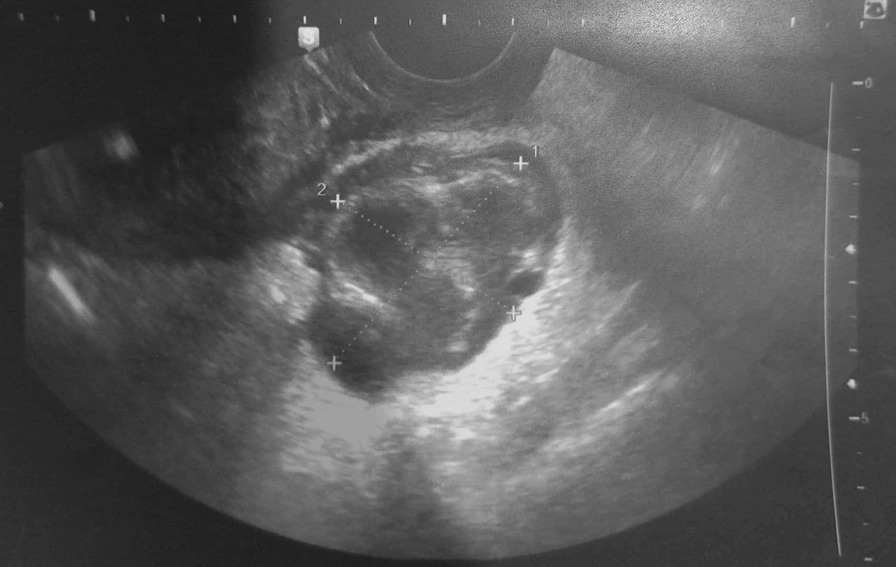
Case presentation: We report the case of a 54-year-old Africain female patient, who presented with sustained high-grade fever, right flank pain, and severe acute storage low-urinary-tract symptoms. Computed tomography showed signs of acute obstructive pyelonephritis with a right tubular juxtauterine mass with complex internal fluid and thick enhancing walls exerting a mass effect on the right ureter. A drainage of the right excretory cavities by a JJ stent was performed. An ultrasound-guided aspiration of the collection was also performed.
Conclusion: A pyosalpinx can then exert a mass effect on the excretory cavities, thus causing an acute obstructive pyelonephritis. A double drainage coupled with an effective antibiotic therapy is then necessary.
Keywords: Drainage; Pelvic inflammatory disease; Pyelonephritis; Pyosalpinx.
© 2023. The Author(s).
Conflict of interest statement
The authors declare that there is no conflict of interest.
Figures
Similar articles
-
Unusual presentation of bilateral pyosalpinx mimicking an ovarian torsion: A case report.Ann Med Surg (Lond). 2020 Feb 26;52:16-18. doi: 10.1016/j.amsu.2020.02.003. eCollection 2020 Apr. Ann Med Surg (Lond). 2020. PMID: 32153774 Free PMC article.
-
Bilateral recurrent pyosalpinx in a sexually inactive 12-year-old girl secondary to rare variant of Mullerian duct anomaly.BMJ Case Rep. 2017 Jun 24;2017:bcr2016218924. doi: 10.1136/bcr-2016-218924. BMJ Case Rep. 2017. PMID: 28647716 Free PMC article.
-
A rare case of large pyosalpinx in an elderly patient with well-controlled type 2 diabetes mellitus: a case report.J Med Case Rep. 2018 Oct 6;12(1):286. doi: 10.1186/s13256-018-1841-6. J Med Case Rep. 2018. PMID: 30290835 Free PMC article.
-
[Imaging in pelvic inflammatory disease].J Radiol. 2008 Jan;89(1 Pt 2):134-41. doi: 10.1016/s0221-0363(08)70385-8. J Radiol. 2008. PMID: 18288039 Review. French.
-
Ultrasound of pelvic inflammatory disease.Ultrasound Q. 2004 Dec;20(4):171-9. doi: 10.1097/00013644-200412000-00003. Ultrasound Q. 2004. PMID: 15602219 Review.
Cited by
-
Double-J Ureteral Stenting in Obstetrics and Gynecology: Pivotal or Problematic?J Clin Med. 2024 Dec 16;13(24):7649. doi: 10.3390/jcm13247649. J Clin Med. 2024. PMID: 39768572 Free PMC article. Review.
References
-
- Moralioglu S, Ozen IQ, Demirogullari B, Basaklar AC. Pyosalpinx and hydrosalpinx in virginal adolescents: report of two cases. W Indian Med J. 2013;62(3):257. - PubMed
-
- Afzal M, Kothari M, Alghasham A, Attal M. Isolated pyosalpinx in a pre-teen with bicornuate uterus. J Pediatr Surg Case Rep. 2021;72:101977. doi: 10.1016/j.epsc.2021.101977. - DOI
Publication types
MeSH terms
LinkOut - more resources
Full Text Sources
Medical
