Review
. 2023 Oct;221(4):409-424.
doi: 10.2214/AJR.23.29104. Epub 2023 Apr 5.

Imaging Follow-Up of Nonsurgical Therapies for Lung Cancer: AJR Expert Panel Narrative Review

Affiliations
Review

Imaging Follow-Up of Nonsurgical Therapies for Lung Cancer: AJR Expert Panel Narrative Review

David J Murphy et al. AJR Am J Roentgenol. 2023 Oct.

Abstract

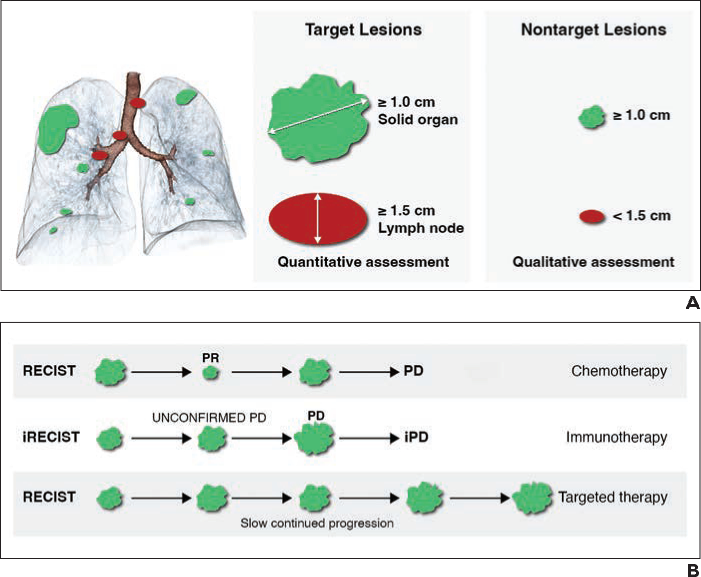
Lung cancer continues to be the most common cause of cancer-related death worldwide. In the past decade, with the implementation of lung cancer screening programs and advances in surgical and nonsurgical therapies, the survival of patients with lung cancer has increased, as has the number of imaging studies that these patients undergo. However, most patients with lung cancer do not undergo surgical re-section, because they have comorbid disease or lung cancer in an advanced stage at diagnosis. Nonsurgical therapies have continued to evolve with a growing range of systemic and targeted therapies, and there has been an associated evolution in the imaging findings encountered at follow-up examinations after such therapies (e.g., with respect to posttreatment changes, treatment complications, and recurrent tumor). This AJR Expert Panel Narrative Review describes the current status of nonsurgical therapies for lung cancer and their expected and unexpected imaging manifestations. The goal is to provide guidance to radiologists regarding imaging assessment after such therapies, focusing mainly on non-small cell lung cancer. Covered therapies include systemic therapy (conventional chemotherapy, targeted therapy, and immunotherapy), radiotherapy, and thermal ablation.

Keywords: RECIST; ablation; imaging follow-up; lung cancer; radiation therapy; response assessment; targeted therapy.


Figures

Fig. 1—
Fig. 1—
Response assessment after nonsurgical therapy for lung cancer using RECIST and immune RECIST (iRECIST). Factors assessed include response of target lesions (percentage change in sum of up to two lesions per organ and five lesions in total), response of nontarget lesions (brain lesions, bone lesions, pleural effusion, ascites, lymphangitic carcinomatosis, leptomeningeal disease, cystic lesions, radiated or ablated lesions, and ground-glass lesions), presence of new lesions, and evaluation time point. A, Schematics show selection of two solid organ target lesions, three lymph node target lesions, and five solid organ nontarget lesions according to response criteria. B, Schematic shows application of response criteria in patients undergoing three different therapies. Selected lesions are followed longitudinally to assess growth or shrinkage over time. PR = partial response, PD = progressive disease, iPD = immune PD.
Fig. 2—
Fig. 2—
65-year-old man with non–small cell lung cancer and epidermal growth factor receptor mutation being treated with tyrosine kinase inhibitor and presenting with left upper lobe mass and left hilar lymphadenopathy. Images show response assessment. A, Axial contrast-enhanced CT image shows left upper lobe mass (target lesion measuring 3.3 cm in longest dimension) and left hilar lymph node (arrow), which is nontarget lesion measuring 1.2 × 1.0 cm. B, Axial contrast-enhanced CT image obtained after two cycles of therapy shows progressive disease. Target lesion is stable. However, left hilar lymph node (arrow) measures 1.6 × 1.7 cm and is considered to represent new target lesion.
Fig. 3—
Fig. 3—
65-year-old man with non–small cell lung cancer and epidermal growth factor receptor mutation being treated with tyrosine kinase inhibitor. Images show response assessment. A, Axial contrast-enhanced CT image obtained before treatment shows paramediastinal mass in right upper lobe. B, Axial contrast-enhanced CT image obtained 2 months after A shows increase in size of tumor with associated adjacent pneumonitis. C, Axial contrast-enhanced CT image obtained 2 months after B shows significant decrease in size of tumor, indicating that prior enlargement represented pseudoprogression. Treatment was not discontinued during these months.
Fig. 4—
Fig. 4—
53-year-old man with lung cancer treated with nivolumab who reported severe fatigue and intermittent low-grade fever. A, Axial fused FDG PET/CT image obtained for routine surveillance shows multiple new symmetric mediastinal and hilar lymph nodes with intense FDG uptake. Sarcoidlike reaction was suspected. Immunotherapy was suspended. B, Axial fused FDG PET/CT image obtained 5 months after A shows resolution of lymphadenopathy, supporting suspected diagnosis of sarcoidlike reaction. Symptoms also resolved between imaging examinations.
Fig. 5—
Fig. 5—
78-year-old man with lung cancer treated by stereotactic body radiation therapy (SBRT; 54 Gy, administered in fractions). A, Axial CT image shows right upper lobe lung cancer (arrow). B, Computed dosimetric axial reconstruction obtained for SBRT planning shows malignancy for which maximal isodose was planned. Numbers in boxes are doses in grays corresponding to isodose lines of same color. C, Axial CT image obtained 2 years 9 months after completion of SBRT shows focal radiation changes (arrow). D, Axial CT image obtained 4 years 2 months after completion of SBRT shows focal nodule within radiation changes, which was suspected to represent recurrence. FDG PET/CT was performed 2 months later. E, CT image from FDG PET/CT shows increase in size of focal nodular component. F, Axial fused FDG PET/CT image shows no associated FDG avidity of focal nodular component. Masslike appearance remained stable for total of 3 years (not shown), confirming finding to represent delayed evolution of fibrotic radiation changes after SBRT.
Fig. 6—
Fig. 6—
49-year-old woman with right lung cancer treated with six cycles of chemotherapy followed by 6 MV of photons in intensity-modulated radiation therapy (IMRT) to total dose of 54 Gy in 36 fractions at 1.5 Gy per fraction given twice daily. A, Axial baseline CT image shows central right lung mass (arrow) and subcarinal nodal metastasis. B, Axial CT image obtained after six cycles of chemotherapy shows complete response. C, Computed dosimetric axial reconstruction obtained for IMRT planning shows malignancy receiving maximal isodose (Iso). Red area on left hilum indicates gross tumor volume (GTV); that is, location and extent of gross tumor; yellow, clinical target volume (CTV), defined as GTV plus margin for subclinical disease spread; green, planning target volume, which is geometric concept that surrounds CTV with additional margin to compensate for uncertainties in planning or treatment delivery. D, Axial CT image obtained 9 months after completion of IMRT shows modified conventional pattern with focal consolidative opacity associated with volume loss, architectural distortion, and traction bronchiectasis confined to region of radiated lung. E, Axial CT image obtained 15 months after completion of IMRT shows increase in soft tissue, bulging margin (arrow), loss of air bronchogram, and disappearance of linear margin; findings are suspicious for tumor recurrence. F, Axial fused FDG PET/CT image shows increase in FDG-avid soft tissue (arrow) consistent with recurrent tumor.
Fig. 7—
Fig. 7—
58-year-old man with left lower lobe squamous cell lung cancer after intensity-modulated radiation therapy (IMRT; 66 Gy, administered in 30 fractions) and initiation of immunotherapy (pembrolizumab). A, Computed dosimetric axial reconstruction obtained for IMRT planning shows primary lung malignancy receiving maximal isodose. Red area on spinal cord (considered critical structure or organ at risk) indicates dose constraints to minimize risk of radiation myelitis. Red area on left hilum indicates gross tumor volume (GTV); that is, location and extent of gross tumor; yellow, clinical target volume (CTV), defined as GTV plus margin for subclinical disease spread; green, planning target volume, which is geometric concept that surrounds CTV with additional margin to compensate for uncertainties in planning or treatment delivery. B, Axial CT image obtained 3 years after completion of IMRT and before start of immunotherapy shows radiation fibrosis in left perihilar region with traction bronchiectasis. C, Axial CT image obtained 5 months after start of immunotherapy (pembrolizumab) shows new consolidative opacities in left perihilar region that conform to radiation treatment plan, consistent with radiation recall pneumonitis. Median time interval for development of radiation recall pneumonitis after initiation of immunotherapy is 61 days (range, 4–520 days).
Fig. 8—
Fig. 8—
79-year-old man with right lower lobe adenocarcinoma treated with percutaneous radiofrequency ablation; cardiovascular comorbidities precluded surgery. A, Axial CT image obtained before procedure shows right lobe nodule. B, Axial CT image obtained at conclusion of ablation procedure shows area of mixed ground-glass opacification and consolidation within periphery of nodule (arrowhead) lacking focal extension of at least 5 mm beyond lesion. C, Axial CT image obtained 1 month after procedure shows cavitation within ablated area, thick nodular posterior wall, and residual surrounding ground-glass opacity. D, Axial CT image obtained 3 months after procedure shows reduction of cavitation and nodular wall thickening and resolution of peripheral ground-glass opacification. Findings are still consistent with physiologic evolution. E, Axial fused FDG PET/CT image obtained 6 months after procedure shows further shrinkage of ablated area with complete resolution of cavitation. Area shows weak tracer uptake that is equivocal for minimal residual disease versus postablation inflammatory changes. (Courtesy of Taralli S, Fondazione Policlinico Universitario A. Gemelli IRCCS, Rome, Italy) F, Axial fused FDG PET/CT image obtained 12 months after procedure shows intense metabolic activity within enlarged nodular lesion at ablation site. Finding is consistent with local recurrence. (Courtesy of Taralli S, Fondazione Policlinico Universitario A. Gemelli IRCCS, Rome, Italy)
Fig. 8—
Fig. 8—
79-year-old man with right lower lobe adenocarcinoma treated with percutaneous radiofrequency ablation; cardiovascular comorbidities precluded surgery. A, Axial CT image obtained before procedure shows right lobe nodule. B, Axial CT image obtained at conclusion of ablation procedure shows area of mixed ground-glass opacification and consolidation within periphery of nodule (arrowhead) lacking focal extension of at least 5 mm beyond lesion. C, Axial CT image obtained 1 month after procedure shows cavitation within ablated area, thick nodular posterior wall, and residual surrounding ground-glass opacity. D, Axial CT image obtained 3 months after procedure shows reduction of cavitation and nodular wall thickening and resolution of peripheral ground-glass opacification. Findings are still consistent with physiologic evolution. E, Axial fused FDG PET/CT image obtained 6 months after procedure shows further shrinkage of ablated area with complete resolution of cavitation. Area shows weak tracer uptake that is equivocal for minimal residual disease versus postablation inflammatory changes. (Courtesy of Taralli S, Fondazione Policlinico Universitario A. Gemelli IRCCS, Rome, Italy) F, Axial fused FDG PET/CT image obtained 12 months after procedure shows intense metabolic activity within enlarged nodular lesion at ablation site. Finding is consistent with local recurrence. (Courtesy of Taralli S, Fondazione Policlinico Universitario A. Gemelli IRCCS, Rome, Italy)

Similar articles

Cited by

References

    1. Sung H, Ferlay J, Siegel RL, et al. Global cancer statistics 2020: GLOBOCAN estimates of incidence and mortality worldwide for 36 cancers in 185 countries. CA Cancer J Clin 2021; 71:209–249 - PubMed
    1. Ettinger DS, Wood DE, Aisner DL, et al. Non–small cell lung cancer, version 3.2022: NCCN clinical practice guidelines in oncology. J Natl Compr Canc Netw 2022; 20:497–530 - PubMed
    1. American Lung Association website. State of lung cancer. www.Lung.org/solc. Published 2021. Accessed Jun 29, 2022
    1. Zhang R, Kang J, Ren S, Xing L, Xu Y. Comparison of stereotactic body radiotherapy and radiofrequency ablation for early-stage non-small cell lung cancer: a systematic review and meta-analysis. Ann Transl Med 2022; 10:104. - PMC - PubMed
    1. Carter BW, Altan M, Shroff GS, Truong MT, Vlahos I. Post-chemotherapy and targeted therapy imaging of the chest in lung cancer. Clin Radiol 2022; 77:e1–e10 - PubMed

MeSH terms