Review
. 2024 Jan 8;30(1):30-41.
doi: 10.4274/dir.2023.232157. Epub 2023 Apr 24.

Earthquakes from a radiological perspective: what is demanded from the radiologists, and what can we do? A pictorial review

Affiliations
Review

Earthquakes from a radiological perspective: what is demanded from the radiologists, and what can we do? A pictorial review

Sonay Aydin et al. Diagn Interv Radiol. .

Abstract

Earthquakes are among the most destructive and unpredictable natural disasters. Various diseases and ailments, such as bone fractures, organ and soft-tissue injuries, cardiovascular diseases, lung diseases, and infectious diseases, can develop in the aftermath of severe earthquakes. Digital radiography, ultrasound, computed tomography, and magnetic resonance imaging are significant imaging modalities utilized for the quick and reliable assessment of earthquake-related ailments to facilitate the planning of suitable therapy. This article examines the usual radiological imaging characteristics observed in individuals from quake-damaged regions and summarizes the strengths and functionality of imaging modalities. In such circumstances, where quick decision-making processes are life-saving and essential, we hope this review will be a practical reference for readers.

Keywords: Earthquakes; compartment syndromes; crush injuries; disasters; embolism; emergencies; multiple trauma; radiology information systems.


Conflict of interest statement

Conflict of interest disclosure

Sonay Aydın, MD, is Section Editor in Diagnostic and Interventional Radiology. He had no involvement in the peer-review of this article and had no access to information regarding its peer-review. Other authors have nothing to disclose.

Figures

Figure 1
Figure 1
Epicenters and magnitudes of the recent earthquakes occurring in the south of Turkey.
Figure 2
Figure 2
Intra-abdominal hemorrhage. The patient was trapped for 12 hours by earthquake debris. Eight-year- old boy, Focused Assessment with Sonography for Trauma ultrasonography images. (a) Axial view of the lower abdominal quadrant, diffuse free fluid with multiple internal echogenicities is seen between the inframezocolic bowel loops (thin arrows); (b, c) in more inferior sections, free fluid (thin arrows) with multiple internal echogenicities in the lower abdomen/pelvic region. Fluid–fluid leveling within the hemorrhage (thick arrows).
Figure 3
Figure 3
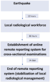
Workload management after the earthquake.
Figure 4
Figure 4
Traumatic pneumothorax, pneumomediastinum the patient was trapped for 19 hours by earthquake debris. Forty-three-year-old male. (a) On the axial images passing through the upper thoracic region, multiple air densities in the mediastinum (thin black arrows, pnomomediastinum), air densities near the left lung upper lobe (asterisk, pneumothorax), and diffuse subcutaneous emphysema more dominantly in the left hemithorax are seen (thick black arrows). (b) Pneumomediastinum (thin black arrows) and diffuse subcutaneous emphysema (thick black arrows) are seen in the axial sections passing through the middle part of the thorax. (c, d) Displaced rib fracture and extension of bone fragment into lung parenchyma on bone window axial section images are seen (thick white arrows). (e) Diffuse subcutaneous emphysema (thick black arrows) is seen in the axial sections passing through the inferior part of the thorax. (f) Pneumomediastinum (thin black arrows), pneumothorax (asterisk) and diffuse subcutaneous emphysema (thick black arrows) are seen in the coronal images.
Figure 5
Figure 5
Diffuse alveolar hemorrhage. The patient was trapped for 22 hours by earthquake debris. Forty-three- year-old male. Diffuse widespread ground glass opacities and consolidations. (a, c, e) Axial images from different levels; (b, d) coronal images from different levels; (f) sagittal.
Figure 6
Figure 6
Type 2 odontoid fracture. The patient was trapped for 9 hours by earthquake debris. Thirty-nine-year- old male. A fragmented and displaced fracture of the odontoid process (arrows) is seen on axial (a) and coronal (b) computed tomography images.
Figure 7
Figure 7
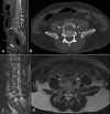
Traumatic listhesis and spinal cord injury. The patient was trapped for 31 hours by earthquake debris. Forty-one-year-old male. Retrolisthesis due to burst fracture of the L5 vertebrae (arrows) on the L5-S1 level. Spinal cord transection is seen on coronal Short tau inversion recovery (STIR) (c, arrows point to the two dissected edges of the spinal cord) (a) coronal computed tomography (CT) image, (b) axial CT image, (c) coronal STIR, (d) axial T2 weighted image.
Figure 8
Figure 8
Different patterns of traumatic abdominal injury. The patient was trapped for 35 hours by earthquake debris. Forty-seven-year-old male (a) American Association for the Surgery of Trauma (AAST) grade III injury in the right lobe-segment VII. (b) Left surrenal hematoma (circle), large perisplenic hematoma (arrows), splenic arterial injury, and corresponding contrast medium extravasation (thick arrow) (AAST grade V injury). (c) Large perisplenic hematoma (arrows), splenic arterial injury, and corresponding contrast medium extravasation (thick arrow). Sudden luminal narrowing in the left renal artery (circle) observed secondarily to blunt trauma. (d) Renal parenchymal infarct (arrows) due to the arterial narrowing shown in d (AAST grade V injury).
Figure 9
Figure 9
Intertrochanteric femur fracture. It was not possible to determine how long the patient was trapped by earthquake debris. Thirty-eight-year-old male. Displaced and comminuted fracture (arrows) in the right femoral intertrochanteric region on the coronal (a) and three-dimensional reconstruction (b) computed tomography images.
Figure 10
Figure 10
Distal tibial fracture. It was not possible to determine how long the patient was trapped by earthquake debris. Thirty-year-old male. (a) Medial cortical depression and internal displaced fracture (arrow) are seen in the distal metaphyseal–diaphyseal part of the tibia on axial (a), coronal (b), and sagittal (c) computed tomography images.
Figure 11
Figure 11
Proximal humeral fracture. It was not possible to determine how long the patient was trapped by earthquake debris. Sixty-year-old female. Fracture is seen in the surgical neck of humerus fracture (arrows) (a: axial view, b: coronal view, c: three-dimensional image).
Figure 12
Figure 12
Multiple lower extremity fractures. The patient was trapped for 23 hours by earthquake debris. Thirty-three-year-old male. Minimally displaced fractures are present in the medial malleolus of the tibia and distal metaphysis of the fibula on X-ray (a), coronal computed tomography (b), and three-dimensional reconstruction (c) images.
Figure 13
Figure 13
Pelvic and humeral fractures. The patient was trapped for 29 hours by earthquake debris. Seventy-six-year-old male. Displaced and comminuted fractures are seen in the left acetabulum and femoral neck (thin arrows) on axial (a) and coronal (c) computed tomography (CT) images. Large intramuscular hematoma due to the fractures is present on axial CT image (b, thick arrow). The fractures extend towards the iliac wing and inferior pubic ramus on the three-dimensional reconstruction image (d, arrow, Tile type A2 fracture).
Figure 14
Figure 14
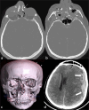
Multiple maxillofacial fractures and accompanying cranial hemorrhage. The patient was trapped for 17 hours by earthquake debris. Forty-four--year-old male. Multiple displaced fractures (thin arrows) are seen in maxillofacial bones in axial (a, b) and 3D reconstruction (c) computed tomography images. Extensive subdural hemorrhage causing a shift in midline structures accompanies the fractures (d, thick arrows).
Figure 15
Figure 15
Rhabdomyolysis. The patient was trapped for 39 hours by earthquake debris. Twenty-year-old male. Rhabdomyolysis due to brachial plexus damage secondary to cervical fracture. Rhabdomyolysis (arrows) is seen as hyperintense on short tau inversion recovery (a) and it enhances on post-contrast T1 weighted image (b). The displaced fracture in the bilateral arch of the C7 vertebrae (arrows) is seen in axial (c) and coronal (d) sections on computed tomography.
Figure 16
Figure 16
Compartment syndrome and rhabdomyolysis. The patient was trapped for 51 hours by earthquake debris. Thirty-four-year-old male. Left lower extremity is larger and subcutaneous edema is present (a-e). Common femoral artery, superficial femoral artery (SFA), and deep femoral artery are normal in the proximal segments of the left lower extremity (a-c). Due to increased intracompartmental pressure, left SFA narrows suddenly (d, g, h, arrows). Semitendinosus (e, circle) and gastrocnemius (f, arrows) muscles are hypodense; there is a ring-enhancing lesion in the popliteus muscle (f, circle) due to rhabdomyolysis.
Figure 17
Figure 17
Acute cerebral infarction. Forty-four-year-old male, diagnosed 50 hours after being rescued from rubble. Ischemic brain parenchyma is seen as a hypodense area on computed tomography (a); corresponding diffusion restriction can be seen on diffusion-weighted imaging (b) and apparent difussion coefficient map (c).
Figure 18
Figure 18
Deep vein thrombosis. Fifty-three-year-old female, diagnosed after rescue after 13 hours in the rubble. Thrombosis is seen in the superficial femoral vein (arrows) on grayscale ultrasound (a), axial (b), and longitudinal color Doppler ultrasonography images (c).
Figure 19
Figure 19
Pulmonary thromboembolism (PTE). The patient was trapped for 41 hours by earthquake debris. Forty-seven-year-old female. Filling defect corresponding to PTE, detected after 72 hours from the rescue from a rubble, is seen on axial (a, arrow) and coronal (b, arrow) images. A peripheral lung necrosis due to PTE is seen on the axial image with parenchymal windowing (c, arrow).
Figure 20
Figure 20
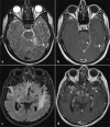
Viral meningitis (a, b) and tuberculous meningitis (c, d). Forty-three-year-old female and 73-year-old male, respectively. The images were obtained after 53 and 83 hours in the rubble, respectively. Viral meningitis: left tentorium cerebelli is seen thickened and hyperintense on T2 weighted image (WI) (a, arrow), left temporal lobe and neighboring dura is hyperintense on T2WI (a, double arrow) (a), increased contrast enhancement is present in both corresponding areas (b). Tuberculous meningitis: faint hyperintensities are present in the bilateral basal areas of the temporal lobes on fluid attenuated inversion recovery (c, arrows); increased contrast enhancement and ring-enhancing lesions can be seen in both corresponding areas (d, arrows).

Similar articles

Cited by

References

    1. Ishihara O, Yoshimura Y. Damages at Japanese assisted reproductive technology clinics by the Great Eastern Japan Earthquake of 2011. Fertil Steril. 2011;95(8):2568–2570. - PubMed
    1. United States Geological Survey (USGS), Earthquakes maps, Accessed February 15, 2023. [Internet]
    1. Anadolu Agency Infographics, Accessed February 15, 2023. [Internet]
    1. Earthquake news, Accessed March 8, 2023. [Internet]
    1. Iyama A, Utsunomiya D, Uetani H, et al. Emergency radiology after a massive earthquake: clinical perspective. Jpn J Radiol. 2018;36(11):641–648. - PubMed