

VEGFA-modified DPSCs combined with LC-YE-PLGA NGCs promote facial nerve injury repair in rats
- PMID: 37095964
- PMCID: PMC10121407
- DOI: 10.1016/j.heliyon.2023.e14626
VEGFA-modified DPSCs combined with LC-YE-PLGA NGCs promote facial nerve injury repair in rats
Abstract
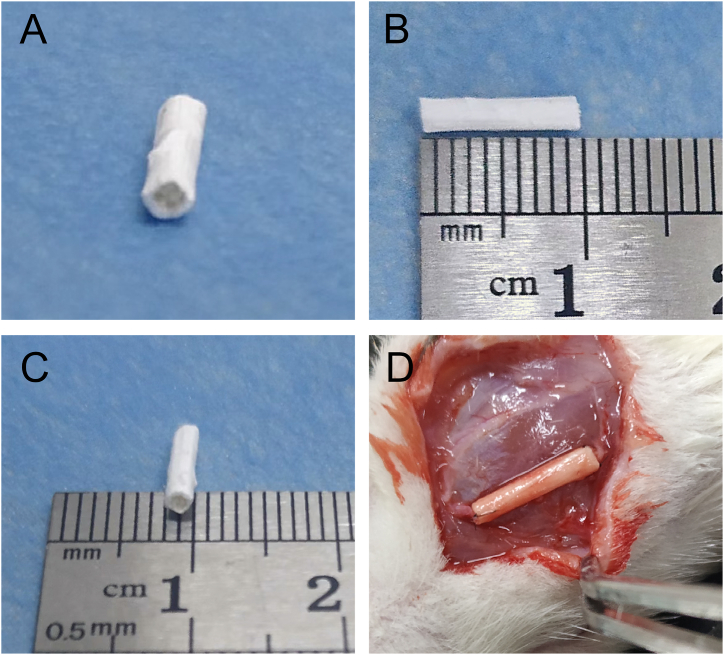
Objective: The aim of this research was to investigate the effect of vascular endothelial growth factor A (VEGFA)-overexpressing rat dental pulp stem cells (rDPSCs) combined with laminin-coated and yarn-encapsulated poly(l-lactide-co-glycolide) (PLGA) nerve guidance conduit (LC-YE-PLGA NGC) in repairing 10 mm facial nerve injury in rats.
Study design: rDPSCs isolated from rat mandibular central incisor were cultured and identified in vitro and further transfected with the lentiviral vectors (Lv-VEGFA). To investigate the role and mechanisms of VEGFA in neurogenic differentiation in vitro, semaxanib (SU5416), Cell Counting Kit-8 (CCK-8), real-time quantitative polymerase chain reaction (qPCR) and Western blotting were performed. Ten-millimeter facial nerve defect models in rats were established and bridged by LC-YE-PLGA NGCs. The repair effects were detected by transmission electron microscopy (TEM), compound muscle action potential (CMAP), immunohistochemistry and immunofluorescence.
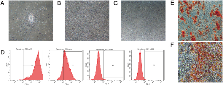
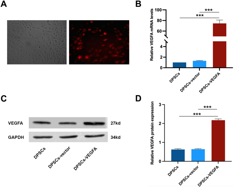
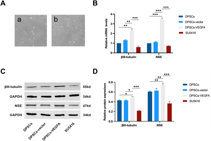
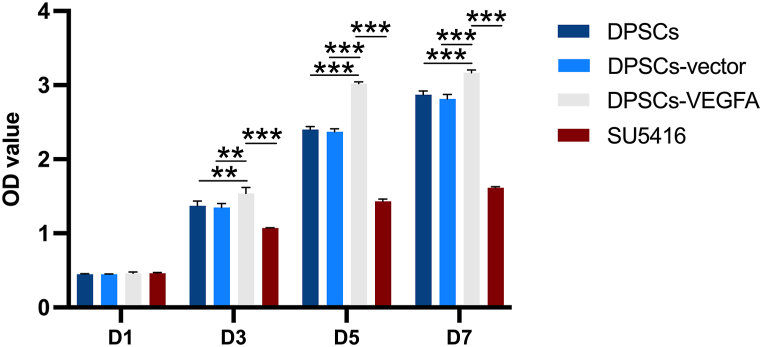
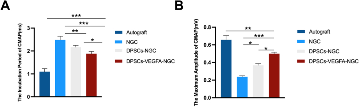
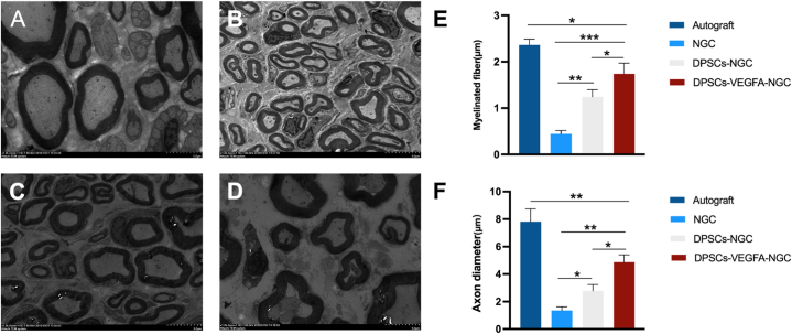
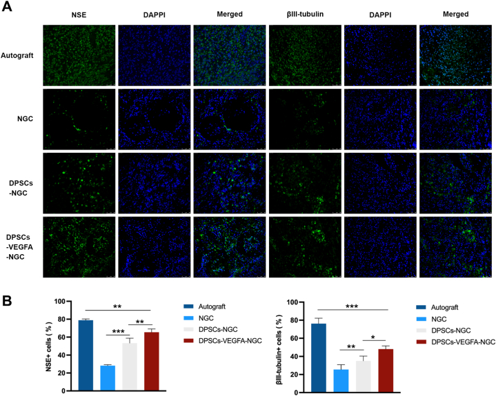
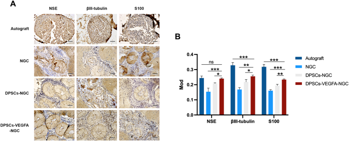
Results: Extracted cells exhibited spindle-shaped morphology, presented typical markers (CD44+CD90+CD34-CD45-), and presented multidirectional differentiation potential. The DPSCs with VEGFA overexpression were constructed successfully. VEGFA enhanced the proliferation and neural differentiation ability of rDPSCs, and the expression of neuron-specific enolase (NSE) and βIII-tubulin was increased. However, these trends were reversed with the addition of SU5416. This suggests that VEGFA mediates the above effects mainly through vascular endothelial growth factor receptor 2 (VEGFR2) binding. The LC-YE-NGC basically meet the requirements of facial nerve repair. For the in vivo experiment, the CMAP latency period was shorter in DPSCS-VEGFA-NGC group in comparison with other experimental groups, while the amplitude was increased. Such functional recovery correlated well with an increase in histological improvement. Further study suggested that VEGFA-modified DPSCs could increase the myelin number, thickness and axon diameter of facial nerve. NSE, βIII-tubulin and S100 fluorescence intensity and immunohistochemical staining intensity were significantly enhanced.
Conclusion: VEGFA-modified rDPSCs combined with LC-YE-PLGA NGCs have certain advantages in the growth and functional recovery of facial nerves in rats.
Keywords: Facial nerve defects; Neural differentiation; Neural tissue engineering; SU5416; VEGFA; rDPSCs.
© 2023 The Authors.
Conflict of interest statement
The authors declare that they have no competing interests.
Figures

Similar articles
-
Laminin-coated nerve guidance conduits based on poly(l-lactide-co-glycolide) fibers and yarns for promoting Schwann cells' proliferation and migration.J Mater Chem B. 2017 May 7;5(17):3186-3194. doi: 10.1039/c6tb03330j. Epub 2017 Apr 12. J Mater Chem B. 2017. PMID: 32263716
-
The involvement of Neuregulin-1 in the process of facial nerve injury repair through the utilization of dental pulp stem cells.BMC Oral Health. 2024 Feb 14;24(1):238. doi: 10.1186/s12903-024-03953-z. BMC Oral Health. 2024. PMID: 38355448 Free PMC article.
-
Chitosan Tubes Inoculated with Dental Pulp Stem Cells and Stem Cell Factor Enhance Facial Nerve-Vascularized Regeneration in Rabbits.ACS Omega. 2022 May 26;7(22):18509-18520. doi: 10.1021/acsomega.2c01176. eCollection 2022 Jun 7. ACS Omega. 2022. PMID: 35694480 Free PMC article.
-
Nanofibrous nerve guidance conduits decorated with decellularized matrix hydrogel facilitate peripheral nerve injury repair.Theranostics. 2021 Jan 1;11(6):2917-2931. doi: 10.7150/thno.50825. eCollection 2021. Theranostics. 2021. PMID: 33456580 Free PMC article.
-
Dental pulp stem cells overexpressing stromal-derived factor-1α and vascular endothelial growth factor in dental pulp regeneration.Clin Oral Investig. 2019 May;23(5):2497-2509. doi: 10.1007/s00784-018-2699-0. Epub 2018 Oct 12. Clin Oral Investig. 2019. PMID: 30315421
Cited by
-
Research status of facial nerve repair.Regen Ther. 2023 Oct 7;24:507-514. doi: 10.1016/j.reth.2023.09.012. eCollection 2023 Dec. Regen Ther. 2023. PMID: 37841661 Free PMC article. Review.
-
Advancements in Spinal Cord Injury Repair: Insights from Dental-Derived Stem Cells.Biomedicines. 2024 Mar 19;12(3):683. doi: 10.3390/biomedicines12030683. Biomedicines. 2024. PMID: 38540295 Free PMC article. Review.
-
KAT2A-mediated succinylation modification of notch1 promotes the proliferation and differentiation of dental pulp stem cells by activating notch pathway.BMC Oral Health. 2024 Mar 31;24(1):407. doi: 10.1186/s12903-024-03951-1. BMC Oral Health. 2024. PMID: 38556862 Free PMC article.
-
Investigation of the effects of Periplaneta americana (L.) extract on ischemic stroke based on combined multi-omics of gut microbiota.Front Pharmacol. 2024 Nov 28;15:1429960. doi: 10.3389/fphar.2024.1429960. eCollection 2024. Front Pharmacol. 2024. PMID: 39679371 Free PMC article.
-
ARMCX3 regulates ROS signaling, affects neural differentiation and inflammatory microenvironment in dental pulp stem cells.Heliyon. 2024 Aug 28;10(17):e37079. doi: 10.1016/j.heliyon.2024.e37079. eCollection 2024 Sep 15. Heliyon. 2024. PMID: 39296219 Free PMC article.
References
LinkOut - more resources
Full Text Sources
Research Materials
Miscellaneous
