. 2023 Apr 8:30:100623.
doi: 10.1016/j.bbih.2023.100623. eCollection 2023 Jul.

Effects of hydrogen gas inhalation on L-DOPA-induced dyskinesia

Affiliations

Effects of hydrogen gas inhalation on L-DOPA-induced dyskinesia

Glauce C Nascimento et al. Brain Behav Immun Health. .

Abstract

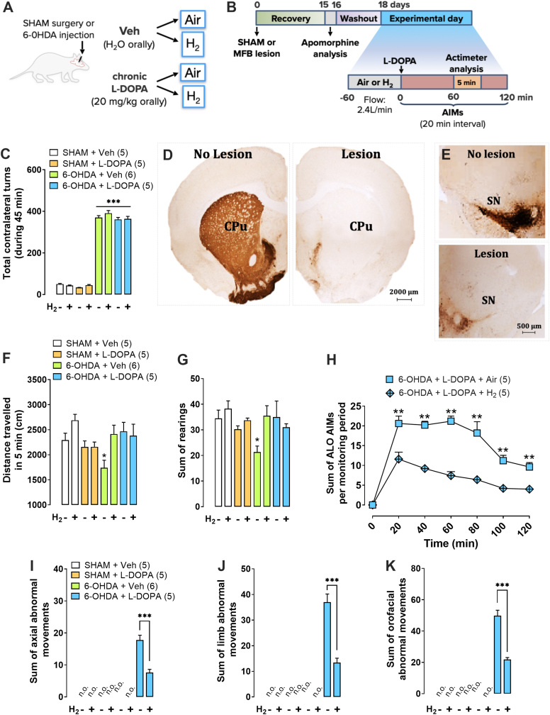
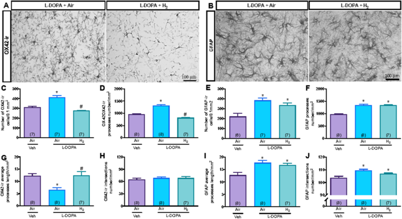
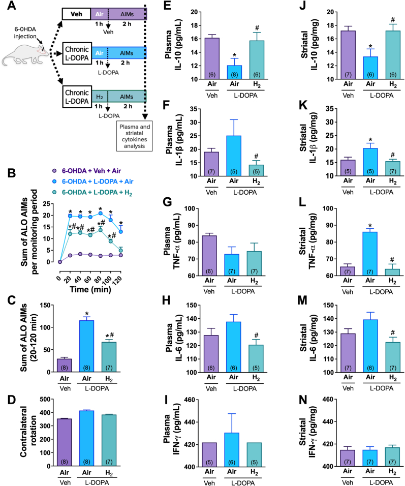
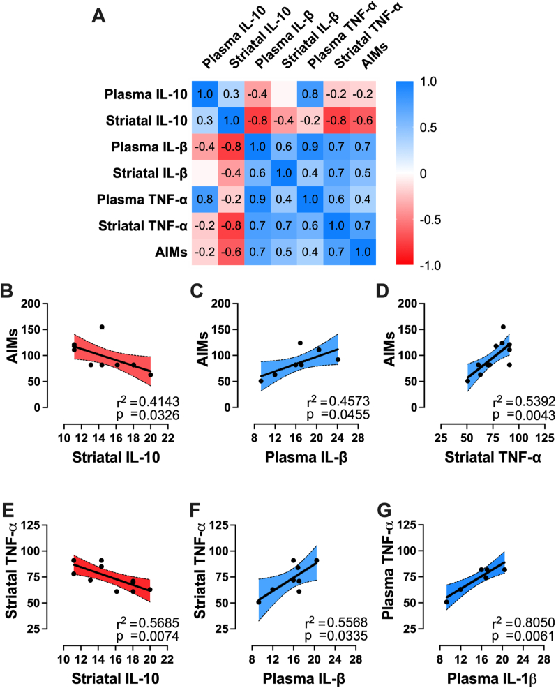
L-3,4-dihydroxyphenylalanine (L-DOPA)-induced dyskinesia is a side effect of Parkinson's disease treatment and it is characterized by atypical involuntary movements. A link between neuroinflammation and L-DOPA-induced dyskinesia has been documented. Hydrogen gas (H2) has neuroprotective effects in Parkinson's disease models and has a major anti-inflammatory effect. Our objective is to test the hypothesis that H2 inhalation reduces L-DOPA-induced dyskinesia. 15 days after 6-hydroxydopamine lesions of dopaminergic neurons were made (microinjection into the medial forebrain bundle), chronic L-DOPA treatment (15 days) was performed. Rats were exposed to H2 (2% gas mixture, 1 h) or air (controls) before L-DOPA injection. Abnormal involuntary movements and locomotor activity were conducted. Striatal microglia and astrocyte was analyzed and striatal and plasma samples for cytokines evaluation were collected after the abnormal involuntary movements analysis. H2 inhalation attenuated L-DOPA-induced dyskinesia. The gas therapy did not impair the improvement of locomotor activity achieved by L-DOPA treatment. H2 inhalation reduced activated microglia in the lesioned striatum, which is consistent with the observed reduced pro-inflammatory cytokines levels. Display of abnormal involuntary movements was positively correlated with plasma IL-1β and striatal TNF-α levels and negatively correlated with striatal IL-10 levels. Prophylactic H2 inhalation decreases abnormal involuntary movements in a preclinical L-DOPA-induced dyskinesia model. The H2 antidyskinetic effect was associated with decreased striatal and peripheral inflammation. This finding has a translational importance to L-DOPA-treated parkinsonian patients' well-being.

Keywords: 6-Hydroxydopamine; Neuroinflammation; Parkinson's disease; Striatum; Systemic inflammation.


Conflict of interest statement

The authors declare that they have no known competing financial interests or personal relationships that could have appeared to influence the work reported in this paper.

Figures

Fig. 1
Fig. 1
Molecular hydrogen (H2) inhalation reduces AIMS without interfering with the anti-parkinsonian effect of L-DOPA. (A, B) Schematic protocol. After Sham surgery or 6-OHDA injection, the rats were analyzed in the apomorphine-induced rotational test for lesion confirmation. Then, Sham or 6-OHDA-lesioned rats were treated chronically (15 days) with L-DOPA or its vehicle. Subsequently before the last L-DOPA injection, Air mixture or H2 inhalation was performed for a 1-h period, followed by the L-DOPA injection and abnormal involuntary movements on axial, limb and orofacial parameters (ALO AIMs) and locomotor activity were measured. (C) 6-OHDA lesion significantly increased total contralateral turns induced by apomorphine. (D) Photomicrographs of coronal brain sections illustrating the loss of TH positive immunolabeling in the striatum fibers (D) and substantia nigra compacta neurons (E), scale bars = 1200 μm. (F, G) H2 treatment does not alter the L-DOPA effect on the distance travelled (F) and rearing (G) in the actimeter. (H) Time course of the appearance of dyskinetic manifestation with 6-OHDA + L-DOPA + Air mixture compared to 6-OHDA + L-DOPA + H2 inhalation (p < 0.001) across time (for each 20-min period). (I, J, K) show the co-administration of L-DOPA and H2 inhalation effects on the axial (I), limb (J) and orofacial (K) AIMs over 120 min. Sham rats treated with vehicle of L-DOPA (SHAM + Veh, white); Sham rats treated chronically with L-DOPA (SHAM + L-DOPA, orange); 6-OHDA lesioned rats treated with vehicle of L-DOPA (6-OHDA + Veh, green); 6-OHDA lesioned rats treated chronically with L-DOPA (6-OHDA + L-DOPA, blue). AIMs were analyzed using 2-Way RM ANOVA analysis with Tukey's multiple comparisons post-hoc test. Distance travelled was analyzed by ordinary One-Way ANOVA with Fisher post-hoc test. Data are reported as mean ± SEM. ***p < 0.001, **p < 0.01, *p < 0.05. A total of 42 rats were tested in this experiment (n = 5/6 per group, the specific amounts of each group are indicated in parentheses). (For interpretation of the references to color in this figure legend, the reader is referred to the Web version of this article.)
Fig. 2
Fig. 2
Molecular hydrogen (H2) inhalation prevented the reactivity of striatal microglia with no impact in striatal astrocytes morphology. Microglia analysis by OX-42-ir staining. (A) representative photomicrographs of striatum slices of lesioned rats treated chronically with L-DOPA inhaling (L-DOPA + H2) or not (L-DOPA) H2. Quantification of OX-42-ir images indicates the number of OX-42-ir cells (B), total number of processes cells (C), average processes length (D), and intersections number between the processes (G). H2 treatment induced a decrease in the number of OX42-ir positive cells, in the average of processes number and length, a without affecting the number of intersections in L-DOPA-treated rats (Fig. H; p > 0.05). Astrocyte analysis by GFAP staining. (B) representative photomicrographs of striatum slices of 6-OHDA-lesioned rats treated chronically with L-DOPA inhaling (L-DOPA + H2) or not (L-DOPA) H2. Quantification of GFAP images indicates the number of GFAP cells (E), the total number of processes cells (F), average processes length (I), and intersections number between the processes (J). H2 treatment caused no alterations in the number of GFP positive cells, processes number, length, and intersections (p > 0.05). 6-OHDA-lesioned rats were represented by the purple bars; 6-OHDA-lesioned rats treated chronically with L-DOPA inhaling Air was represented by the blue bars, and 6-OHDA-lesioned rats treated chronically with L-DOPA and inhaling H2 were represented by the green bars. Data were statistically analyzed by ordinary One-Way ANOVA with Fisher post-hoc test. Bars are represented as mean ± SEM. #p < 0.05 vs. L-DOPA + Air, *p < 0.05 vs. Veh + Air groups. A total of 23 rats were tested in this experiment (n = 7/8 per group, the specific amounts of each group are indicated in parentheses). (For interpretation of the references to color in this figure legend, the reader is referred to the Web version of this article.)
Fig. 3
Fig. 3
Molecular hydrogen (H2) inhalation improves peripheral and striatal inflammation in L-DOPA-treated 6-OHDA-lesioned rats. (A) Schematic timeline for cytokine measurements. (B) Time course of the appearance of dyskinetic manifestation of the animals used for cytokines measurement (6-OHDA + Veh + Air mixture; 6-OHDA + L-DOPA + Air mixture; 6-OHDA + L-DOPA + H2 inhalation; p < 0.001) across time (for each 20-min period). (C) Effect of the co-administration of L-DOPA and H2 inhalation effects on the sum of axial, limb and orofacial AIMs over 120 min of these animals. (D) These animals were tested also in the apomorphine test, 6-OHDA lesion significantly increased total contralateral turns induced by apomorphine. Plasma and striatal levels of IL-10 (E and J), IL-1β (F and K), TNF-α (G and L), IL-6 (H and M) and IFN-γ (I and N) 2 h after L-DOPA administration in rats inhaling H2 (L-DOPA + H2) or not (L-DOPA + Air). 6-OHDA-lesioned rats were represented by the purple bars; 6-OHDA-lesioned rats treated chronically with L-DOPA inhaling Air were represented by the blue bars, and 6-OHDA-lesioned rats treated chronically with L-DOPA and inhaling H2 were represented by the green bars. H2 inhalation caused an increase in plasma and striatal levels of IL-10 (p < 0.05) and a decrease in plasma and striatal levels of IL-1β (p < 0.05), striatal levels of TNF-α (p < 0.05) and plasma and striatal levels of IL-6 (p < 0.05) in L-DOPA treated rats. All cytokine measurements were statistically analyzed by ordinary One-Way ANOVA with Fisher's post-hoc test. Bars are represented as mean ± SEM. #p < 0.05 vs. L-DOPA + Air (blue bars), *p < 0.05 vs. Veh + Air (purple bars) groups. A total of 22 rats were tested in this experiment (n = 7/8 per group, the specific amounts of each group are indicated in parentheses). (For interpretation of the references to color in this figure legend, the reader is referred to the Web version of this article.)
Fig. 4
Fig. 4
AIMS were negatively correlated with striatal IL-10 and positively correlated with plasma IL-1β and striatal TNF-α levels. (A) Correlation matrix of plasma and striatal IL-10, IL-1β, TNF-α levels, and AIMs. The correlation coefficient (r) of each correlation is inside its correspondent box ranging from −1 (darkest red gradient) to +1 (darkest blue gradient). A two-tailed p-value was chosen to evaluate the statistical significance of each correlation. The coefficient of determination (r2) between a statistically correlated pair of variables was calculated and a best-fit linear regression line (dark full line in the plot) was built for each two variables in one panel (panels B–G). Correlation plot was set at 95% confidence interval (shadowed area with the exactly gradient of the correspondent correlation coefficient - r) surrounded by dark dashed lines. There was a statistical correlation between: AIMS and striatal IL-10 levels (B); AIMS with plasma IL-1β (C) and AIMs with striatal TNF-α (D) levels; striatal TNF-α levels with striatal IL-10 (E), plasma IL-1β with striatal TNF-α (F), and plasma TNF-α (G) levels. (For interpretation of the references to color in this figure legend, the reader is referred to the Web version of this article.)

Similar articles

Cited by

References

    1. Amorim M.R., et al. Increased lipopolysaccharide-induced hypothermia in neurogenic hypertension is caused by reduced hypothalamic PGE 2 production and increased heat loss. J. Physiol. 2020;598:4663–4680. - PubMed
    1. Amri F., Ghouili I., Amri M., Carrier A., Masmoudi-Kouki O. Neuroglobin protects astroglial cells from hydrogen peroxide-induced oxidative stress and apoptotic cell death. J. Neurochem. 2017;140:151–169. - PubMed
    1. Bariotto-Dos-Santos K., Ribeiro D.L., Guimarães R.P., Padovan-Neto F.E. Rating L-DOPA-induced dyskinesias in the unilaterally 6-OHDA-lesioned rat model of Parkinson's disease. J. Vis. Exp. 2021:176. doi: 10.3791/62924. Oct 4. - DOI - PubMed
    1. Barnum C.J., et al. Exogenous corticosterone reduces l-DOPA-induced dyskinesia in the hemi-parkinsonian rat: role for interleukin-1β. Neuroscience. 2008;156:30–41. - PMC - PubMed
    1. Barriola S., Pérez-Cerdá F., Matute C., Bribián A., López-Mascaraque L. A clonal NG2-glia cell response in a mouse model of multiple sclerosis. Cells. 2020;9:1279. - PMC - PubMed

LinkOut - more resources