. 2023 Apr 10:27:200-215.
doi: 10.1016/j.bioactmat.2023.04.002. eCollection 2023 Sep.

In situ self-assembled organoid for osteochondral tissue regeneration with dual functional units

Affiliations

In situ self-assembled organoid for osteochondral tissue regeneration with dual functional units

Zhen Yang et al. Bioact Mater. .

Abstract

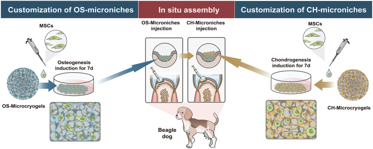
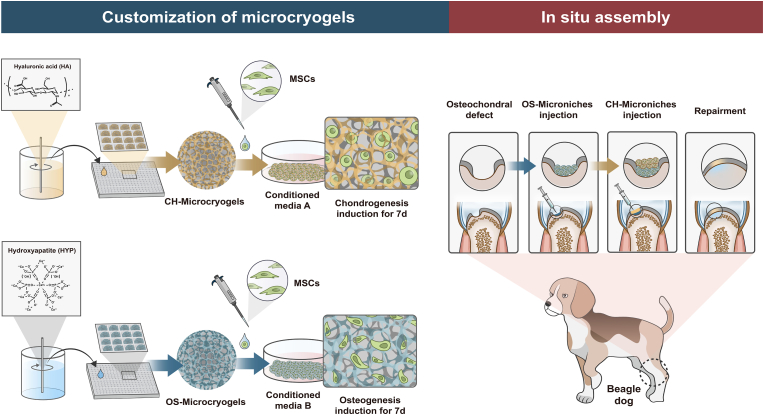
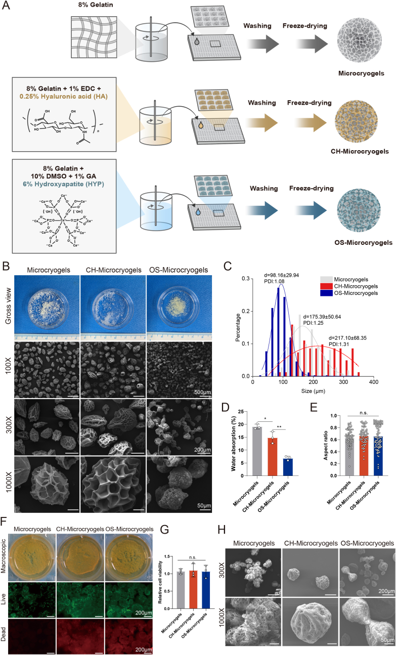
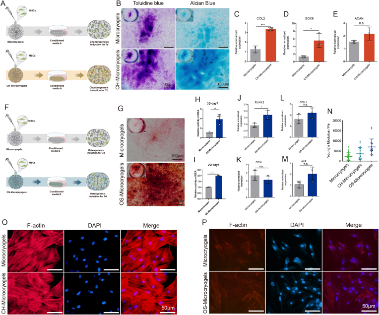
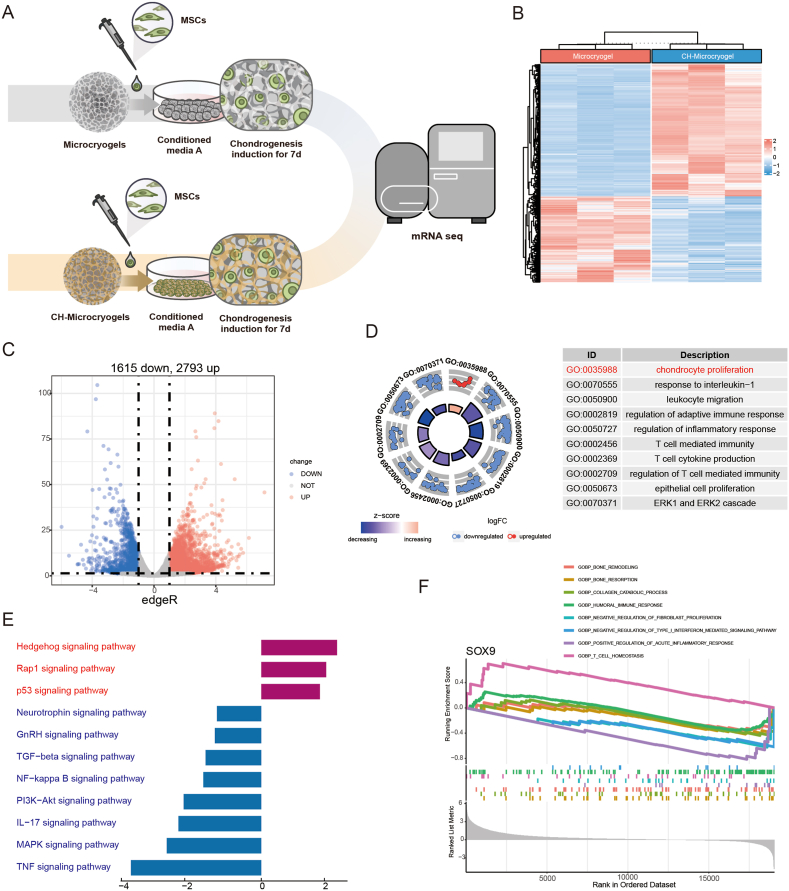
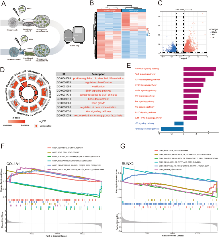
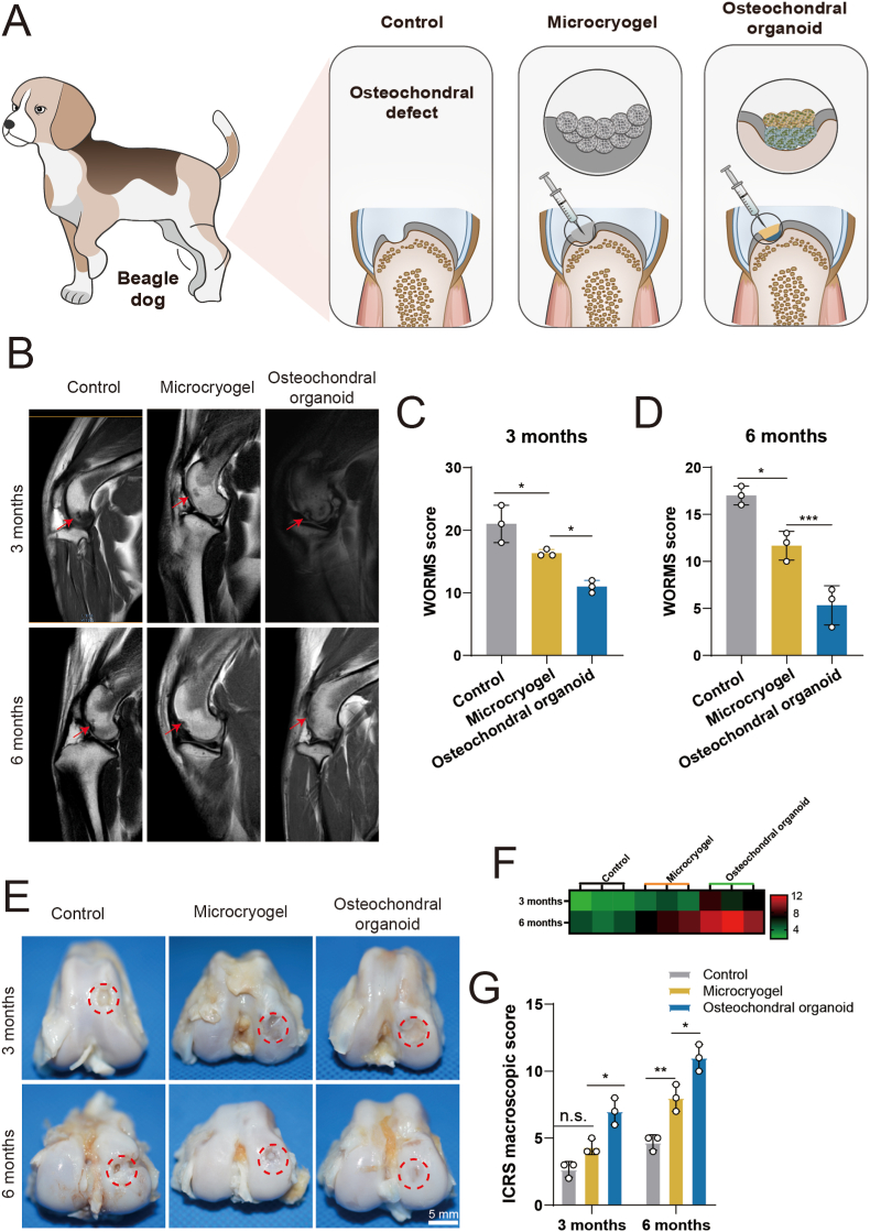
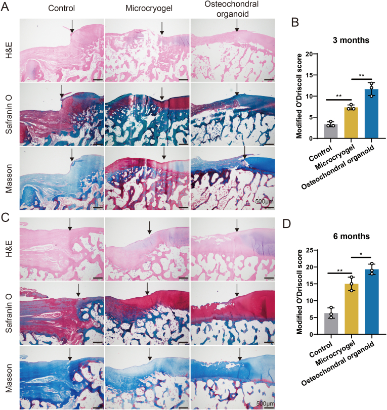
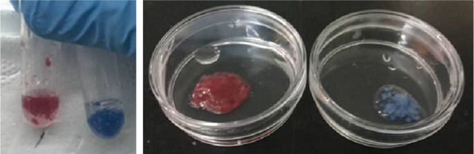
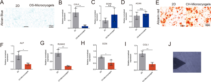
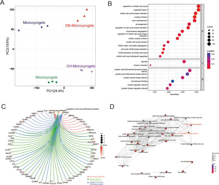
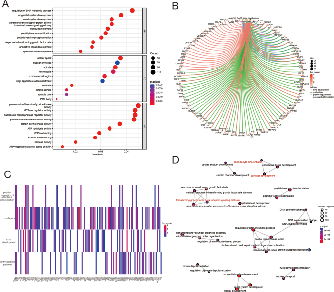
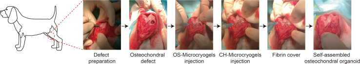
The regeneration of hierarchical osteochondral units is challenging due to difficulties in inducing spatial, directional and controllable differentiation of mesenchymal stem cells (MSCs) into cartilage and bone compartments. Emerging organoid technology offers new opportunities for osteochondral regeneration. In this study, we developed gelatin-based microcryogels customized using hyaluronic acid (HA) and hydroxyapatite (HYP), respectively for inducing cartilage and bone regeneration (denoted as CH-Microcryogels and OS-Microcryogels) through in vivo self-assembly into osteochondral organoids. The customized microcryogels showed good cytocompatibility and induced chondrogenic and osteogenic differentiation of MSCs, while also demonstrating the ability to self-assemble into osteochondral organoids with no delamination in the biphasic cartilage-bone structure. Analysis by mRNA-seq showed that CH-Microcryogels promoted chondrogenic differentiation and inhibited inflammation, while OS-Microcryogels facilitated osteogenic differentiation and suppressed the immune response, by regulating specific signaling pathways. Finally, the in vivo engraftment of pre-differentiated customized microcryogels into canine osteochondral defects resulted in the spontaneous assembly of an osteochondral unit, inducing simultaneous regeneration of both articular cartilage and subchondral bone. In conclusion, this novel approach for generating self-assembling osteochondral organoids utilizing tailor-made microcryogels presents a highly promising avenue for advancing the field of tissue engineering.

Keywords: Mesenchymal stem cells; Microcryogels; Organoid; Osteochondral regeneration; Self-assembly.


Conflict of interest statement

The authors declare no competing interests.

Figures

Image 1
Graphical abstract
Fig. 1
Fig. 1
Schematic diagram of the study design.
Fig. 2
Fig. 2
Microscopic observation and cytocompatibility analysis of control microcryogels, CH-Microcryogels, and OS-Microcryogels. (A) Schematic of fabricating customized microcryogels. (B) Macroscopic view and SEM images of customized microcryogels at different magnifications (100X, 300X and 1000X). (C) Diameter distribution and polydispersity index (PDI) of customized microcryogels. (D) Water absorption rate of customized microcryogels. (E) Aspect ratio evaluation of customized microcryogels. (F) Macroscopic view and live/dead staining of MSCs cultured in customized microcryogels for 7 d. Representative 3D reconstruction images show live (green) cells and dead (red) cells. (G) Quantitative analysis of cell viability in customized microcryogels (n = 4). (H) SEM images of customized microcryogels after MSCs were seeded and grown for 14 d. (n.s. represents no significant difference).
Fig. 3
Fig. 3
In vitro chondrogenic and osteogenic pre-differentiation induction of MSC-seeded microcryogels. (A) Schematic of the chondrogenic induction process. (B) Toluidine blue and Alcian blue staining of control microcryogels and CH-Microcryogels after 7 d of chondrogenic induction. Relative gene expression of (C) COL2, (D) SOX9, and (E) ACAN. (F) Schematic of the osteogenic induction process. (G) Alizarin red staining of control microcryogels and OS-Microcryogels after 7 d of osteogenic induction. (H) Quantitative detection of ALP in MSCs after 7 d of osteogenic induction on control microcryogels and OS-Microcryogels. (I) Quantitative detection of ALP in MSCs on 2D culture plates. Relative gene expression of (J) RUNX2, (K) OCN, (L) COL1, and (M) ALP. (N) Atomic force microscopy (AFM) of microcryogels after 7 d of chondrogenic and osteogenic induction. F-actin and DAPI staining of MSCs cultured in microcryogels after 7 d of (O) chondrogenic induction and (P) osteogenic induction. (*p < 0.05, **p < 0.01, ***p < 0.005, n. s. represents no significant difference, n = 3).
Fig. 4
Fig. 4
In vitro self-assembly of osteochondral organoids over 14 d. (A) Schematic of in vitro self-assembly process of osteochondral organoids. (B) Gross observation of self-assembled osteochondral organoids. (C) The self-assembled osteochondral organoid was incised in the axial position to allow separate analysis of the chondrogenic and osteogenic components. (D) SEM image of self-assembled osteochondral organoids, showing the CH-Microcryogel part on the left of the dotted line and the OS-Microcryogel part on the right. (E) Cell tracking of self-assembled osteochondral organoids after 14 d of osteochondral induction. Red indicates MSCs in the CH-Microcryogel part, and green indicates MSCs in the OS-Microcryogel part. (F) Live/dead staining of the chondrogenic and osteogenic parts of self-assembled osteochondral organoids. (G) Relative chondrogenic gene expression of CH-Microcryogels and CH-organoids (chondrogenic part of osteochondral organoid). (H) Relative osteogenic gene expression of OS-Microcryogels and OS-organoids (osteogenic part of osteochondral organoid). (I) Macroscopic observations and H&E staining of CH-Microcryogels and OS-Microcryogels 1 week after subcutaneous implantation in nude rats (yellow arrows indicate new blood vessels). (J) Stress-strain curve of osteochondral organoid together with CH-organoid and OS-organoid components. (K) Young's modulus of osteochondral organoid together with CH-organoid and OS-organoid components.
Fig. 5
Fig. 5
mRNA-seq and DEG analyses of MSCs grown in control microcryogels and CH-Microcryogels after 7 d of chondrogenic induction. (A) Schematic of chondrogenic induction before mRNA-seq. (B) Heatmap distribution of control microcryogels and CH-Microcryogels. (C) Volcano diagram of control microcryogels and CH-Microcryogels (1615 downregulated DEGs, 2793 upregulated DEGs). (D) The significant GO of DEGs indicated that the effects of CH-Microcryogels on the regulation of MSCs were associated with chondrocyte proliferation and immunosuppression. (E) KEGG enrichment analysis of targeted genes indicated that the effects of CH-Microcryogels on the regulation of MSCs were associated with the hedgehog, rap1, TGF-β, PI3K-Akt, and MAPK signaling pathways. (F) GSEA of SOX9 in GO terms of bone remodeling, bone resorption, collagen catabolic process, humoral immune response, negative regulation of fibroblast proliferation, negative regulation of type I interferon mediated signaling pathway, positive regulation of acute inflammatory response, and T-cell homeostasis.
Fig. 6
Fig. 6
mRNA-seq and DEG analyses of MSCs grown in control microcryogels and OS-Microcryogels after 7 d of osteogenic induction. (A) Schematic of osteogenic induction before mRNA-seq. (B) Heatmap distribution of control microcryogels and OS-Microcryogels. (C) Volcano diagram of control microcryogels and OS-Microcryogels (2185 downregulated DEGs, 3213 upregulated DEGs). (D) The significant GO of upregulated DEGs indicated that the effects of OS- Microcryogels on the regulation of MSCs were associated with osteoblast differentiation, ossification, bone development, bone growth, and bone mineralization. (E) KEGG enrichment analyses of targeted genes indicated that the effects of OS-Microcryogels on the regulation of MSCs were associated with the PI3K-Akt, FoxO, TGF-β, MAPK, and Wnt signaling pathways. (F) GSEA of COL1A1 in GO terms of activation of MAPK activity, bone cell development, positive regulation of macrophage migration, regulation of macrophage migration, transforming growth factor-beta production and vascular associated smooth muscle contraction. (G) GSEA of RUNX2 in the GO terms of positive regulation of osteoclast differentiation, response to transforming growth factor-beta, etc.
Fig. 7
Fig. 7
In vivo analysis of osteochondral regeneration using in situ self-assembled organoids, including MRI and macroscopic evaluation of repair tissues. (A) Schematic of the in vivo experiment in beagle dogs. (B) MRI evaluation of the repair tissue at 3 and 6 months post-surgery. Red arrows indicate the region of repair tissue. (C, D) WORMS scoring of repair tissue at 3 and 6 months. (E) Macroscopic evaluation of repair tissue at 3 and 6 months. Red circles indicate the defect area. (F) Heatmap of ICRS macroscopic scores. (G) ICRS macroscopic scores of repair tissue at 3 and 6 months. (*p < 0.05, **p < 0.01, ***p < 0.005, n. s. represents no significant difference, n = 3).
Fig. 8
Fig. 8
Histological evaluation of repair tissue in osteochondral defects. (A, C) H&E, safranin O and Masson staining of repair tissue at 3 and 6 months post-surgery. Black solid arrows indicate the repair interface. (B, D) Modified O'Driscoll scores for histological evaluation of cartilage repair after 3 and 6 months (n = 3). (*p < 0.05, **p < 0.01).
figs1
figs1
General view of CH-Microcryogels and OS-Microcryogels suspended in PBS.
figs2
figs2
Alcian blue staining of MSCs cultured on 2D plates and OS-Microcryogels after 7 days of chondrogenic induction. (B) Relative gene expression of COL2 related to chondrogenic differentiation. (C) Relative gene expression of SOX9 related to chondrogenic differentiation. (D) Relative gene expression of ACAN related to chondrogenic differentiation. (E) Alizarin red staining of 2D plates and CH-Microcryogels after 7 days of osteogenic induction. (F) Relative gene expression of ALP related to osteogenic differentiation. (G) Relative gene expression of RUNX2 related to osteogenic differentiation. (H) Relative gene expression of OCN related to osteogenic differentiation. (I) Relative gene expression of COL1 related to osteogenic differentiation. (J) Atomic force microscope (AFM) detection of microcryogels.
figs3
figs3
mRNA-seq and differentially expressed gene (DEG) analyses of MSCs of microgels and CH microgels after 7 days of chondrogenic induction. (A) PCA of mRNA-seq. (B) The bubble chart represents the significantly enriched pathways identified in the GO analysis. (C) Circular plot of DEGs mainly associated with the biological process of chondrocyte proliferation, positive regulation of cell cycle process, and regulation of cell cycle G2/M phase transition. (D) The PPI network of DEGs.
figs4
figs4
mRNA-seq and DEG analyses of MSCs of microgels and OS-Microcyrogels after 7 days of osteogenic induction. (A) The bubble chart represents the significantly enriched pathways identified in the GO analysis. (B) Circular plot of DEGs mainly associated with the biological process of bone development, ossification, and positive regulation of osteoblast differentiation. (C) Heatmap of DEGs mainly associated with the biological process of positive regulation of osteoblast differentiation, ossification, bone development, and BMP signaling pathway. (D) The PPI network of DEGs.
figs5
figs5
The higher magnification image of H&E staining of OS-Microcryogels 1 week after subcutaneous implantation in nude rats (yellow arrows indicate new blood vessels).
figs6
figs6
Surgical process of in situ assembly of microniches.

References

    1. Ansari S., Khorshidi S., Karkhaneh A. Engineering of gradient osteochondral tissue: from nature to lab. Acta Biomater. 2019;87:41–54. - PubMed
    1. Niu X., Li N., Du Z., Li X. Integrated gradient tissue-engineered osteochondral scaffolds: challenges, current efforts and future perspectives. Bioact. Mater. 2023;20:574–597. - PMC - PubMed
    1. Lee J., Lee S., Huh S.J., Kang B.J., Shin H. Directed regeneration of osteochondral tissue by hierarchical assembly of spatially organized composite spheroids. Adv. Sci. 2022;9(3) - PMC - PubMed
    1. Seo S.J., Mahapatra C., Singh R.K., Knowles J.C., Kim H.W. Strategies for osteochondral repair: focus on scaffolds. J. Tissue Eng. 2014;5 - PMC - PubMed
    1. Zhang B., Huang J., Narayan R.J. Gradient scaffolds for osteochondral tissue engineering and regeneration. J. Mater. Chem. B. 2020;8(36):8149–8170. - PubMed

LinkOut - more resources