. 2023 Apr 20;7(5):e874.
doi: 10.1097/HS9.0000000000000874. eCollection 2023 May.

Identification of Adult Patients With Classical Dyskeratosis Congenita or Cryptic Telomere Biology Disorder by Telomere Length Screening Using Age-modified Criteria

Affiliations

Identification of Adult Patients With Classical Dyskeratosis Congenita or Cryptic Telomere Biology Disorder by Telomere Length Screening Using Age-modified Criteria

Mareike Tometten et al. Hemasphere. .

Abstract

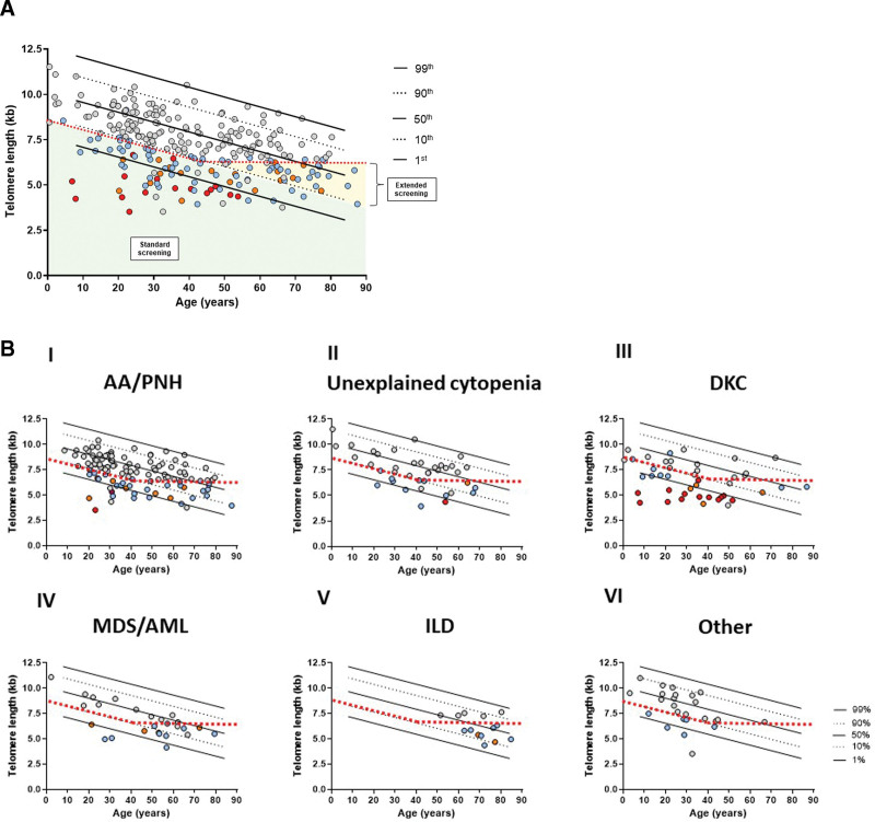
Telomere biology disorders (TBD) result from premature telomere shortening due to pathogenic germline variants in telomere maintenance-associated genes. In adults, TBD are characterized by mono/oligosymptomatic clinical manifestations (cryptic TBD) contributing to severe underdiagnosis. We present a prospective multi-institutional cohort study where telomere length (TL) screening was performed in either newly diagnosed patients with aplastic anemia (AA) or if TBD was clinically suspected by the treating physician. TL of 262 samples was measured via flow-fluorescence in situ hybridization (FISH). TL was considered suspicious once below the 10th percentile of normal individuals (standard screening) or if below 6.5 kb in patients >40 years (extended screening). In cases with shortened TL, next generation sequencing (NGS) for TBD-associated genes was performed. The patients referred fell into 6 different screening categories: (1) AA/paroxysmal nocturnal hemoglobinuria, (2) unexplained cytopenia, (3) dyskeratosis congenita, (4) myelodysplastic syndrome/acute myeloid leukemia, (5) interstitial lung disease, and (6) others. Overall, TL was found to be shortened in 120 patients (n = 86 standard and n = 34 extended screening). In 17 of the 76 (22.4%) standard patients with sufficient material for NGS, a pathogenic/likely pathogenic TBD-associated gene variant was identified. Variants of uncertain significance were detected in 17 of 76 (22.4%) standard and 6 of 29 (20.7%) extended screened patients. Expectedly, mutations were mainly found in TERT and TERC. In conclusion, TL measured by flow-FISH represents a powerful functional in vivo screening for an underlying TBD and should be performed in every newly diagnosed patient with AA as well as other patients with clinical suspicion for an underlying TBD in both children and adults.


Conflict of interest statement

THB and FB have a long-ranging scientific collaboration with Repeat Dx., Vancouver, Canada. All the other authors have no conflicts of interest to disclose.

Figures

Figure 1.
Figure 1.
Flow-chart of screening work-up. All patients included in the Aachen TBD registry (ATR) were screened with flow-FISH. Telomeres were considered critically short when (1) lymphocyte TL was <10th percentile or if lymphocyte TL was >10th but granulocyte TL less than first percentile (standard screening) or (2) age was >40 y and lymphocyte TL <6.5 kb (extended screening). Patients with critically short TL underwent NGS with analysis of TBD-related genes. +n = 7 patients had commercial NGS. ++n = 1 patient had commercial NGS. *Patient had 1 more variant (VUS). **Likely pathogenic includes TERC variants that were not classified according to the (ACMG) criteria (see Methods section). ***Three patients had >1 variant (VUS). ****One patient had 2 variants. ACMG = American College of Medical Genetics and Genomics; kb = kilobase; NGS = next generation sequencing; TBD = telomere biology disorder; TL = telomere length; VUS = variant of uncertain significance.
Figure 2.
Figure 2.
Telomere length measurement of the entire registry cohort. (A). Lymphocyte TL (given in kb) of 261 patients: first, 10th, 50th, 90th, and 99th percentiles are indicated. Blue, orange, and red dots show 105 patients with TL under 10th percentile (standard screening) or <6.5 kb if aged >40 y (extended screening), respectively, in whom NGS was performed. Of those, detection of variants is indicated as follows: Red dots indicate patients with pathogenic and likely pathogenic variants (n = 17), including TERC variants that were not classified according to the ACMG criteria (see Methods section). Orange dots represent patients with VUS (n = 24). Blue dots indicate no variant found in NGS. Gray dots indicate patients without NGS screening. Red dotted line indicates TL threshold as follows: <10th percentile until 40 y; <6.5 kb beyond 40 y. (B). Distribution of the TL (kb) within the respective diagnosis categories; AA/PNH (I), unexplained cytopenia (II), DKC (III), MDS/AML (IV), ILD (V), and other (VI). First, 10th, 90th, and 99th percentiles are indicated. Red dotted line indicates TL threshold as follows: <10th percentile until 40 y; <6.5 kb beyond 40 y. AA = aplastic anemia; ACMG = American College of Medical Genetics and Genomics; AML = acute myeloid leukemia; DKC = dyskeratosis congenital; ILD = interstitial lung disease; kb = kilobase; MDS = myelodysplastic syndrome; NGS = next generation sequencing; PNH = paroxysmal nocturnal hemoglobinuria; TL = telomere length; VUS = variants of uncertain significance.
Figure 3.
Figure 3.
Absolute number of cases within the respective diagnosis categories. NGS screening is shown including patients not screened, because of not fulfilling the criteria. Absolute numbers of variants are shown after classification according to the ACMG. Likely pathogenic variants include TERC variants that were not classified according to the ACMG criteria (see Methods section). *One patient had each 1 pathogenic variant and 1 VUS. +One patient had 2 VUS. #Two patients had each 1 likely pathogenic variant and 2 VUS; 1 patient had 1 likely pathogenic variant and 1 VUS. AA = aplastic anemia; ACMG = American College of Medical Genetics and Genomics; AML = acute myeloid leukemia; DKC = dyskeratosis congenita; ILD = interstitial lung disease; LP = likely pathogenic; MDS = myelodysplastic syndrome; NGS = next generation sequencing; P = pathogenic; PNH = paroxysmal nocturnal hemoglobinuria; VUS = variant of uncertain significance.
Figure 4.
Figure 4.
Distribution of detected genetic variants in TBD-associated genes. (A) Pathogenic/likely pathogenic variants in standard screening patients. (B) Variants of uncertain significance in standard screening patients. (C) Variants of uncertain significance in extended screening patients. LP = likely pathogenic; N = number; P = pathogenic; TBD = telomere biology disorder; VUS = variant of uncertain significance.

References

    1. Martinez P, Blasco MA. Telomeric and extra-telomeric roles for telomerase and the telomere-binding proteins. Nat Rev Cancer. 2011;11:161–176. - PubMed
    1. Townsley DM, Dumitriu B, Young NS. Bone marrow failure and the telomeropathies. Blood. 2014;124:2775–2783. - PMC - PubMed
    1. Tummala H, Walne A, Dokal I. The biology and management of dyskeratosis congenita and related disorders of telomeres. Expert Rev Hematol. 2022;15:685–696. - PubMed
    1. Calado RT, Young NS. Telomere diseases. N Engl J Med. 2009;361:2353–2365. - PMC - PubMed
    1. Tometten M, Kirschner M, Isfort S, et al. . Transient elastography in adult patients with cryptic dyskeratosis congenita reveals subclinical liver fibrosis: a retrospective analysis of the Aachen telomere biology disease registry. Orphanet J Rare Dis. 2021;16:395. - PMC - PubMed