

doi: 10.1177/10711007231165765.
Epub 2023 Apr 25.
A Stepwise Minimally Invasive Sinus Tarsi Approach to Open Reduction and Internal Fixation of Displaced Intra-articular Calcaneal Fractures: Technique Tip
Affiliations
- PMID: 37096690
- PMCID: PMC10248302
- DOI: 10.1177/10711007231165765
Item in Clipboard
A Stepwise Minimally Invasive Sinus Tarsi Approach to Open Reduction and Internal Fixation of Displaced Intra-articular Calcaneal Fractures: Technique Tip
Foot Ankle Int.
2023 Jun.
No abstract available
Keywords: calcaneus fracture; internal fixation; open reduction; sinus tarsi approach.
Conflict of interest statement
The author(s) declared no potential conflicts of interest with respect to the research, authorship, and/or publication of this article. ICMJE forms for all authors are available online.
Figures
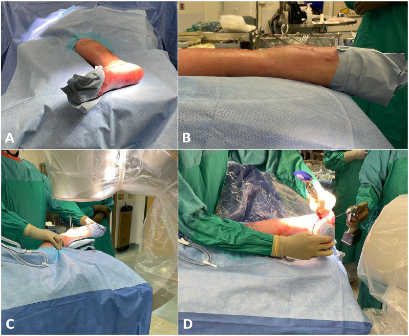
Patient positioning and C-arm intensifier setup. (A) The patient is
placed in the lateral decubitus position with the operative side (left)
up. (B) A bump is placed under the foot to elevate it and make it
horizontal. The C-arm intensifier is positioned obliquely in order to
obtain a (C) true lateral view as well as a (D) calcaneal axial
view.
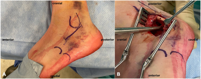
Superior views of a left foot. (A) Skin landmarks and incision. (B)
Lateral wall blunt dissection. Careful dissection of the lateral wall as
well as both proximal and distal edges of the calcaneal tuberosity in
preparation for plate positioning. It is important to dissect proximal
to the peroneal tendons (*), as this will make inferior retraction
easier.
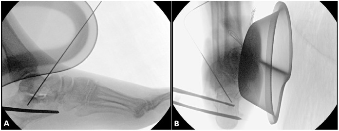
Fluoroscopic views (A, lateral view; B, axial view) of a left foot
displaced intra-articular calcaneal fracture showing medial wall
reduction. Once the medial wall is elevated against the talar chondral
facet, a 1.6-mm Kirschner wire is used to pin it in place through a
transtalar trajectory started from the superolateral talar cortex. A
Howarth elevator was used in this case for reduction.
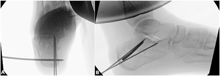
Fluoroscopic views (A, axial view; B, lateral view) of a left foot
displaced intra-articular calcaneal fracture showing calcaneal
tuberosity reduction. Two 1.6-mm Kirschner wires are used to fix the
calcaneal tuberosity to the previously fixed medial wall. The axial view
is used first to guide the wire trajectory, followed by the lateral
view, which confirms the divergent pattern for a stable reduction. The
fluoroscopic views used to illustrate this step and the following steps
are taken from a separate operative case.
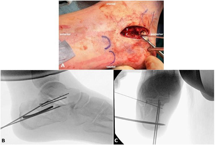
Subtalar joint reduction of a left foot displaced intra-articular
calcaneal fracture. (A) Intraoperative superior view after retraction of
the peroneal tendons (*). The calcaneal facet (C) is elevated against
the talar facet (T) and pinned to the medial calcaneal part. (B, C)
Fluoroscopic views (B, lateral view; C, axial view) confirming joint
reduction. One or two 2.7-mm lag screws can be used for fixation. A
cannulated design was used in this case. The lateral view (B) is
important to confirm subtalar joint reduction and the axial view (C) to
confirm the screw length.
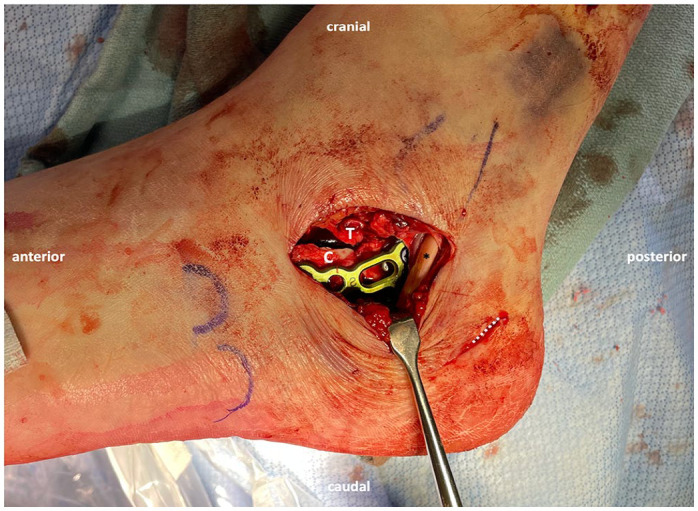
Intraoperative superior view of the subtalar joint of a left foot
displaced intra-articular calcaneal fracture after retraction of the
peroneal tendons (*). The plate is buttressing the lateral wall of the
calcaneus (C), with its middle part overlying the lag screw. The white
dashed line indicates the stab wound incision used first to introduce
the transcalcaneal reduction pin, and second to insert the screws
targeting the calcaneal tuberosity.
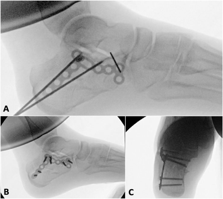
Fluoroscopic views (A, B, lateral views; C, axial view) of a left foot
displaced intra-articular calcaneal fracture showing internal fixation
with a calcaneal locking plate. (A) Correct plate positioning is
confirmed with a lateral view, and a temporary wire fixing the anterior
end of the plate allows fine control of the plate position prior to
insertion of the first compression screw. Anatomic reduction is achieved
with restored calcaneal height and subtalar joint (B), as well as a
restored calcaneal axis (C).
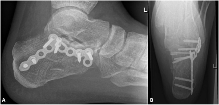
Postoperative radiographic views. A, lateral; B, axial.
References
-
- Ågren PH, Wretenberg P, Sayed-Noor AS. Operative versus nonoperative treatment of displaced intra-articular calcaneal fractures. A prospective, randomized, controlled multicenter trial. J Bone Joint Surg Am. 2013;95(15):1351-1357. - PubMed
-
- Ågren PH, Mukka S, Tullberg T, Wretenberg P, Sayed-Noor AS. Factors affecting long-term treatment results of displaced intraarticular calcaneal fractures: a post hoc analysis of a prospective, randomized, controlled multicenter trial. J Orthop Trauma. 2014;28(10):564-568. - PubMed
-
- Berberian W, Sood A, Karanfilian B, Najarian R, Lin S, Liporace F. Displacement of the sustentacular fragment in intra-articular calcaneal fractures. J Bone Joint Surg Am. 2013;95(11):995-1000. - PubMed
-
- Bruce J, Sutherland A. Surgical versus conservative interventions for displaced intra-articular calcaneal fractures. Cochrane Database Syst Rev. 2013;(1):CD008628. - PubMed
-
- Buckley R, Tough S, McCormack R, et al.. Operative compared with nonoperative treatment of displaced intra-articular calcaneal fractures. A prospective, randomized, controlled multicenter trial. J Bone Joint Surg Am. 2002;84(10):1733-1744. - PubMed
MeSH terms
LinkOut - more resources
Full Text Sources
Medical
