. 2023 Nov;56(11):e13486.
doi: 10.1111/cpr.13486. Epub 2023 Apr 25.

Generation of Rh D-negative blood using CRISPR/Cas9

Affiliations

Generation of Rh D-negative blood using CRISPR/Cas9

Lei Xu et al. Cell Prolif. 2023 Nov.

Abstract

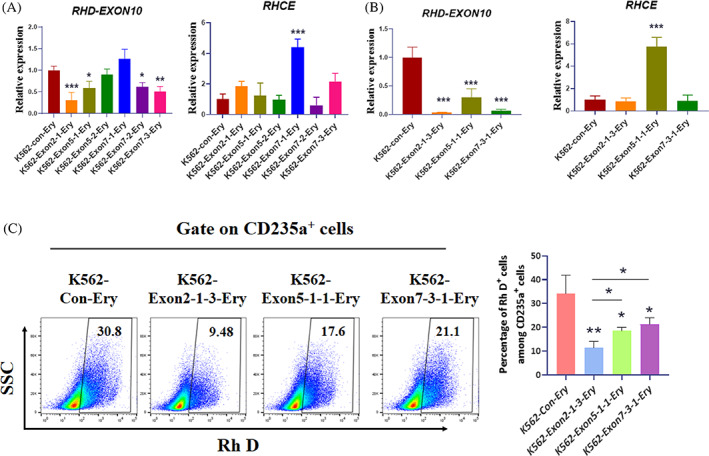
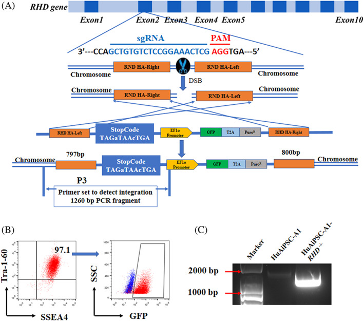
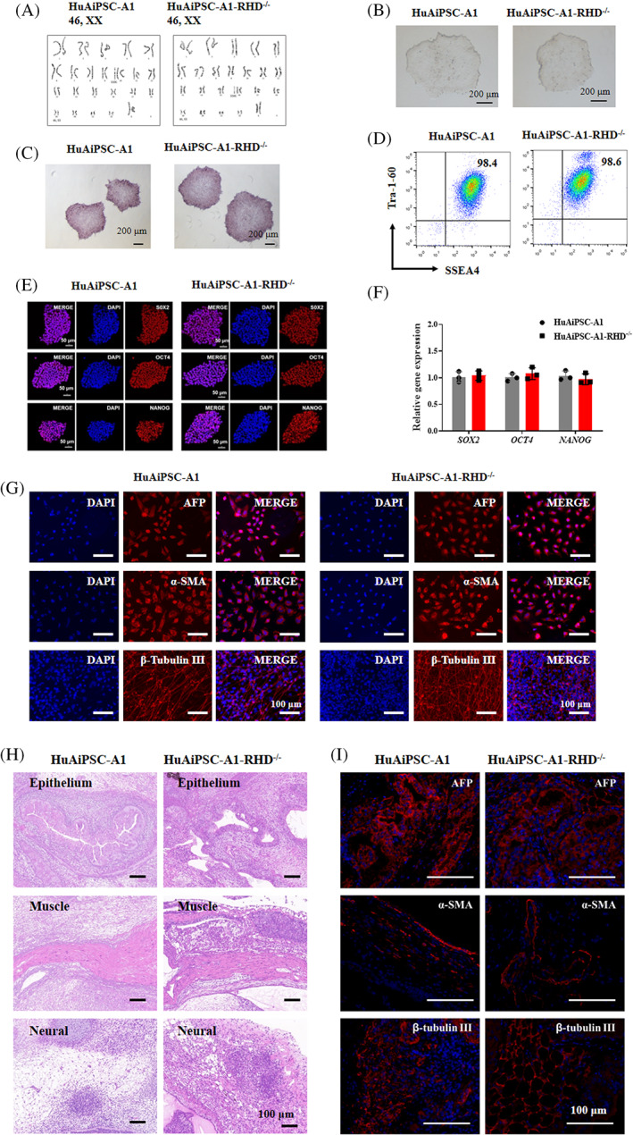
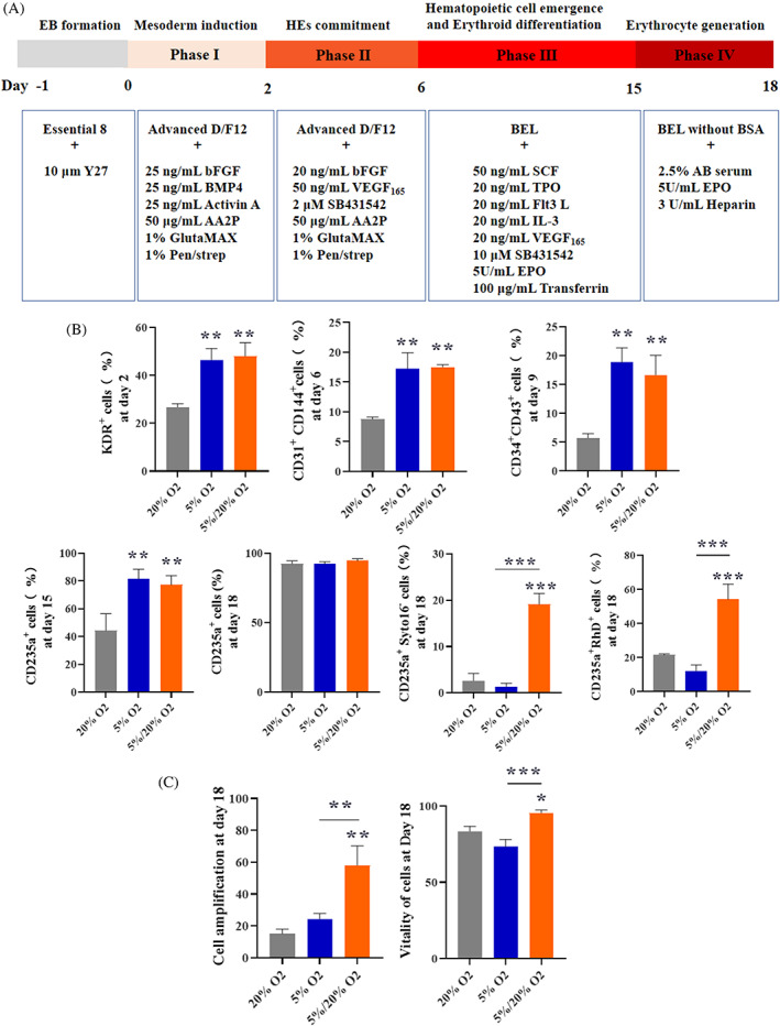
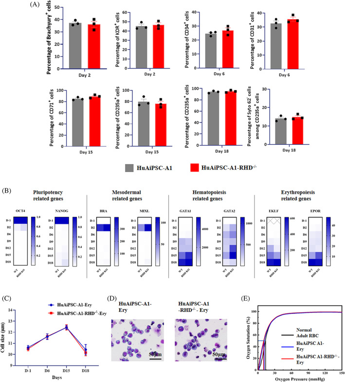
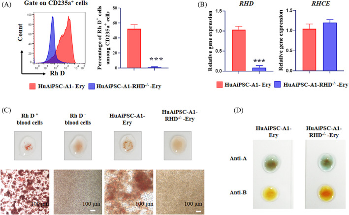
Blood supply shortages, especially the shortage of rare blood types, threaten the current medical system. Research on stem cells has shed light on in vitro blood cell manufacturing. The in vitro production of universal red blood cells (RBCs) from induced pluripotent stem cells (iPSCs) has become the focus of transfusion medicine. To obtain O-type Rh D-negative blood, we developed O-type Rh D-negative human (h)iPSCs using homology-directed repair (HDR)-based CRISPR/Cas9. HuAiPSCs derived from human umbilical arterial endothelial cells and showing haematopoietic differentiation preferences were selected for gene modification. Guide RNAs (gRNAs) were selected, and a donor template flanked by gRNA-directed homologous arms was set to introduce a premature stop code to RHD exon 2. CRISPR/Cas9 gene editing has resulted in the successful generation of an RHD knockout cell line. The HuAiPSC-A1-RHD-/- cell line was differentiated into haematopoietic stem/progenitor cells and subsequently into erythrocytes in the oxygen concentration-optimized differentiation scheme. HuAiPSC-A1-RHD-/- derived erythrocytes remained positive for the RBC markers CD71 and CD235a. These erythrocytes did not express D antigen and did not agglutinate in the presence of anti-Rh D reagents. In conclusion, taking the priority of haematopoietic preference hiPSCs, the HDR-based CRISPR/Cas9 system and optimizing the erythroid-lineage differentiation protocol, we first generated O-type Rh D-negative universal erythrocytes from RHD knockout HuAiPSCs. Its production is highly efficient and shows great potential for clinical applications.


Conflict of interest statement

The authors declare no conflict of interest.

Figures

FIGURE 1
FIGURE 1
Selecting guide RNAs (gRNAs) targeting the human RHD gene. K562 cells stably transfected with LentiCRISPR v2‐gRNAs were analysed for the knockout effect on the RHD gene. (A) Quantitative reverse‐transcription‐polymerase chain reaction (qRT‐PCR) was performed to detect the expression of RHD and RHCE genes in K562‐con‐Ery, K562‐exon 2‐1‐Ery, K562‐exon 5‐1‐Ery, K562‐exon 5‐2‐Ery, K562‐exon 7‐1‐Ery, K562‐exon 7‐2‐Ery and K562‐exon 7‐3‐Ery cells. RHD‐exon 10 was designed to indicate the expression of the RHD gene using exon 10 qRT‐PCR. The results were obtained from three independent replicate experiments, with GAPDH used as the internal reference and the K562‐con‐Ery group set as 1. Results are expressed as mean ± standard deviation, *p < 0.05, **p < 0.01 and ***p < 0.001. (B and C) K562‐exon 2‐1‐Ery, K562‐exon 5‐1‐Ery and K562‐exon 7‐3‐Ery cells were subjected to single cell cloning, qRT‐PCR analysis of the expression of the RHD and RHCE genes (B), and FCM assay the expression of Rh D protein (C). Results were obtained from three independent replicate experiments and are expressed as mean ± standard deviation, *p < 0.05, **p < 0.01 and ***p < 0.001. Ery, Erythrocyte.
FIGURE 2
FIGURE 2
Generation of the RHD knockout human induced pluripotent stem cells (hiPSC) line. (A) Schematic overview of the gene targeting strategy to knockout RHD using the CRISPR/Cas9 system based on HDR. Guide RNA targeted to exon 2 was selected for RHD knockout. Dark blue boxes indicate exons. sgRNA target sites and PAM sequences are shown in blue and red letters, respectively. The donor construct comprised a strong stop code, an EF1α promoter, two screening tag (GFP and puromycin) and two homology arms. (B) FACS SSEA4+Tra‐1‐60+GFP+ cells. (C) Polymerase chain reaction (PCR) confirms the knockout of RHD in the HuAiPSC‐A1‐RHD−/− cells. Properly targeted clones were validated using a PCR strategy with specific primer sets (P3) located in the right homology arm and stop codon, respectively.
FIGURE 3
FIGURE 3
Characterization of the RHD knockout human induced pluripotent stem cells (hiPSC) line, HuAiPSC‐A1‐RHD−/−. (A). The HuAiPSC‐A1‐RHD−/− cells show normal karyotype (46, XX). (B). HuAiPSC‐A1‐RHD−/− cells exhibit normal morphology. (C–F) The HuAiPSC‐A1‐RHD−/− cells exhibit positive alkaline phosphatase activity, express hiPSC surface markers (SSEA‐4 and Tra‐1‐60), hiPSC pluripotency factors (SOX2, OCT4 and NANOG), and hiPSC pluripotency genes (SOX2, OCT4 and NANOG). PCR reactions were normalized to GAPDH and plotted relative to expression levels in HuAiPSC‐A1. Error bars indicate ± standard deviation of triplicates. n.s. indicates non‐statistically significant differences. (G–I) The HuAiPSC‐A1‐RHD−/− cells maintained pluripotency. (G) Immunofluorescence staining revealed that the HuAiPSC‐A1‐RHD−/− cells are capable of differentiating to the endodermal (alpha fetoprotein, AFP), mesodermal (alpha smooth muscle actin, α‐SMA) and ectodermal (β‐Tubulin III) lineages after EB induction in vitro. (H and I) To further evaluate the pluripotency of the HuAiPSC‐A1‐RHD−/− cells, we performed in vivo teratoma formation assays. We injected HuAiPSC‐A1 and HuAiPSC‐A1‐RHD−/− cells intramuscularly into NOD/SCID mice. Histological examination showed that the tumour contained tissues corresponding to the three embryonic germ layers, including epithelium (endoderm), muscle (mesoderm) and neural (ectoderm). Immunofluorescence staining revealed that tumour contains three germ tissue layers, including endodermal (AFP), mesodermal (α‐SMA) and ectodermal (β‐Tubulin III) lineages.
FIGURE 4
FIGURE 4
The efficient generation of erythroblasts from HuAiPSC‐A1‐RHD−/− cells using a four‐step differentiation strategy. (A) Schematic diagram showing induction of human induced pluripotent stem cells (hiPSCs) to erythroid differentiation. (B) Flow cytometry analysis of expression of stage‐specific markers during induction of erythroid‐lineage in the 20% O2, 5% O2 and 5/20% O2 groups. Results were derived from three independent replicate experiments and are expressed as mean ± standard deviation, **p < 0.01 and ***p < 0.001. (C) Erythroid cell expansion fold and cell viability obtained in the 20% O2, 5% O2 and 5/20% O2 groups. Results were obtained from three independent replicate experiments and are expressed as mean ± standard deviation, *p < 0.05, **p < 0.01 and ***p < 0.001.
FIGURE 5
FIGURE 5
The RHD knockout does not affect the differentiation and function of HuAiPSC‐A1. (A, B) HuAiPSC‐A1 and HuAiPSC‐A1‐RHD−/− cells were induced to differentiation for 18 days and subjected to FCM and qRT‐PCR analysis, at the indicated time point. (A) The expression of the PS (PS)/early mesoderm marker (Brachyury+ and KDR+) on Day 2, HE marker (CD31+) and early haematopoietic markers (CD34+) on Day 6, early erythroid progenitor marker (CD71+) and mature erythroid cell marker (CD235a+) on Day 15, and mature erythroid cell marker (CD235a+) and enucleated cell indicator (Syto62) on Day 18 were tested using FCM. Results were obtained from three independent replicate experiments and are expressed as mean ± standard deviation, with n.s. indicating that the differences are not statistically significant. (B) The expression level of pluripotency genes (OCT4 and SOX2), mesoderm genes (BRA and MIXL), haematopoietic related genes (GATA1 and RUNX1) and erythroid related genes (EKLF and EPOR) were detected using qRT‐PCR. The results were derived from three independent replicate experiments with GAPDH as an internal reference and wild‐type group set as 1 for statistical analysis. (C–E) HuAiPSC‐A1 and HuAiPSC‐A1‐RHD−/− cells were induced for differentiation for 18 days and performed to cell size (C), cell morphology (D) and oxygen‐carrying capacity (E) detection. Ery, Erythrocyte; RHD KO, HuAiPSC‐A1‐RHD−/−; WT, HuAiPSC‐A1.
FIGURE 6
FIGURE 6
D antigen and RHD gene expression in HuAiPSC‐A1‐RHD−/−‐Erythrocytes. HuAiPSC‐A1 and HuAiPSC‐A1‐RHD−/− were induced towards erythroid cells for 18 days. (A). FCM was used to analyse the percentage of Rh D antigen‐positive cells in the population of CD235a‐positive cells. Typical FCM diagrams (left) and data statistics (right) of three independent replicate experiments. Results are expressed as mean ± standard deviation, ***p < 0.001. (B) HuAiPSC‐A1‐ and HuAiPSC‐A1‐RHD−/−‐induced erythrocytes were analysed for the expression of RHD and RHCE genes using qRT‐PCR. Results were obtained from three independent replicate experiments, with GAPDH as an internal reference and the HuAiPSC‐A1‐Ery group set as 1 for statistical analysis, results are expressed as mean ± standard deviation, ***p < 0.001, and n.s. indicates that non‐statistically significant differences. (C). Absence of Rh D antigen‐mediated agglutination in erythrocytes derived from the RHD knockout hiPSC line HuAiPSC‐A1‐RHD−/−. HuAiPSC‐A1 and HuAiPSC‐A1‐RHD−/− cells were induced to differentiation for 18 days and subjected to an agglutination test using anti‐Rh D blood grouping reagents in 96‐well plates and on glass slides. Rh D‐positive and Rh D‐negative human peripheral blood cells were used as the controls. A representative photograph and photomicrographs of each cell line are shown. Scale bar, 100 μm. (D). Absence of A or B antigen‐mediated agglutination in erythrocytes derived from HuAiPSC‐A1‐RHD−/− cells. HuAiPSC‐A1 and HuAiPSC‐A1‐RHD−/− cells were induced to differentiation for 18 days and subjected to an agglutination test using anti‐A or anti‐B blood grouping reagents. Ery, Erythrocyte.

Similar articles

Cited by

References

    1. Focosi D, Amabile G. Induced pluripotent stem cell‐derived red blood cells and platelet concentrates: from bench to bedside. Cell. 2017;7(1):2. - PMC - PubMed
    1. Shah S, Huang X, Cheng L. Concise review: stem cell‐based approaches to red blood cell production for transfusion. Stem Cells Transl Med. 2014;3(3):346‐355. - PMC - PubMed
    1. Lim ZR, Vassilev S, Leong YW, et al. Industrially compatible transfusable iPSC‐derived RBCs: progress, challenges and prospective solutions. Int J Mol Sci. 2021;22(18):9808. - PMC - PubMed
    1. Sahu S, Hemlata VA. Adverse events related to blood transfusion. Indian J Anaesth. 2014;58(5):543‐551. - PMC - PubMed
    1. Mujahid A, Dickert FL. Blood group typing: from classical strategies to the application of synthetic antibodies generated by molecular imprinting. Sensors. 2015;16(1):51. - PMC - PubMed