

Novel Mg 2+ binding sites in the cytoplasmic domain of the MgtE Mg 2+ channels revealed by X-ray crystal structures
- PMID: 37097058
- PMCID: PMC10200709
- DOI: 10.3724/abbs.2023067
Novel Mg 2+ binding sites in the cytoplasmic domain of the MgtE Mg 2+ channels revealed by X-ray crystal structures
Abstract
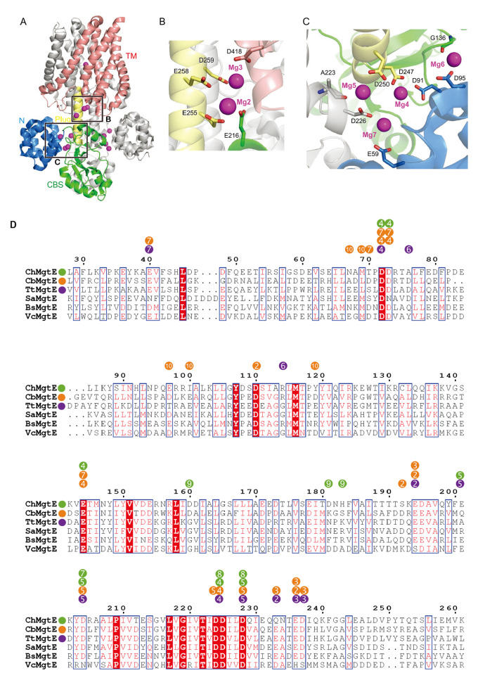
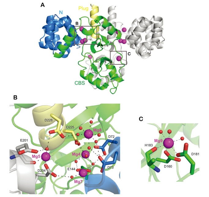
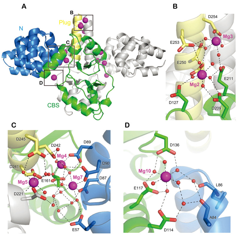
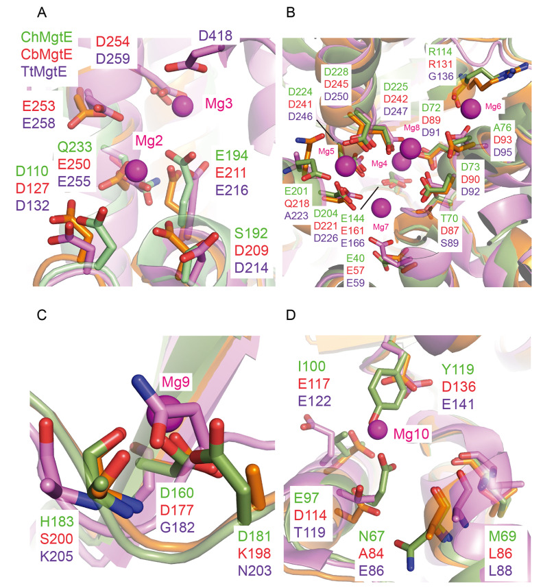
MgtE is a Mg 2+-selective channel regulated by the intracellular Mg 2+ concentration. MgtE family proteins are highly conserved in all domains of life and contribute to cellular Mg 2+ homeostasis. In humans, mutations in the SLC41 proteins, the eukaryotic counterparts of the bacterial MgtE, are known to be associated with various diseases. The first MgtE structure from a thermophilic bacterium, Thermus thermophilus, revealed that MgtE forms a homodimer consisting of transmembrane and cytoplasmic domains with a plug helix connecting the two and that the cytoplasmic domain possesses multiple Mg 2+ binding sites. Structural and electrophysiological analyses revealed that the dissociation of Mg 2+ ions from the cytoplasmic domain induces structural changes in the cytoplasmic domain, leading to channel opening. Thus, previous works showed the importance of MgtE cytoplasmic Mg 2+ binding sites. Nevertheless, due to the limited structural information on MgtE from different species, the conservation and diversity of the cytoplasmic Mg 2+ binding site in MgtE family proteins remain unclear. Here, we report crystal structures of the Mg 2+-bound MgtE cytoplasmic domains from two different bacterial species, Chryseobacterium hispalense and Clostridiales bacterium, and identify multiple Mg 2+ binding sites, including ones that were not observed in the previous MgtE structure. These structures reveal the conservation and diversity of the cytoplasmic Mg 2+ binding site in the MgtE family proteins.
Keywords: crystal structure; ion channels; magnesium; metal homeostasis; regulation.
Figures
Similar articles
-
Crystal structure of the MgtE Mg2+ transporter.Nature. 2007 Aug 30;448(7157):1072-5. doi: 10.1038/nature06093. Epub 2007 Aug 15. Nature. 2007. PMID: 17700703
-
The structure of MgtE in the absence of magnesium provides new insights into channel gating.PLoS Biol. 2021 Apr 27;19(4):e3001231. doi: 10.1371/journal.pbio.3001231. eCollection 2021 Apr. PLoS Biol. 2021. PMID: 33905418 Free PMC article.
-
ATP-dependent modulation of MgtE in Mg2+ homeostasis.Nat Commun. 2017 Jul 27;8(1):148. doi: 10.1038/s41467-017-00082-w. Nat Commun. 2017. PMID: 28747715 Free PMC article.
-
The structure and regulation of magnesium selective ion channels.Biochim Biophys Acta. 2013 Nov;1828(11):2778-92. doi: 10.1016/j.bbamem.2013.08.002. Epub 2013 Aug 15. Biochim Biophys Acta. 2013. PMID: 23954807 Review.
-
The unique nature of mg2+ channels.Physiology (Bethesda). 2008 Oct;23:275-85. doi: 10.1152/physiol.00019.2008. Physiology (Bethesda). 2008. PMID: 18927203 Free PMC article. Review.
Cited by
-
A distal convoluted tubule-specific isoform of murine SLC41A3 extrudes magnesium.Acta Physiol (Oxf). 2025 Mar;241(3):e70018. doi: 10.1111/apha.70018. Acta Physiol (Oxf). 2025. PMID: 39931759 Free PMC article.
References
-
- Hartwig A. Role of magnesium in genomic stability. Mutat Res Fundamental Mol Mech Mutagenesis. . 2001;475:113–121. doi: 10.1016/s0027-5107(01)00074-4. - DOI - PubMed
-
- Nierhaus KH. Mg 2+, K +, and the Ribosome . J Bacteriol. . 2014;196:3817–3819. doi: 10.1128/JB.02297-14. - DOI - PMC - PubMed
-
- Saris NEL, Mervaala E, Karppanen H, Khawaja JA, Lewenstam A. Magnesium. Clinica Chim Acta. . 2000;294:1–26. doi: 10.1016/S0009-8981(99)00258-2. - DOI - PubMed
MeSH terms
Substances
LinkOut - more resources
Full Text Sources
