. 2023 Jun 15;11(3):e0000923.
doi: 10.1128/spectrum.00009-23. Epub 2023 Apr 25.

Cell Entry of Avian Reovirus Modulated by Cell-Surface Annexin A2 and Adhesion G Protein-Coupled Receptor Latrophilin-2 Triggers Src and p38 MAPK Signaling Enhancing Caveolin-1- and Dynamin 2-Dependent Endocytosis

Affiliations

Cell Entry of Avian Reovirus Modulated by Cell-Surface Annexin A2 and Adhesion G Protein-Coupled Receptor Latrophilin-2 Triggers Src and p38 MAPK Signaling Enhancing Caveolin-1- and Dynamin 2-Dependent Endocytosis

Wei-Ru Huang et al. Microbiol Spectr. .

Abstract

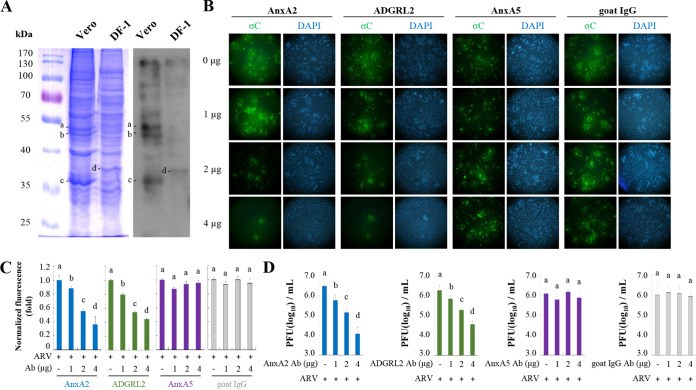
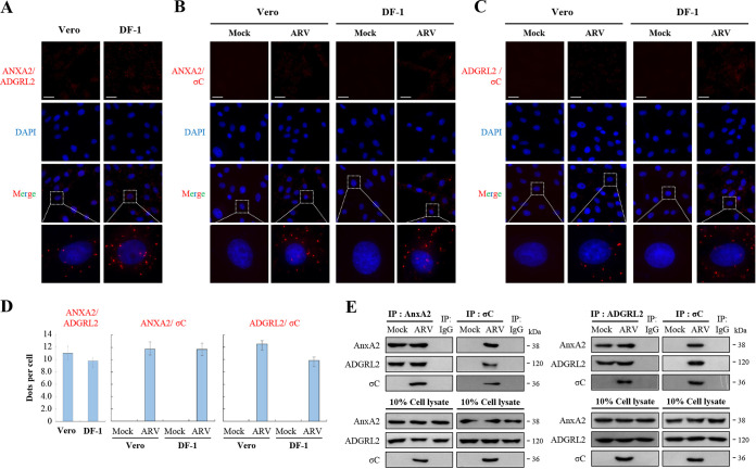
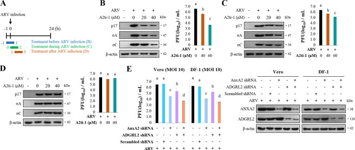
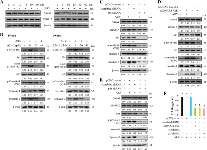
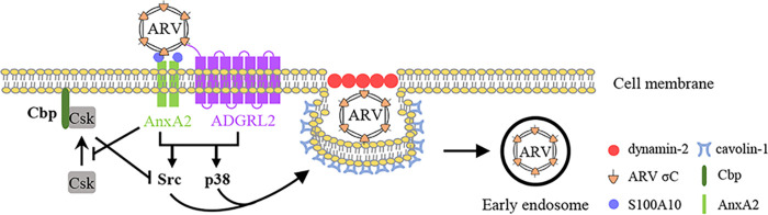
The specifics of cell receptor-modulated avian reovirus (ARV) entry remain unknown. By using a viral overlay protein-binding assay (VOPBA) and an in-gel digestion coupled with liquid chromatography-tandem mass spectrometry (LC-MS/MS), we determined that cell-surface annexin A2 (AnxA2) and adhesion G protein-coupled receptor Latrophilin-2 (ADGRL2) modulate ARV entry. Direct interaction between the ARV σC protein and AnxA2 and ADGRL2 in Vero and DF-1 cells was demonstrated in situ by proximity ligation assays. By using short hairpin RNAs (shRNAs) to silence the endogenous AnxA2 and ADGRL2 genes, ARV entry could be efficiently blocked. A significant decrease in virus yields and the intracellular specific signal for σC protein was observed in Vero cells preincubated with the specific AnxA2 and ADGRL2 monoclonal antibodies, indicating that AnxA2 and ADGRL2 are involved in modulating ARV entry. Furthermore, we found that cells pretreated with the AnxA2/S100A10 heterotetramer (A2t) inhibitor A2ti-1 suppressed ARV-mediated activation of Src and p38 mitogen-activated protein kinase (MAPK), demonstrating that Src and p38 MAPK serve as downstream molecules of cell-surface AnxA2 signaling. Our results reveal that suppression of cell-surface AnxA2 with the A2ti-1 inhibitor increased Csk-Cbp interaction, suggesting that ARV entry suppresses Cbp-mediated relocation of Csk to the membrane, thereby activating Src. Furthermore, reciprocal coimmunoprecipitation assays revealed that σC can interact with signaling molecules, lipid raft, and vimentin. The current study provides novel insights into cell-surface AnxA2- and ADGRL2-modulated cell entry of ARV which triggers Src and p38 MAPK signaling to enhance caveolin-1-, dynamin 2-, and lipid raft-dependent endocytosis. IMPORTANCE By analyzing results from VOPBA and LC-MS/MS, we have determined that cell-surface AnxA2 and ADGRL2 modulate ARV entry. After ARV binding to receptors, Src and p38 MAPK signaling were triggered and, in turn, increased the phosphorylation of caveolin-1 (Tyr14) and upregulated dynamin 2 expression to facilitate caveolin-1-mediated and dynamin 2-dependent endocytosis. In this work, we demonstrated that ARV triggers Src activation by impeding Cbp-mediated relocation of Csk to the membrane in the early stages of the life cycle. This work provides better insight into cell-surface AnxA2 and ADGRL2, which upregulate Src and p38MAPK signaling pathways to enhance ARV entry and productive infection.

Keywords: ADGRL2; Csk-Cbp interaction; Src; annexin A2; avian reovirus; caveolin-1; dynamin 2; p38 MAPK; σC.


Conflict of interest statement

The authors declare no conflict of interest.

Figures

FIG 1
FIG 1
Viral overlay protein-binding assay (VOPBA) analysis of avian reovirus (ARV) binding to Vero and DF-1 cell membrane proteins. (A) SDS-PAGE shows the Vero and DF-1 cell membrane proteins in the left panel. VOPBA was used to identify membrane proteins that bind to ARV as shown in the right panel. Binding of ARV to membrane proteins is shown at approximately 32 and 50 kDa, indicated by bands a, b, c, and d. Band a, ADGRL2; band b, AnxA5; bands c and d, AnxA2. These proteins were further identified by liquid chromatography-tandem mass spectrometry (LC-MS/MS) analysis. (B) Detection of internalized σC protein in ARV by antibody blocking assays. Confluent monolayers of Vero cells, grown in 8-well chambers for 24 h, were pre-incubated or not with anti-AnxA2, -AnxA5, and-ADGRL2 antibodies at the indicated concentrations for 1 h at 37°C. Goat-IgG was used as a negative control. After extensive washing, cells were incubated with ARV at a multiplicity of infection (MOI) of 10 for 1 h, and the cells were fixed and stained with DAPI (4′,6-diamidino-2-phenylindole; blue) and antibodies specific for σC (green). (C) Fluorescence signals in panel B were quantified with ImageJ software. The amount of fluorescence in the mock control group was considered to be 1-fold. Duncan’s multiple range test (MDRT) using SPSS software was used to analyze the statistical significance of all data. (D) Virus yields were determined as treatments from panel B. Significance between treatments was determined by MDRT using SPSS software (version 20.0). Means with common lowercase letters (a, b, c, d) indicate no significant difference at P < 0.05. Each value is the mean (with standard error, SE) from three independent experiments.
FIG 2
FIG 2
Proximity ligation assays (PLA) for cell-surface AnxA2 and ADGRL2 in Vero and DF-1 cells. (A to C) PLAs for ARV σC protein and cell surface AnxA2 and ADGRL2 receptors in Vero and DF-1 cells. To analyze the interactions of AnxA2/ADGRL2 (A), σC/AnxA2 (B), and σC/ADGRL2 (C), interactions between Vero and DF-1 cells were detected using a Duolink commercial kit (Sigma-Aldrich, cat no. DUO 92008) based on in situ PLA based on the manufacturer’s instructions. Enlarged images correspond to the region indicated by the white box in the merged image. Representative images are from three independent experiments. (D) The fluorescence bright spots in Fig. 2A, B, and C were quantified, with each bar graph (mean ± SE) representing the average value obtained from about 100 cells in 5 randomly selected regions. (E) To confirm the interaction of ARV σC protein with AnxA2, ADGRL2 or AnxA2/ADGRL2 in Vero cells, the cell membrane fraction was isolated for reciprocal co-immunoprecipitation assays. IgG was used as a negative control. Predicted size (kDa) of each protein is labeled to the right of gels and blots in each figure. All original/uncropped blots and images from this study are provided in Fig. S8 in the supplemental material.
FIG 3
FIG 3
Inhibition of cell-surface AnxA2 and ADGRL2 suppressed virus replication. (A) Experimental design for pulse treatment with the A2ti-1 inhibitor in ARV-infected cells during the early stage of the viral life cycle. (B to D) Vero cells were pretreated with inhibitor A2ti-1 before (B), during (C), and after (D) infection for 2 h. Cells were washed to remove the drug and further incubated with ARV at a MOI of 10 until 24 h. The expression levels of viral proteins p17, σA, and σC were analyzed by Western blotting with the respective antibodies. Virus titers were also determined. All experiments were conducted in triplicate, and data are presented as the means ± SE. (E) Since ADGRL2 inhibitor was not available, we used a short hairpin RNA (shRNA) to deplete the ADGRL2 gene and determined the virus yield. Cells were transfected with the indicated shRNAs for 6 h followed by infection with virus at a MOI of 10 for 24 h. Protein levels were normalized to that of β-actin. Levels of the indicated proteins in the mock treatment were considered 1-fold. Immunoblots in panels B, C, D, and E were quantitated by densitometric analysis using ImageJ software and normalized to that of β-actin. Data are shown in Fig. S3A to D in the supplemental material.
FIG 4
FIG 4
Src and p38MAPK are key downstream molecules of cell-surface AnxA2. (A) Levels of AnxA2 and ADGRL2 were examined in ARV-infected Vero and DF-1 cells at the indicated time points. (B) Vero cells were pretreated with inhibitor A2ti-1 for 1 h and then infected with ARV at a MOI of 10 at the indicated time points. Expression levels of the indicated proteins were analyzed by Western blotting with the respective antibodies, quantitated by densitometric analysis using ImageJ, and normalized to that of actin. The levels of indicated proteins in the mock group are considered 1-fold. The predicted size of each protein (kDa) is labeled to the right of gels and blots in each figure. Numbers below each lane indicate the relative fold of the control level for each specific protein in mock-treated cells. (C to E) Vero cells were transfected with pcDNA3.1-Csk plasmid and shRNAs for 6 h followed by infection with ARV at a MOI of 10 for 24 h. Cell lysates were collected 24 h postinfection and immunoblotted with the respective antibodies. Protein levels were normalized to that of β-actin. The levels of the indicated proteins in the mock treatment are considered 1-fold. Image shown is from a single experiment and is representative of at least three separate experiments. (F) To examine whether the Csk-Src pathway and p38 MAPK regulate virus propagation, we performed overexpression of Csk or knockdown of Src and p38 MAPK. Virus titer was determined. Each value is the mean (with SE) from three independent experiments.
FIG 5
FIG 5
Suppression of lipid raft, Src, and p38 MAPK inhibits ARV entry. (A) Experimental design for pulse treatment with inhibitors. (B) Vero cells were treated with Csk inhibitor (5 μM), MβCD (3.2 mM), p38 MAPK inhibitor (5 μM), and Src inhibitor (5 μM) during different time windows before, during, and after ARV infection at a MOI of 10. The cells were washed to remove the drug and further incubated for 24 h. Immunofluorescence signals were detected at 24 h postinfection using a monoclonal antibody against σC protein and observed by fluorescence microscopy to visualize viral protein expression (green). Cell nuclei were stained with DAPI (blue). (C) Fluorescence signals in panel B were quantified with ImageJ software. The amount of fluorescence in the mock control group was considered to be 1-fold. MDRT was used to analyze the statistical significance of all data.
FIG 6
FIG 6
ARV reduces Cbp-Csk interaction during the early stage of its life cycle. (A) Vero cells were infected with ARV at a MOI of 10. Cell lysates were collected at the indicated time points. In co-immunoprecipitation experiments, cells lysates were immunoprecipitated with Cbp or Csk antibodies and analyzed by Western blotting assays with the indicated antibodies. (B) Densitometry analysis results for Western blotting represent the amount of protein association in panel B. Mock-treated cells were considered 1-fold. Signals for all blots were quantified using Image J software. All experiments were conducted independently in triplicate. Student’s t test was used to analyze the statistical significance of all data. (C) Left panel: Vero cells were pretreated with inhibitor A2ti-1 before infection for 2 h. Cells were washed to remove the drug and further incubated with ARV at a MOI of 10 for 30 min. Cell lysates were immunoprecipitated with Cbp or Csk antibodies and analyzed by Western blotting assays with the indicated antibodies. Right panel: Western blots (left panel) were quantitated by densitometric analysis using ImageJ and normalized to β-actin. Right panel is expressed as folds representing the amount of protein and protein association. Mock cells were considered 1-fold. Each value is the mean (with SE) from three independent experiments. Student’s t test was used to analyze the statistical significance of all data. (D) Vero cells were pretreated with CSK inhibitor before infection for 2 h. Cells were washed to remove the drug and further incubated with ARV at a MOI of 10 for 24 h. The expression levels of the indicated proteins were analyzed by Western blotting with the respective antibodies. Image shown is from a single experiment and is representative of at least three separate experiments.
FIG 7
FIG 7
Schematic diagram showing the binding of ARV to cell-surface AnxA2 and ADGRL2, which activates cellular signaling pathways to enhance caveolin-1- and dynamin 2-dependent endocytosis. This study indicates that AnxA2 and ADGRL2 mediate cell entry of ARV. ARV σC protein binding to cell-surface AnxA2 and ADGRL2 of Vero and DF-1 cells activates Src and p38 MAPK signaling to increase caveolin-1 phosphorylation and upregulate dynamin 2 expression, thereby facilitating ARV entry through caveolin-1-, dynamin 2-, and lipid-raft-dependent endocytosis. Importantly, ARV activates Src by impeding Cbp-mediated relocation of Csk to the membrane during the early stage of its life cycle. Arrows (→) indicate activation; bars (⊥) indicate repression.

Similar articles

Cited by

References

    1. Rosenberger JK, Sterner FJ, Botts S, Lee KP, Margolin A. 1989. In vitro and in vivo characterization of avian reoviruses. I. Pathogenicity and antigenic relatedness of several avian reovirus isolates. Avian Dis 33:535–544. doi:10.2307/1591118. - DOI - PubMed
    1. Hofstad MS. 1978. Diseases of poultry. Iowa State University Press, Ames, Iowa.
    1. Kozak RA, Hattin L, Biondi MJ, Corredor JC, Walsh S, Xue-Zhong M, Manuel J, McGilvray ID, Morgenstern J, Lusty E, Cherepanov V, McBey BA, Leishman D, Feld JJ, Bridle B, Nagy É. 2017. Replication and oncolytic activity of an avian orthoreovirus in human hepatocellular carcinoma cells. Viruses 9:90. doi:10.3390/v9040090. - DOI - PMC - PubMed
    1. Chiu HC, Huang WR, Liao TL, Chi PI, Nielsen BL, Liu JH, Liu HJ. 2018. Mechanistic insights into avian reovirus p17-modulated suppression of cell cycle CDK-cyclin complexes and enhancement of p53 and cyclin H interaction. J Biol Chem 293:12542–12562. doi:10.1074/jbc.RA118.002341. - DOI - PMC - PubMed
    1. Cai R, Meng G, Li Y, Wang W, Diao Y, Zhao S, Feng Q, Tang Y. 2019. The oncolytic efficacy and safety of avian reovirus and its dynamic distribution in infected mice. Exp Biol Med (Maywood) 244:983–991. doi:10.1177/1535370219861928. - DOI - PMC - PubMed

Publication types

LinkOut - more resources