. 2023 Aug 7;220(8):e20222090.
doi: 10.1084/jem.20222090. Epub 2023 Apr 25.

CD30 co-stimulation drives differentiation of protective T cells during Mycobacterium tuberculosis infection

Affiliations

CD30 co-stimulation drives differentiation of protective T cells during Mycobacterium tuberculosis infection

Taylor W Foreman et al. J Exp Med. .

Abstract

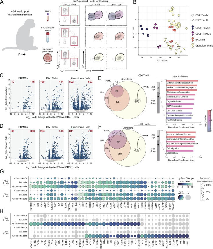
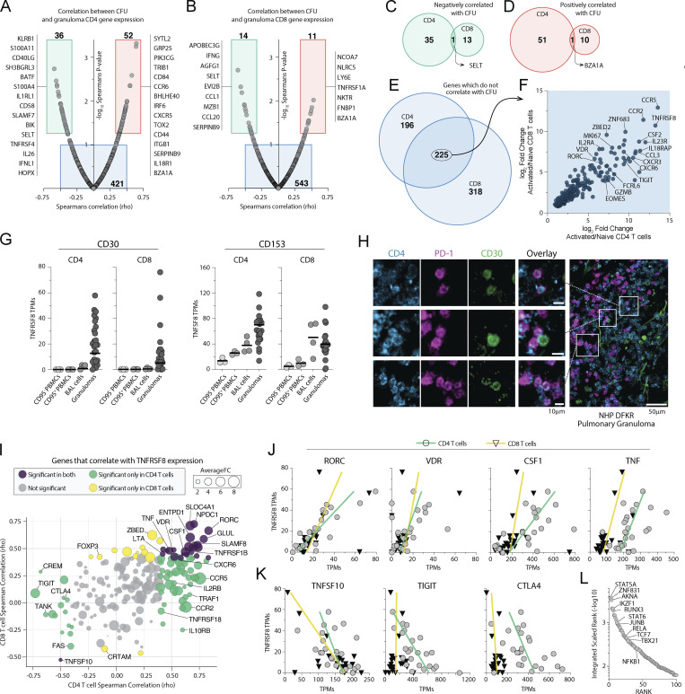
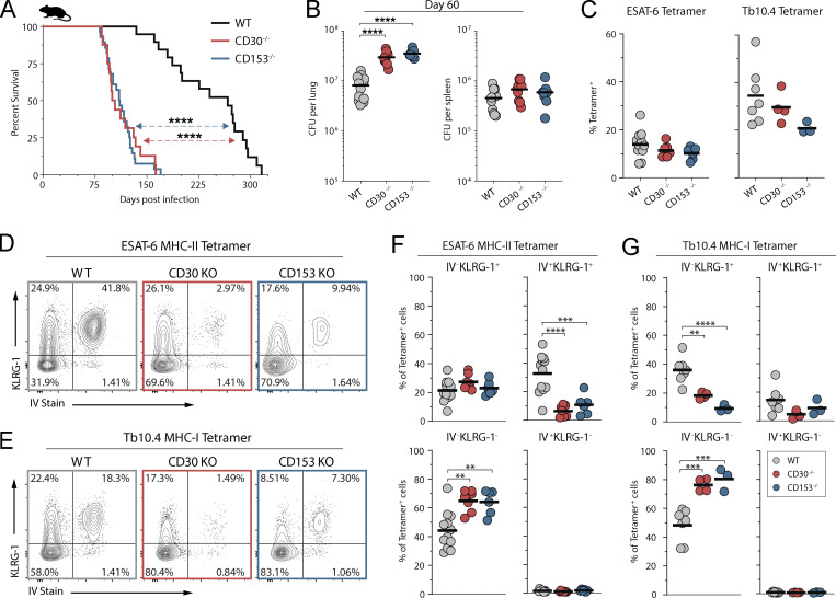
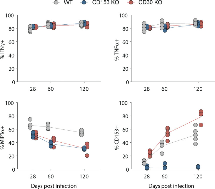
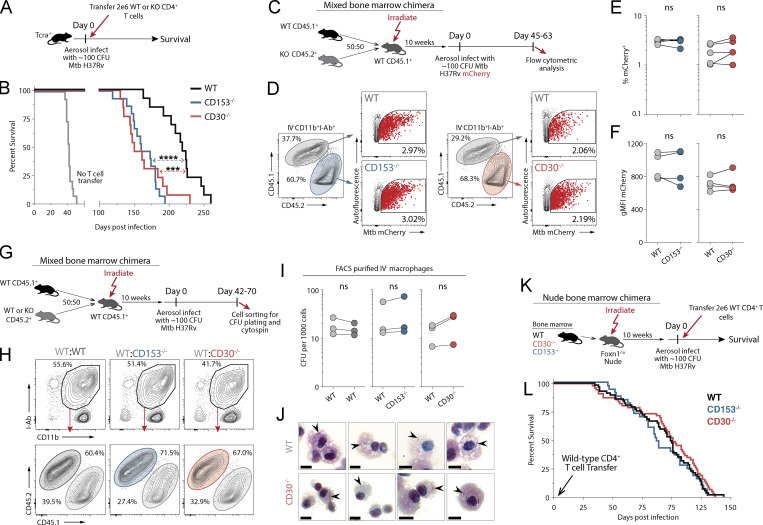
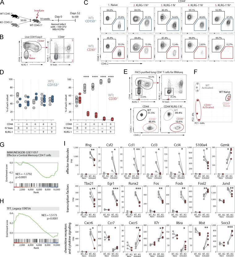
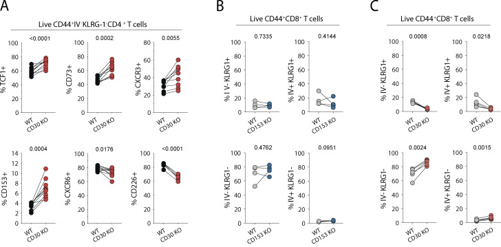
Control of Mycobacterium tuberculosis (Mtb) infection requires generation of T cells that migrate to granulomas, complex immune structures surrounding sites of bacterial replication. Here we compared the gene expression profiles of T cells in pulmonary granulomas, bronchoalveolar lavage, and blood of Mtb-infected rhesus macaques to identify granuloma-enriched T cell genes. TNFRSF8/CD30 was among the top genes upregulated in both CD4 and CD8 T cells from granulomas. In mice, CD30 expression on CD4 T cells is required for survival of Mtb infection, and there is no major role for CD30 in protection by other cell types. Transcriptomic comparison of WT and CD30-/- CD4 T cells from the lungs of Mtb-infected mixed bone marrow chimeric mice showed that CD30 directly promotes CD4 T cell differentiation and the expression of multiple effector molecules. These results demonstrate that the CD30 co-stimulatory axis is highly upregulated on granuloma T cells and is critical for protective T cell responses against Mtb infection.


Conflict of interest statement

Disclosures: T.W. Foreman, K.D. Kauffman, M.A. Sallin, and D.L. Barber reported a patent to CD153 and/or CD30 in infection, application no. 62/633,816, pending. No other disclosures were reported.

Figures

Figure 1.
Figure 1.
Gene expression pattern in rhesus macaque Mtb granuloma T cells. (A) Study schematic showing CD4 and CD8 T cells FACS-purified from blood, BAL, and 23 pulmonary granulomas isolated from four rhesus macaques infected with a low dose of Mtb for subsequent transcriptomic analysis. (B) Principal component analysis of the top 7,000 genes expressed. (C and D) Log2 fold change of gene expression of activated over naive T cells from (C) CD4 and (D) CD8 T cells with number of genes indicated in red. (E and F) Proportional Venn diagrams of significant genes differentially regulated from the three tissue compartments with GSEA of the upregulated genes specific only to granulomas from (E) CD4 and (F) CD8 T cells. (G and H) Curated list of genes demonstrated as relative percent expression with indicated significant fold change shown by color to demonstrate genes (G) upregulated in activated T cells compared to (H) genes significantly upregulated only in T cells isolated from granulomas.
Figure 2.
Figure 2.
TNFRSF8 (CD30) is highly upregulated in granuloma CD4 and CD8 T cells. (A and B) Gene expression correlation with individual granuloma CFU demonstrating genes positively and negatively correlated with bacterial burden in (A) CD4 and (B) CD8 T cells with list of curated genes displayed next to box indicating significance. (C–E) Venn diagrams of genes shared between CD4 and CD8 T cells which (C) negatively correlate, (D) positively correlate, or (E) do not correlate with granuloma CFU. (F) Log2 fold change of the genes which are independent of granuloma bacterial burden and shared between CD4 and CD8 T cells. (G) TPM expression levels of both CD30 and CD153 in blood, BAL, and granulomas. (H) Immunohistochemical staining for CD30 expression on distinct subset of CD4 T cells in a granuloma. Scale bars represent 10 μm in small panels and 50 μm in the zoomed image. (I) Gene correlations with Tnfrsf8 expression in both CD4 and CD8 T cells showing average fold change between both CD4 and CD8 T cells from granulomas. (J and K) Linear correlations of genes (J) positively correlated and (K) negatively correlated with CD30 expression. (L) Transcription factor enrichment analysis of genes significantly correlated with Tnfrsf8 expression.
Figure 3.
Figure 3.
CD30-deficient mice are susceptible to Mtb infection. (A) Survival curve of intact knockout mice after aerosol infection with ∼100 CFU of Mtb (n = 16–28 per group, three independent experiments shown together). (B) Bacterial burden in the lungs and spleens of mice on day 60 after infection (n = 10–12 per group). (C) Frequency of live CD4+CD44+ T cells that are ESAT-6 tetramer+ or live CD8+CD44+ T cells that are Tb10.4 tetramer+. (D and E) Example flow cytometry plots of T cells showing iv stain and expression of KLRG-1 in (D) ESAT-6 tetramer+ CD4 T cells and (E) Tb10.4 tetramer+ CD8 T cells. (F and G) Frequency of (F) ESAT-6 tetramer+ CD4 T cells and (G) Tb10.4 tetramer+ CD8 T cells expressing KLRG-1 within the vasculature versus parenchyma; (C–G) two independent experiments shown together with n = 3–12 per group. Statistical analysis was calculated by (A) Kaplan–Meier survival curve with log-rank test or (B, C, F, and G) one-way ANOVA with Tukey correction for multiple comparisons. **, P < 0.01; ***, P < 0.001; ****, P < 0.0001.
Figure S1.
Figure S1.
WT, CD153−/−, and CD30−/− mice were euthanized at days 28, 60, and 120 after aerosol infection and T cells restimulated for 5 h with ESAT-61-20 peptide in the presence of brefeldin/monensin. Data shown are gated on CD4+CD154+ T cells to compare cytokine expression of antigen-specific CD4 T cells.
Figure 4.
Figure 4.
T cells require CD30 to mediate protection against Mtb infection in mice. (A and B) Experiment schematic for CD4 T cell adoptive transfer (A) and the survival curve of Tcra−/− mice who received WT, CD30−/−, or CD153−/− CD4 T cells at the time of infection (B). Data represent three independent experiments with n = 4–5 per group per experiment. (C) Experiment schematic for MBMC mice to study the intrinsic roles of CD30 and CD153. (D) Example flow cytometry plots of live IVCD11b+I-Ab+ macrophages from mice infected with mCherry-reporter Mtb. (E and F) Frequency of (E) and gMFI of (F) mCherry+ macrophages in MBMC mice in WT:CD30−/− and WT:CD153−/− chimeras. Data shown are representative of three independent experiments with n = 4–5 per group. (G) Experiment schematic for MBMC mice to study bacterial burdens on a per cell basis. (H) Example flow cytometry plots of live IVCD11b+I-Ab+ macrophages FACS-sorted for CFU plating. (I) Quantification of the number of bacteria per 1,000 macrophages. Each point represents four to five mice pooled per group showing three independent experiments. (J) FACS-purified macrophages from MBMC mice were cytospin-mounted for subsequent acid-fast bacilli staining, demonstrating no increase in bacterial levels per cell. Arrowheads point to bacilli. Scale bars represent 5 μm. (K and L) Experiment schematic (K) for bone marrow reconstitution of athymic nude mice with subsequent CD4 T cell adoptive transfer at the time of infection with the (L) survival curve of mice where all hematopoietic cells except CD4 T cells are from WT, CD30−/−, or CD153−/− bone marrow donors. Data are representative of six independent experiments with n = 3–14 mice per group per experiment, for a total of 33–55 mice per group. Statistical analysis was calculated by (B and L) Kaplan–Meier survival curve with log-rank test or (E, F, and L) paired t tests. ***, P < 0.001; ****, P < 0.0001
Figure S2.
Figure S2.
Quantification of bacterial loads in FACS-purified cells from Mtb-infected MBMC mice. (A) Gating schema for FACS purification of parenchymal CD11b+I-Ab+ macrophages and CD11b+I-Ab myeloid cells to plate for bacterial burden on a per cell basis. (B) Quantification of the number of bacteria per 1,000 parenchymal CD11b+I-Ab myeloid cells. Each point represents four to five mice pooled per group, showing independent experiments. (C) Quantification of acid-fast bacilli from cytospin slides of sorted macrophages. (B and C) Statistical analysis was calculated using paired t tests.
Figure 5.
Figure 5.
CD30 signaling drives CD4 T cell differentiation and effector molecule expression during Mtb infection. (A) Experiment schematic showing MBMC mice to study the T cell–intrinsic roles of CD30 and CD153 in responses to Mtb infection. (B and C) Example flow cytometry plots and gating of CD4 T cell populations for analysis comparing WT:KO ratios within each of these populations. (D) Frequency of WT:CD153−/− and WT:CD30−/− cells within each of these T cell populations. Data shown are representative of four independent experiments with n = 4–10 mice per group per experiment. (E) Example flow cytometry plots for FACS purification of live IVKLRG-1 CD44+CD4+ or CD44CD4+ T cells for gene expression profiling. (F) Principal component analysis of the top 3,000 genes expressed in these populations from WT or CD30−/− CD4 T cells. Data shown are representative of three independent experiments with n = 5 mice per experiment pooled after FACS sorting. (G and H) GSEA of genes differentially expressed between WT and KO activated T cells in comparison to two gene sets showing WT cells highly enriched for STAT5a signaling, while KO cells are enriched for less effector-like gene expression. (I) Manually curated gene sets of CD30-regulated T cell effector molecules, transcription factors, chemokine receptors, and cytokine signaling molecules. Statistical analysis was calculated by (D) multiple t tests per row or (I) t tests. *, P < 0.05; **, P < 0.01; ***, P < 0.001; ****, P < 0.0001.
Figure S3.
Figure S3.
Phenotype of CD4 and CD8 T cells lacking CD30 and CD153. (A) Flow cytometric quantification of CD4 T cells from WT:CD30−/− MBMC mice euthanized 62–69 d after aerosol infection. Data are from two independent experiments with n = 5 each. (B and C) Comparison of CD8 T cell differentiation by IV stain and KLRG-1 expression in (B) WT:CD153−/− and (C) WT:CD30−/− MBMC mice. Statistical analysis was calculated using paired t tests with P value indicated above each graph.

Comment in

References

    1. Acosta-Rodriguez, E.V., Rivino L., Geginat J., Jarrossay D., Gattorno M., Lanzavecchia A., Sallusto F., and Napolitani G.. 2007. Surface phenotype and antigenic specificity of human interleukin 17-producing T helper memory cells. Nat. Immunol. 8:639–646. 10.1038/ni1467 - DOI - PubMed
    1. Burel, J.G., Lindestam Arlehamn C.S., Khan N., Seumois G., Greenbaum J.A., Taplitz R., Gilman R.H., Saito M., Vijayanand P., Sette A., and Peters B.. 2018. Transcriptomic Analysis of CD4(+) T Cells Reveals Novel Immune Signatures of Latent Tuberculosis. J. Immunol. 200:3283–3290. 10.4049/jimmunol.1800118 - DOI - PMC - PubMed
    1. Cai, Y., Wang Y., Shi C., Dai Y., Li F., Xu Y., Zhang P., Kong F., Deng G., Wen Z., et al. . 2022. Single-cell immune profiling reveals functional diversity of T cells in tuberculous pleural effusion. J. Exp. Med. 219:219. 10.1084/jem.20211777 - DOI - PMC - PubMed
    1. Chen, Y., Zander R.A., Wu X., Schauder D.M., Kasmani M.Y., Shen J., Zheng S., Burns R., Taparowsky E.J., and Cui W.. 2021. BATF regulates progenitor to cytolytic effector CD8+ T cell transition during chronic viral infection. Nat. Immunol. 22:996–1007. 10.1038/s41590-021-00965-7 - DOI - PMC - PubMed
    1. Clemmensen, H.S., Dube J.Y., McIntosh F., Rosenkrands I., Jungersen G., Aagaard C., Andersen P., Behr M.A., and Mortensen R.. 2021. In Vivo Antigen Expression Regulates CD4 T Cell Differentiation and Vaccine Efficacy against Mycobacterium tuberculosis Infection. mBio. 12. e00226-21. 10.1128/mBio.00226-21 - DOI - PMC - PubMed

Publication types