. 2023 Oct;16(5):1021-1031.
doi: 10.1007/s12265-023-10384-8. Epub 2023 Apr 25.

Hif-1α/Slit2 Mediates Vascular Smooth Muscle Cell Phenotypic Changes in Restenosis of Bypass Grafts

Affiliations

Hif-1α/Slit2 Mediates Vascular Smooth Muscle Cell Phenotypic Changes in Restenosis of Bypass Grafts

Sen Li et al. J Cardiovasc Transl Res. 2023 Oct.

Abstract

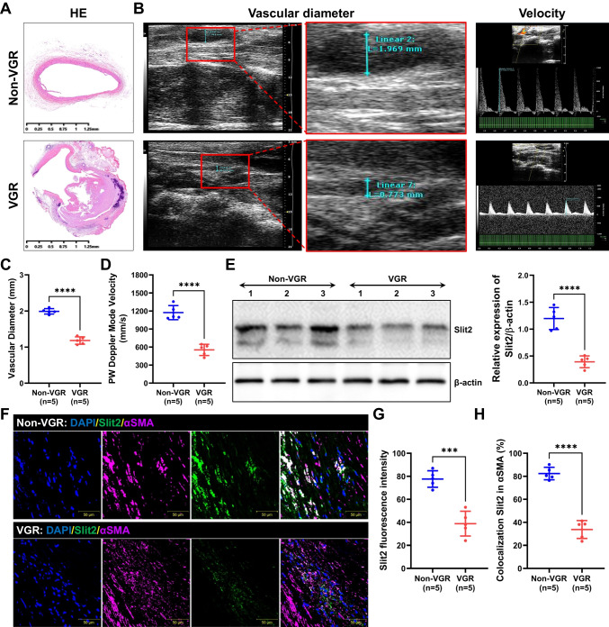
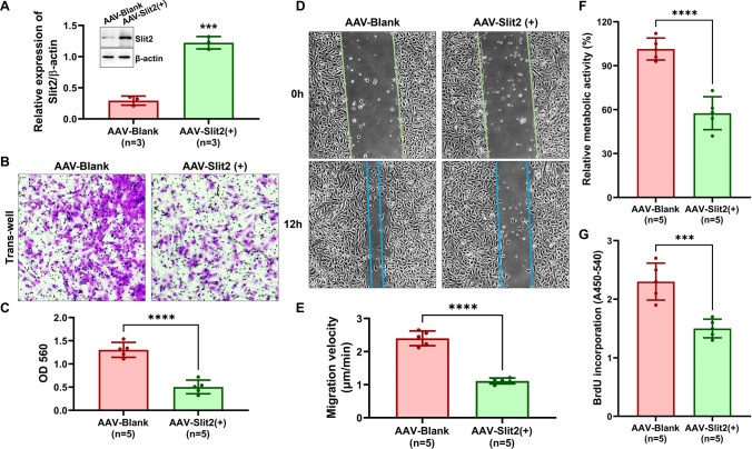
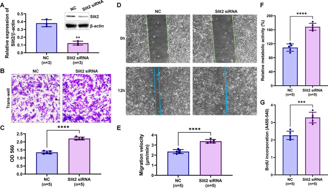
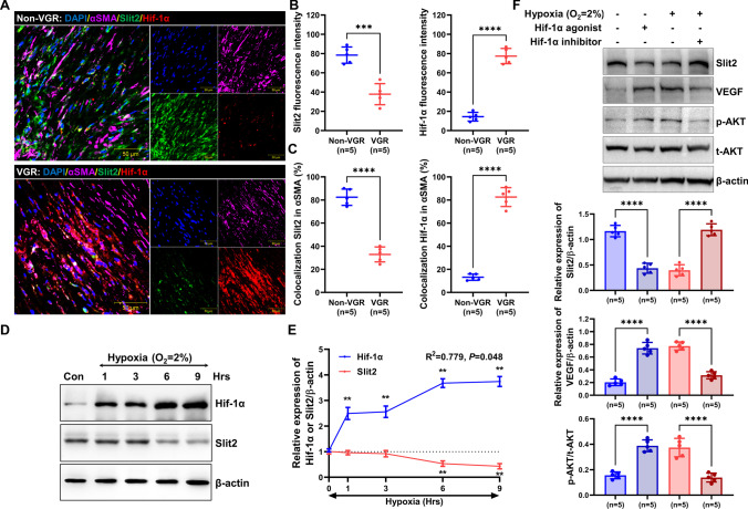
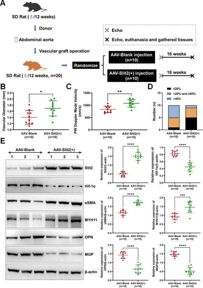
Vascular smooth muscle cells (VSMCs) are involved in restenosis of bypass grafts and cause artery graft occlusion. This study aimed to explore the role of Slit2 in phenotypic switching of VSMCs and its effect on restenosis of vascular conduits. An animal model of vascular graft restenosis (VGR) was produced in SD rats and assessed by echocardiography. The expression of Slit2 and Hif-1α was measured in vivo and in vitro. After Slit2 overexpression, the migration and proliferation of VSMCs were detected in vitro, and the restenosis rates and phenotype of VSMCs were tested in vivo. The arteries of the VGR model presented significant stenosis, and Slit2 was decreased in VSMCs of the VGR model. In vitro, Slit2 overexpression inhibited the migration and proliferation of VSMCs, but Slit2 knockdown promoted migration and proliferation. Hypoxia induced Hif-1α but reduced Slit2, and Hif-1α negatively regulated Slit2 expression. Moreover, Slit2 overexpression weakened the rate of VGR and maintained the patency of artery bypass grafts, which suppressed the phenotypic switching of VSMCs. Slit2 inhibited the synthetic phenotype transformation to inhibit the migration and proliferation of VSMCs and delayed the VGR via Hif-1α.

Keywords: Bypass graft; Hif-1α; Restenosis; Slit2; Smooth muscle cell.


Conflict of interest statement

The authors declare no competing interests.

Figures

Fig. 1
Fig. 1
The expression and location of Slit2 in vascular graft restenosis in a rat model. The SD rats underwent abdominal aortic transplantation, and the VGR and non-VGR arteries were collected to measure the expression and location of Slit2. A The HE staining of VGR and non-VGR arteries. B Images of echocardiography. CD The vascular diameter and PW velocity were measured (n = 5, ****P < 0.0001 with Student’s t test). E Western blotting was used to detect the level of Slit2 (β-actin was used as an internal control, n = 5, ****P < 0.0001 with Student’s t test). F Staining for DAPI (blue), Slit2 (green), and αSMA (pink) was performed. G The fluorescence intensity of Slit2 (n = 5, ***P < 0.001 with Student’s t test). H The ratio of colocalization of Slit2 in α-SMA (n = 5, ****P < 0.0001 with Student’s t test)
Fig. 2
Fig. 2
Slit2 overexpression regulated the migration and proliferation of VSMCs. AAV-blank and AAV-Slit2( +) were transfected into VSMCs, and migration was detected by Transwell and wound-healing assays. Proliferation was measured by CCK8 and BrdU assays. A Western blotting was used to detect the efficiency of Slit2 overexpression with AAV-Slit2 ( +) transfected into VSMCs (β-actin was used as an internal control, n = 3, ***P < 0.001 with Student’s t test). BC The effect of Slit2 overexpression on VSMC migration by Transwell assays (n = 5, ***P < 0.001 with Student’s t test). DE Wound healing was used to test the migration velocity of VSMCs (n = 5, ***P < 0.001 with Student’s t test). FG The proliferation of VSMCs was detected by CCK8 or BrdU assays (n = 5, ***P < 0.001, ****P < 0.0001 with Student’s t test)
Fig. 3
Fig. 3
Slit2 knockdown regulated the migration and proliferation of VSMCs. Negative control (NC) and Slit2 siRNA were transfected into VSMCs, and migration was detected by Transwell and wound-healing assays. Proliferation was measured by CCK8 and BrdU assays. A Western blotting was used to detect the efficiency of Slit2 knockdown with Slit2 siRNA transfected into VSMCs (β-actin was used as an internal control, n = 3, **P < 0.01 with Student’s t test). BC The effect of Slit2 knockdown on VSMC migration by Transwell assays (n = 5, ****P < 0.0001 with Student’s t test). DE Wound healing was used to test the migration velocity of VSMCs (n = 5, ****P < 0.0001 with Student’s t test). FG The proliferation of VSMCs was detected by CCK8 or BrdU assays (n = 5, ***P < 0.001, ****P < 0.0001 with Student’s t test)
Fig. 4
Fig. 4
Hypoxia downregulated Slit2 expression via Hif-1α. A DAPI (blue), Slit2 (green), Hif-1α (red), and αSMA (pink) were labeled on the arteries of the VGR and non-VGR groups. B The fluorescence intensity of Slit2 and Hif-1α (n = 5, ***P < 0.001, ****P < 0.0001 with Student’s t test). C The ratio of colocalization of Slit2 and Hif-1α in α-SMA (n = 5, ****P < 0.0001 with Student’s t test). D and E VSMCs were incubated with hypoxia for 1, 3, 6, and 9 h, and the expression of Slit2 and Hif-1α was detected by western blotting. β-actin was used as an internal control (n = 5, **P < 0.01 with Dunnett’s multiple comparison test). F VSMCs were treated with agonist or inhibitor under normoxia or hypoxia, and the expression was tested by western blotting (β-actin was used as an internal control, n = 5, ****P < 0.0001 with one-way ANOVA)
Fig. 5
Fig. 5
Overexpression of Slit2 delayed VGR and phenotypic switching of VSMCs in rats. AAV-blank and AAV-Slit2( +) were injected into the rats with abdominal aortic transplantation. The patency of aorta bypass grafts was observed by echocardiography, and phenotype switching of VSMCs was detected by western blotting. A Flowchart of the animal experiments. B and C The vascular diameter and PW velocity were measured (n = 10, *P < 0.05, **P < 0.01 with Student’s t test). D The number of different stenosis rates in the rats with abdominal aortic transplantation. E The Slit2, Hif-1α, contractile phenotype (αSMA and MYH11), and synthetic phenotype (OPN and MGP) were tested by western blots (β-actin was used as an internal control, n = 10, ***P < 0.001, ****P < 0.0001 with Student’s t test)

Similar articles

Cited by

References

    1. Shukla N, Jeremy JY. Pathophysiology of saphenous vein graft failure: a brief overview of interventions. Curr Opin Pharmacol. 2012;12(2):114–120. doi: 10.1016/j.coph.2012.01.001. - DOI - PubMed
    1. Gaudino M, Antoniades C, Benedetto U, Deb S, Di Franco A, Di Giammarco G, et al. Mechanisms, consequences, and prevention of coronary graft failure. Circulation. 2017;136(18):1749–1764. doi: 10.1161/CIRCULATIONAHA.117.027597. - DOI - PubMed
    1. Lin CJ, Cocciolone AJ, Wagenseil JE. Elastin, arterial mechanics, and stenosis. Am J Physiol Cell Physiol. 2022;322(5):C875–C886. doi: 10.1152/ajpcell.00448.2021. - DOI - PMC - PubMed
    1. Allahverdian S, Chaabane C, Boukais K, Francis GA, Bochaton-Piallat ML. Smooth muscle cell fate and plasticity in atherosclerosis. Cardiovasc Res. 2018;114(4):540–550. doi: 10.1093/cvr/cvy022. - DOI - PMC - PubMed
    1. Shi N, Chen SY. Mechanisms simultaneously regulate smooth muscle proliferation and differentiation. J Biomed Res. 2014;28(1):40–46. doi: 10.7555/JBR.28.20130130. - DOI - PMC - PubMed

Publication types

LinkOut - more resources