. 2023 May 22;8(10):e163041.
doi: 10.1172/jci.insight.163041.

Impaired angiogenesis in diabetic critical limb ischemia is mediated by a miR-130b/INHBA signaling axis

Affiliations

Impaired angiogenesis in diabetic critical limb ischemia is mediated by a miR-130b/INHBA signaling axis

Henry S Cheng et al. JCI Insight. .

Abstract

Patients with peripheral artery disease (PAD) and diabetes compose a high-risk population for development of critical limb ischemia (CLI) and amputation, although the underlying mechanisms remain poorly understood. Comparison of dysregulated microRNAs from diabetic patients with PAD and diabetic mice with limb ischemia revealed the conserved microRNA, miR-130b-3p. In vitro angiogenic assays demonstrated that miR-130b rapidly promoted proliferation, migration, and sprouting in endothelial cells (ECs), whereas miR-130b inhibition exerted antiangiogenic effects. Local delivery of miR-130b mimics into ischemic muscles of diabetic mice (db/db) following femoral artery ligation (FAL) promoted revascularization by increasing angiogenesis and markedly improved limb necrosis and amputation. RNA-Seq and gene set enrichment analysis from miR-130b-overexpressing ECs revealed the BMP/TGF-β signaling pathway as one of the top dysregulated pathways. Accordingly, overlapping downregulated transcripts from RNA-Seq and miRNA prediction algorithms identified that miR-130b directly targeted and repressed the TGF-β superfamily member inhibin-β-A (INHBA). miR-130b overexpression or siRNA-mediated knockdown of INHBA induced IL-8 expression, a potent angiogenic chemokine. Lastly, ectopic delivery of silencer RNAs (siRNA) targeting Inhba in db/db ischemic muscles following FAL improved revascularization and limb necrosis, recapitulating the phenotype of miR-130b delivery. Taken together, a miR-130b/INHBA signaling axis may provide therapeutic targets for patients with PAD and diabetes at risk of developing CLI.

Keywords: Angiogenesis; Endothelial cells; Mouse models; Noncoding RNAs; Vascular Biology.


Figures
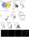
Figure 1
Figure 1. Identification of miR–130b-3p in experimental PAD in diabetic mice and in patients with PAD and diabetes.
(A) Venn diagram indicating number of miRNAs commonly dysregulated from human and mouse miRNA-Seq analysis (F, Fontaine; Db, diabetes). (B) Normalized counts of miR-130b in patients with PAD and with or without diabetes or limb events (n = 6–7). (C) Normalized counts of miR-130b in plasma from a separate cohort of patients with PAD compared with plasma from healthy individuals (n = 6). (D) (Left) Volcano plot highlighting miR-130b in muscles from patients with PAD compared with healthy individuals. (Right) Normalized counts of miR-130b in muscles of patients with PAD compared with healthy individuals (n = 7). (E) miR-130b expression normalized to U6 in ischemic gastrocnemius of db/+ and db/db mice at different time points after FAL. Comparison between groups at specific time points by unpaired Student’s t test (d0, n = 11–12; d3, n = 7–12; d11, n = 5–6; d31, n = 4–7). (FH) Relative expression of miR-130b normalized to U6 in HUVECs under different conditions: after 72 hours of D-glucose compared with mannitol control (n = 5–6) (F); after 3 or 24 hours of VEGF-A stimulation (n = 3) (G); or after 4, 16, and 24 hours of 2% hypoxia compared with normoxia control (n = 3-4) (H), performed with 2-way ANOVA. (I) Representative immunofluorescence images of human skeletal muscles from patients with diabetes and PAD stained for miR–130b-3p (green), CD31 (red), DAPI (blue), and merged, with arrows indicating colocalization. Scale bar: 50 μm. Statistics performed using unpaired 2-tailed Student’s t test unless stated otherwise. *P < 0.05, **P < 0.01.
Figure 2
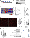
Figure 2. Endothelial miR-130b promotes angiogenesis, migration, and proliferation.
HUVECs transfected with miR-130b or nonspecific (NS) mimics (m) or inhibitors (i) prior to cellular assays. (A) (Top) Representative images of 3D EC spheroid assay. (Bottom) Quantification of mean and cumulative sprout length and number of sprouts. Scale bar: 200 μm. (B) (Top) Representative images of scratch assay of partitioned ECs separated by a cleared area (blue). (Bottom) Images captured every hour to determine area under the curve (n = 12). (C) (Left) Representative images of ECs (purple) emerged through transwell. (Right) Normalized number of ECs in transwell migration assay (n = 3). Original magnification ×4. (D) BrdU incorporation in ECs after 5 days (n = 8). All statistics performed with unpaired 2-tailed Student’s t test. *P < 0.05, **P < 0.01, ***P < 0.001, ****P < 0.0001.
Figure 3
Figure 3. Endothelial miR-130b promotes proliferative transcriptomic networks.
HUVECs transfected with miR-130b mimics compared with NS control mimics for 48 hours (n = 3) before RNA-Seq. (A) Volcano plot highlighting 2,199 downregulated and 2,310 upregulated genes in miR-130b–overexpressing ECs compared with NS mimic controls. (B) Pathway analysis of 2,310 upregulated genes organized by adjusted P values. (C) (Left) Volcano plot highlighting predicted miR-130b targets (yellow). (Right) List of top 10 downregulated predicted targets of miR-130b organized by log2 fold change.
Figure 4
Figure 4. miR-130b targets INHBA to mediate angiogenic changes in ECs.
(A) Protein abundance of INHBA in HUVECs overexpressing miR-130a or miR-130b. Densitometry normalized to β-actin (n = 3). (B) Relative expression of INHBA normalized to HPRT in HUVECs overexpressing miR-130b (n = 3). (C) (Top) Schematic of binding areas between miR-130b and INHBA 3′UTR. (Bottom) Relative luciferase units (RLU) of WT INHBA 3′UTR and MUT INHBA 3′UTR luciferase reporter assay with NS mimic or miR-130b mimic (n = 4–6). (D) (Left) Representative images of 3D EC spheroid assay. (Right) Quantification of mean and cumulative sprout length and number of sprouts. Scale bar: 200 μm. (E) Scratch assay of partitioned ECs captured every hour to determine area under the curve (n = 5). (F) Dependency of miR-130b for INHBA: 3D EC spheroid assay with combination of miR-130b inhibitors and siRNA targeting INHBA (n = 5-7). (G) miR-130b mimic or si-INHBA induce IL-8 expression by ELISA (n = 3). All statistics performed with unpaired 2-tailed Student’s t test. *P < 0.05, **P < 0.01, ****P < 0.0001.
Figure 5
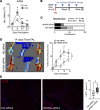
Figure 5. In vivo miR-130b delivery improves revascularization in diabetic mice experiencing hindlimb ischemia.
(A) Schema showing experimental setup and intramuscular (I.M.) injection regimen. (B) Necrosis score of ischemic foot 2 weeks after FAL. (C) (Left) Representative LDI images of hindlimbs immediately after FAL and 14 days later. (Right) Quantification of blood flow (surgical limb/contralateral limb) by LDI images, normalized to measurement immediate after surgery, 2-way ANOVA (n = 8–10). (D) Quantification of blood flow following ameroid constrictor transplantation, 2-way ANOVA (n = 10). (E) (Left) Representative immunofluorescent images of ischemic gastrocnemius stained for SMA (green), CD31 (red), and DAPI (blue). Scale bar: 100 μm. (Right) Quantification of CD31+ areas per field of view (5 images per sample, n = 4); statistics performed with unpaired 2-tailed Student’s t test. (F and G) Relative expression of Inhba (F) and Cxcl15 (G) normalized to Gapdh from CD31+ ECs and CD31 non-ECs from ischemic gastrocnemius given NS or miR-130b mimics (n = 4). (H) Pathway analysis of CD31+ ECs compartment given NS or miR-130b mimics (n = 3). *P < 0.05, **P < 0.01.
Figure 6
Figure 6. In vivo delivery of siRNA targeting Inhba improves revascularization in diabetic hindlimb ischemia.
(A) Inhba expression normalized to Gapdh in ischemic gastrocnemius of db/+ and db/db mice at different time points after FAL. Comparison between groups at specific time points by unpaired 2-tailed Student’s t test (d0, n = 11–12; d3, n = 7–12; d11, n = 5–6; d31, n = 4–7). (B) Schema showing experimental setup and intramuscular injection regimen. (C) Necrosis score of ischemic foot 2 weeks after FAL. (D) (Left) Representative LDI images of hindlimbs 14 days after FAL surgeries. (Right) Quantification of blood flow (surgical limb/contralateral limb) by LDI images, normalized to measurement immediate after surgery, 2-way ANOVA (n = 8–10). (E) (Left) Representative immunofluorescent images of ischemic gastrocnemius stained for CD31 (red) and DAPI (blue). Scale bar: 100 μm. (Right) Quantification of CD31+ areas per field of view (n = 6–8). Statistics performed with unpaired 2-tailed Student’s t test. *P < 0.05, **P < 0.01.
Figure 7
Figure 7. miR-130b/INHBA axis observed in nondiabetic patients with PAD and CLI.
(A) Relative expression of Inhba normalized to Gapdh (left) and miR-130b normalized to U6 (right) comparing sham control with db/+ gastrocnemius 11 days after FAL (n = 6–12). (B) Normalized counts of miR-130b (left) and INHBA (right) from RNA-Seq of skeletal muscles from patients without PAD, with PAD, and with more severe PAD (n = 7). (C) Normalized counts of INHBA from RNA-Seq of skeletal muscles from patients without PAD (n = 15), patients with PAD with intermittent claudication (n = 20), and patients with CLI (n = 16). All statistics performed with 1-way ANOVA with multiple comparisons. *P < 0.05, **P < 0.01, ****P < 0.0001.

Similar articles

Cited by

References

    1. Fowkes FG, et al. Comparison of global estimates of prevalence and risk factors for peripheral artery disease in 2000 and 2010: a systematic review and analysis. Lancet. 2013;382(9901):1329–1340. doi: 10.1016/S0140-6736(13)61249-0. - DOI - PubMed
    1. Uccioli L, et al. Critical limb ischemia: current challenges and future prospects. Vasc Health Risk Manag. 2018;14:63–74. doi: 10.2147/VHRM.S125065. - DOI - PMC - PubMed
    1. Marso SP, Hiatt WR. Peripheral arterial disease in patients with diabetes. J Am Coll Cardiol. 2006;47(5):921–929. doi: 10.1016/j.jacc.2005.09.065. - DOI - PubMed
    1. Avogaro A, et al. Endothelial dysfunction in diabetes: the role of reparatory mechanisms. Diabetes Care. 2011;34 Suppl 2(suppl 2):S285–S290. - PMC - PubMed
    1. Hazarika S, et al. Impaired angiogenesis after hindlimb ischemia in type 2 diabetes mellitus: differential regulation of vascular endothelial growth factor receptor 1 and soluble vascular endothelial growth factor receptor 1. Circ Res. 2007;101(9):948–956. doi: 10.1161/CIRCRESAHA.107.160630. - DOI - PubMed

Publication types