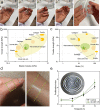
. 2023 May 8;24(5):2264-2277.
doi: 10.1021/acs.biomac.3c00152. Epub 2023 Apr 25.

Self-Assembly of Nanocellulose Hydrogels Mimicking Bacterial Cellulose for Wound Dressing Applications

Affiliations

Self-Assembly of Nanocellulose Hydrogels Mimicking Bacterial Cellulose for Wound Dressing Applications

Linn Berglund et al. Biomacromolecules. .

Abstract

The self-assembly of nanocellulose in the form of cellulose nanofibers (CNFs) can be accomplished via hydrogen-bonding assistance into completely bio-based hydrogels. This study aimed to use the intrinsic properties of CNFs, such as their ability to form strong networks and high absorption capacity and exploit them in the sustainable development of effective wound dressing materials. First, TEMPO-oxidized CNFs were separated directly from wood (W-CNFs) and compared with CNFs separated from wood pulp (P-CNFs). Second, two approaches were evaluated for hydrogel self-assembly from W-CNFs, where water was removed from the suspensions via evaporation through suspension casting (SC) or vacuum-assisted filtration (VF). Third, the W-CNF-VF hydrogel was compared to commercial bacterial cellulose (BC). The study demonstrates that the self-assembly via VF of nanocellulose hydrogels from wood was the most promising material as wound dressing and displayed comparable properties to that of BC and strength to that of soft tissue.


Conflict of interest statement

The authors declare no competing financial interest.

Figures

Figure 1
Figure 1
(a–f) Schematic presentation of the processing routes from the two starting materials (a) wood powder and (b) pulp, to self-assembly via vacuum-assisted filtration of CNF suspension into hydrogels. Optical microscopy (OM) images of (a) wood powder scale bar: 500 μm, (d) pulp fibers scale bar: 100 μm, photographs of (b,e) CNF gel (1 wt %), and (c,f) assembled hydrogel (40 g m–2) of W-CNF and P-CNF, respectively, after immersion for 48 h in water. (g) Water absorption capacity of the hydrogels (40 g m–2). (h) Representative tensile stress–strain curves of the two CNF hydrogels at 40 g m–2, tested after 1000% absorption.
Figure 2
Figure 2
(a) Water absorption capacity of the vacuum-filtered W-CNF hydrogels at different grammages. (b) Representative tensile stress–strain curves of the W-CNF-VF hydrogels at different grammages. *120 g m–2 hydrogel could not be tested in tensile.
Figure 3
Figure 3
(a) Setup for the CNF hydrogel self-assembly via suspension casting (W-CNF-SC) and vacuum-assisted filtration (W-CNF-VF) techniques. A freeze-dried hydrogel grammage of 120 g m–2 visualized using scanning electron microscopy (SEM) (b) before absorption and (c) after absorption to 1000% for W-CNF-SC and W-CNF-VF, respectively. (d) Visualization in 2D and 3D X-ray microtomography (XRT) reconstruction of a part of the cross-sections after 1000% absorption. Scale bar: 100 μm.
Figure 4
Figure 4
Hydrogels by the SC and VF of grammage (10 g m–2). (a) Water absorption capacity and (b) representative stress–strain curves. Absorption capacity after cyclic drying and re-swelling. (c) W-CNF-SC and (d) W-CNF-VF hydrogels and (e) representative photographs in the hydrogel state and dried state.
Figure 5
Figure 5
(a) Photographs of W-CNF-VF at a grammage of 10 g m–2 and BC hydrogels applied on hand. (b) XRT 2D reconstruction scale bar: 100 μm, (c) XRT 3D reconstruction scale bar: 100 μm and (d) SEM image scale bar: 1 μm of BC hydrogel cross section. W-CNF-VF and BC (e) absorption capacity in water, (f) weight loss, and (g) WVTR. TGA/derivative thermogravimetric curves for (h) W-CNF-VF and (i) BC samples. (j) Representative stress–strain curves.
Figure 6
Figure 6
W-CNF-VF hydrogel after self-assembly. (a) Photographs upon handling, Ashby plot of (b) strength as a function of elastic modulus, and (c) strength as a function of strain compared to the soft tissue and its building blocks. (d) Photograph after application on finger and arm in the form of a hydrogel strip. (e) Effects of hydrogel on fibroblast proliferation (n = 5), inset: laser-formed hydrogel into a spiral shape.

References

    1. Tapiwa Chamanga E. Clinical Management of Non-Healing Wounds. Nursing Standard 2018, 32, 48–63. 10.7748/ns.2018.e10829. - DOI - PubMed
    1. Ogueri K. S.; Laurencin C. T. Nanofiber Technology for Regenerative Engineering. ACS Nano 2020, 14, 9347–9363. 10.1021/acsnano.0c03981. - DOI - PMC - PubMed
    1. Bacakova L.; Pajorova J.; Bacakova M.; Skogberg A.; Kallio P.; Kolarova K.; Svorcik V. Versatile Application of Nanocellulose: From Industry to Skin Tissue Engineering and Wound Healing. Nanomaterials 2019, 9, 164.10.3390/nano9020164. - DOI - PMC - PubMed
    1. Basu A.; Celma G.; Strømme M.; Ferraz N. In Vitro and in Vivo Evaluation of the Wound Healing Properties of Nanofibrillated Cellulose Hydrogels. ACS Appl. Bio Mater. 2018, 1, 1853–1863. 10.1021/acsabm.8b00370. - DOI - PubMed
    1. Du H.; Liu W.; Zhang M.; Si C.; Zhang X.; Li B. Cellulose Nanocrystals and Cellulose Nanofibrils Based Hydrogels for Biomedical Applications. Carbohydr. Polym. 2019, 209, 130–144. 10.1016/j.carbpol.2019.01.020. - DOI - PubMed

Publication types