. 2023 Aug;69(2):172-181.
doi: 10.1165/rcmb.2021-0526OC.

Rhinovirus C15 Attenuates Relaxation and cAMP Production in Human Airways and Smooth Muscle

Affiliations

Rhinovirus C15 Attenuates Relaxation and cAMP Production in Human Airways and Smooth Muscle

Eric B Gebski et al. Am J Respir Cell Mol Biol. 2023 Aug.

Abstract

Rhinoviruses (RVs) evoke as many as 85% of acute asthma exacerbations in children and 50% in adults and can induce airway hyperresponsiveness and decrease efficacy of current therapeutics to provide symptom relief. Using human precision-cut lung slices (hPCLSs), primary human air-liquid interface-differentiated airway epithelial cells (HAECs), and human airway smooth muscle (HASM) as preclinical experimental models, we demonstrated that RV-C15 attenuates agonist-induced bronchodilation. Specifically, airway relaxation to formoterol and cholera toxin, but not forskolin (Fsk), was attenuated following hPCLS exposure to RV-C15. In isolated HASM cells, exposure to conditioned media from RV-exposed HAECs decreased cellular relaxation in response to isoproterenol and prostaglandin E2, but not Fsk. Additionally, cAMP generation elicited by formoterol and isoproterenol, but not Fsk, was attenuated following HASM exposure to RV-C15-conditioned HAEC media. HASM exposure to RV-C15-conditioned HAEC media modulated expression of components of relaxation pathways, specifically GNAI1 and GRK2. Strikingly, similar to exposure to intact RV-C15, hPCLS exposed to UV-inactivated RV-C15 showed markedly attenuated airway relaxation in response to formoterol, suggesting that the mechanism(s) of RV-C15-mediated loss of bronchodilation is independent of virus replication pathways. Further studies are warranted to identify soluble factor(s) regulating the epithelial-driven smooth muscle loss of β2-adrenergic receptor function.

Keywords: asthma; bronchodilation; exacerbations; rhinovirus.


Figures

Figure 1.
Figure 1.
Rhinovirus (RV)-C15 attenuates formoterol (Form)-induced relaxation of human small airways in human precision-cut lung slices (hPCLSs) derived from donors without asthma. hPCLSs (n = 10–18 distinct donors) were stimulated with RV-C15 (105 or 106 plaque-forming units [pfu], 48 h) or control buffer, and airways were contracted with carbachol and then dilated to a dose response of Form (10−9 to 10−5 M). Dose–response curves were generated (A), and maximal bronchodilation (B), integrated area under the curve (C), and log of the effective concentration to induce 50% airway relaxation (D) of Form were calculated from the curves and plotted. Data were analyzed using a one-way ANOVA (*P < 0.05), with subsequent Student’s t tests comparing RV-stimulated versus control buffer (*P < 0.05; n = 10–18 distinct donors). LogEC50 = log of the effective concentration to induce 50% airway relaxation.
Figure 2.
Figure 2.
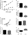
RV-C15 attenuates cholera toxin, but has little effect on forskolin (Fsk),-induced relaxation of human small airways in hPCLSs. hPCLSs were exposed to control buffer or RV-C15 (106, 48 h). Airways were constricted with carbachol and then dilated to a dose response of cholera toxin (0.01–10 μg/ml) or Fsk (10−9 to 10−5 M). Dose–response curves of the data were plotted (A and D), and parameters were derived from the curves and compared, including maximal bronchodilation (B and E), integrated area under the curve (C and F), and log of the effective concentration to induce 50% airway relaxation (G). Data were analyzed using a two-way Student’s t test (A–C, n = 5 distinct donors, *P < 0.05, RV-C15 vs. control; D–G, n = 4 distinct donors, P value not significant). CTX = cholera toxin.
Figure 3.
Figure 3.
RV-C15 exposure of human air–liquid interface–differentiated airway epithelial cells (HAECs) attenuates isoproterenol (Iso)-induced and prostaglandin E2 (PGE2)–induced, but not Fsk-induced, human airway smooth muscle (HASM) relaxation. HAECs were exposed to RV-C15 (106 pfu, 48 h). Conditioned basolateral media was transferred to serum-starved HASM, and cell stiffness was measured following carbachol (20 μM) exposure followed by stimulation with Iso (10 μM; n = 83–181 individual cell measurements), PGE2 (1 μM; n = 123–171 individual cell measurements), or Fsk (10 μM; n = 94–122 individual cell measurements) derived from a lung donor without asthma. Data are presented as mean ± SEM (****P < 0.0001 and *P < 0.05, significant by Student’s t test).
Figure 4.
Figure 4.
RV-C15 exposure of HAECs attenuates Iso-induced and Form-induced, but not Fsk-induced, cAMP production in HASM. HAECs were exposed to RV-C15 (106 pfu, 48 h). Conditioned basolateral media was transferred to serum-starved HASM to Iso (1 μM), Form (1 μM), or Fsk (10 μM) was used to induce production of cAMP, as measured by ELISA. Data are representative of (A) n = 6, (B) n = 7, and (C) n = 5 distinct donors (*P < 0.05, Student’s t test).
Figure 5.
Figure 5.
RV-C15 significantly increases GNAI1, PDE4D, and GRK2, but has little effect on ADRB2, GNAS, GNAI2, or GNAI3 expression. HAECs were exposed to RV-C15 (106 pfu, 48 h). Basolateral conditioned media was transferred to serum-starved HASM, and mRNA transcript expression was measured by TaqMan quantitative PCR. Expression of all transcripts was first normalized to GABARAP as a loading control, then normalized to the control buffer–stimulated condition. Data are expressed as mean ± SEM for n = 4–5 distinct HASM donor cell lines (*P < 0.05 vs. control, one-tailed Student’s t test). P values are as follows versus control: ADRB2, P = 0.11; GNAS, P = 0.07; GNAI2, P = 0.24; and GNAI3, P = 0.18.

Comment in

References

    1. Calışkan M, Bochkov YA, Kreiner-Møller E, Bønnelykke K, Stein MM, Du G, et al. Rhinovirus wheezing illness and genetic risk of childhood-onset asthma. N Engl J Med . 2013;368:1398–1407. - PMC - PubMed
    1. Jartti T, Gern JE. Role of viral infections in the development and exacerbation of asthma in children. J Allergy Clin Immunol . 2017;140:895–906. - PMC - PubMed
    1. Reddel H, Ware S, Marks G, Salome C, Jenkins C, Woolcock A. Differences between asthma exacerbations and poor asthma control. Lancet . 1999;353:364–369. - PubMed
    1. Reddel HK, Jenkins C, Quirce S, Sears MR, Bateman ED, O’Byrne PM, et al. Effect of different asthma treatments on risk of cold-related exacerbations. Eur Respir J . 2011;38:584–593. - PubMed
    1. Rueter K, Bizzintino J, Martin AC, Zhang G, Hayden CM, Geelhoed GC, et al. Symptomatic viral infection is associated with impaired response to treatment in children with acute asthma. J Pediatr . 2012;160:82–87. - PubMed

Publication types