. 2023 Apr 25;29(1):59.
doi: 10.1186/s10020-023-00630-9.

Mesenchymal stem cells reversibly de-differentiate myofibroblasts to fibroblast-like cells by inhibiting the TGF-β-SMAD2/3 pathway

Affiliations

Mesenchymal stem cells reversibly de-differentiate myofibroblasts to fibroblast-like cells by inhibiting the TGF-β-SMAD2/3 pathway

Ruohao Xu et al. Mol Med. .

Abstract

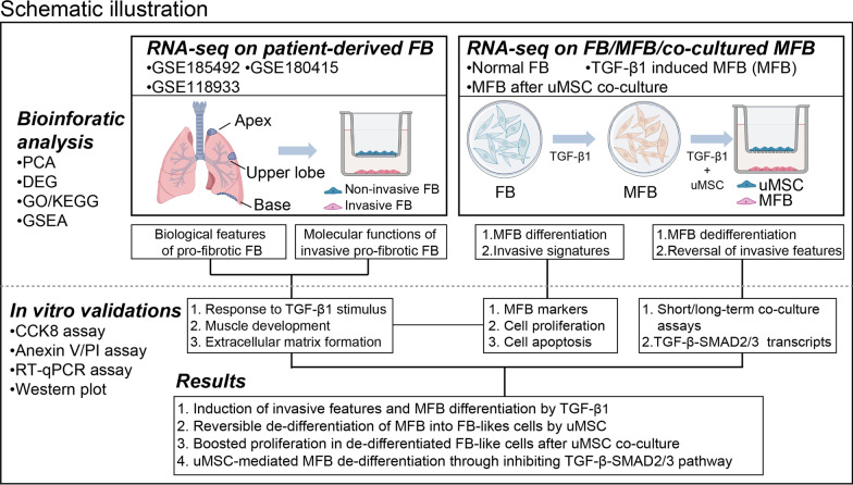
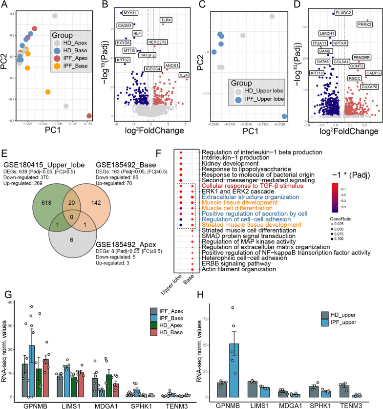
Background: Myofibroblasts (MFB), one of the major effectors of pathologic fibrosis, mainly derived from the activation of fibroblast to myofibroblast transition (FMT). Although MFBs were historically considered terminally differentiated cells, their potential for de-differentiation was recently recognized and implied with therapeutic value in treating fibrotic diseases, for instance, idiopathic pulmonary fibrosis (IPF) and post allogeneic hematopoietic stem cell transplantation bronchiolitis obliterans (BO). During the past decade, several methods were reported to block or reverse MFB differentiation, among which mesenchymal stem cells (MSC) have demonstrated potential but undetermined therapeutic values. However, the MSC-mediated regulation of FMT and underlying mechanisms remained largely undefined.

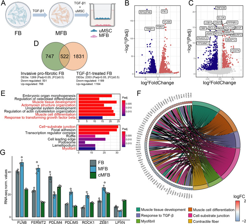
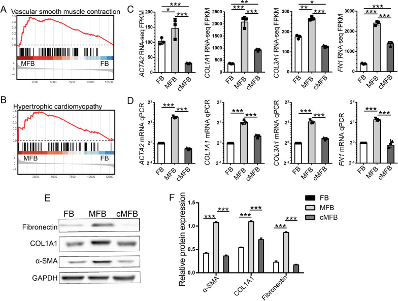
Method: By identifying TGF-β1 hypertension as the pivotal landmark during the pro-fibrotic FMT, TGF-β1-induced MFB and MSC co-culture models were established and utilized to investigate regulations by MSC on FMT in vitro. Methods including RNA sequencing (RNA-seq), Western blot, qPCR and flow cytometry were used.

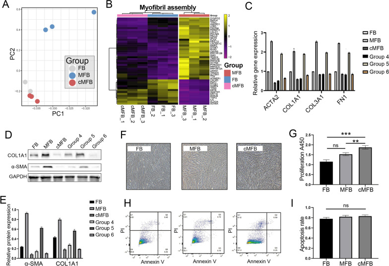
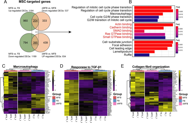
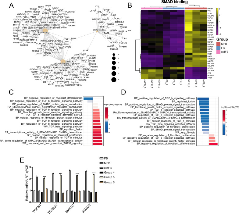
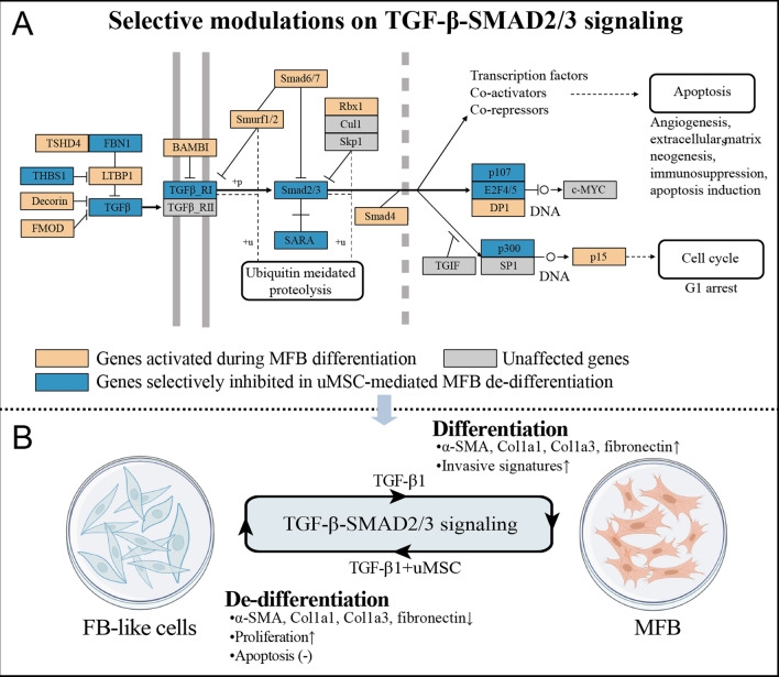
Result: Our data revealed that TGF-β1 readily induced invasive signatures identified in fibrotic tissues and initiated MFB differentiation in normal FB. MSC reversibly de-differentiated MFB into a group of FB-like cells by selectively inhibiting the TGF-β-SMAD2/3 signaling. Importantly, these proliferation-boosted FB-like cells remained sensitive to TGF-β1 and could be re-induced into MFB.

Conclusion: Our findings highlighted the reversibility of MSC-mediated de-differentiation of MFB through TGF-β-SMAD2/3 signaling, which may explain MSC's inconsistent clinical efficacies in treating BO and other fibrotic diseases. These de-differentiated FB-like cells are still sensitive to TGF-β1 and may further deteriorate MFB phenotypes unless the pro-fibrotic microenvironment is corrected.

Keywords: De-differentiation; Fibroblast; Mesenchymal stem cell; Myofibroblast; TGF-β1.


Conflict of interest statement

The authors declare no competing interests.

Figures

Fig. 1
Fig. 1
Schematic illustration of the present study. PCA principal component analysis, DEG differential expressed gene, GO gene ontology analysis, KEGG Kyoto Encyclopedia of Genes and Genomes Atlas, GSEA gene set enrichment analysis, RNA-seq RNA sequencing, uMSC Umbilical cord-derived mesenchymal stem cells, TGF-β1 transforming growth factor-β1, FB fibroblast, MFB myofibroblast
Fig. 2
Fig. 2
Identifications of transcriptomic profiles of lung FB samples from patients with IPF and healthy donors (HD). A Principal component analysis on IPF/HD-derived FB samples from the apex (n = 12) and basal lung sites (n = 12, GSE185492). B Volcano plot displaying identified DEGs in IPF-FB compared with HD-FB from the basal lung site (GSE185492) by log2 foldchange (x-axis) and adjust P-value (y-axis). Blue dots represent DEGs significantly down-regulated; red dots represent DEGs significantly upregulated; grey dots represent genes not significantly regulated. C Principal component analysis on IPF/HD-derived FB samples from the upper lobe lung site (n = 9, GSE180415). D Volcano plot displaying identified DEGs in IPF-FB compared with HD-FB from the upper lobe lung site (GSE180415) by log2 foldchange (x-axis) and adjust P-value (y-axis). Blue dots represent DEGs significantly down-regulated; red dots represent DEGs significantly upregulated; grey dots represent genes not significantly regulated. E Venn diagrams depicting the number of DEGs specifically or mutually regulated in IPF-FB samples from different lung anatomical sites. F Enriched analysis by EGO using DEGs identified in IPF-FB samples from upper lobe lung and basal lung sites. G, H RNA-seq expression normalized value (fragments per kilobase of exon model per million mapped fragments, FPKM) for transcripts encode for genes related to response to TGF-β1 stimulus and muscle tissue development in each RNA-seq dataset
Fig. 3
Fig. 3
In vitro administration of TGF-β1 induced invasive IPF-FB signatures in normal lung FB samples. A Schematic representation of in vitro TGF-β1 induction and subsequent uMSC co-culture models. Normal lung FB was treated with ether 10 ng/mL of TGF-β1 or DMSO before being subjected to subsequent RNA-seq and in vitro assays. B Volcano plot displaying identified DEGs in MFB compared with untreated FB by log2 foldchange (x-axis) and adjust P-value (y-axis). C Volcano plot displaying identified DEGs in invasive IPF-FB (n = 9) compared with non-invasive IPF-FB (n = 9, GSE118933) by log2 foldchange (x axis) and adjust P value (y axis). D Venn diagrams depicting the number of DEGs specifically or mutually regulated in invasive IPF-FB and MFB samples. E Enriched gene ontology analysis (EGO) concerning biological process (BP), molecular function (MF), and cellular component (CC) of DEGs identified in MFB (MFB vs. FB). F Circos plot displaying the most significantly enriched EGO terms and related DEGs participating in each process. Symbols of DEGs are displayed on the left side of the graph with their fold change value mapped by color (red represents up-regulation and blue represents down-regulation). G RNA-seq expression normalized value (FPKM) for transcripts encode for genes related to TGF-β signaling, muscle/fibrin development in FB, iMFB, and co-cultured MFB
Fig. 4
Fig. 4
Co-culturing with uMSC inhibited TGF-β-induced expression of MFB-specific markers. A, B Gene set enrichment analysis (GSEA) indicates activation of vascular smooth muscle contraction and hypertrophy cardiomyopathy functions in TGF-β1-induced MFB. C, D RNA-seq gene expression (FPKM) and qPCR validations for MFB markers, including ACTA2 (encoding for a-SMA), COL1A1, COL3A1, and FN1 in FB, MFB, and co-cultured MFB samples. E, F Western blot assays on fibronectin, COL1A1, and a-SMA proteins in FB, MFB, and co-cultured MFB samples. FPKM, fragments per kilobase of exon model per million mapped fragments. * < 0.05; **P < 0.01; ***P < 0.001
Fig. 5
Fig. 5
Identification of the cellular properties and reversibility of uMSC-mediated MFB de-differentiation. A Principal component analysis (PCA) on transcriptomes of FB, MFB, and co-cultured MFB samples. B Unsupervised clustering heatmap of 45 gene-panel involved in myofibril assembly on FB, MFB, and co-cultured MFB samples. C RT-qPCR assays on the expression of ACTA2 (encoding for a-SMA), COL1A1, COL3A1, and FN1 in cells from the 6 group settings: (1) Untreated FB; (2) MFB; (3) Short-term co-cultured MFB (FB-like cells); (4) Long-term co-cultured MFB; (5) FB-like cells treated with TGF-β1 without uMSC feeder layer; (6) TGF-β1-treated FB-like cells with additional uMSC feeder layer. D, E Western blot assays on FN1, COL1A1, and a-SMA proteins in cells from the 6 group settings: (1) Untreated FB; (2) MFB; (3) Short-term co-cultured MFB (FB-like cells); (4) Long-term co-cultured MFB; (5) FB-like cells treated with TGF-β1 without uMSC feeder layer; (6) TGF-β1-treated FB-like cells with additional uMSC feeder layer. F, G The proliferation of FB, MFB, and co-cultured MFB by light microscopy and CCK-8 assays. H, I Apoptosis FB, MFB and co-cultured MFB by Annexin V/PI assays. * < 0.05; **P < 0.01; ***P < 0.001
Fig. 6
Fig. 6
Annotation of the targeted genes during uMSC-mediated MFB de-differentiation. A Venn diagrams depicting the number of DEGs regulated in MFB (MFB vs. FB) and then reversely regulated in co-cultured MFB (co-cultured MFB vs. MFB). B Enriched gene ontology analysis (EGO) concerning biological process (BP), molecular function (MF), and cellular component (CC) using the targeted DEGs. CE Unsupervised clustering heatmap on genes participating in macroautophagy, response to TGF-β1, and collagen fibril organization in FB, MFB, and co-cultured MFB samples
Fig. 7
Fig. 7
Reversible modulations on the TGF-β-SMAD2/3 signaling pathway in the uMSC-mediated MFB de-differentiation. A Network illustration of actin-binding, cadherin binding, SMAD binding, and associated DEGs. B Unsupervised clustering heatmap on genes participating in SMAD binding in FB, MFB, and co-cultured MFB samples. C GSEA analysis on regulated genes identified in MFB (MFB vs. FB). D GSEA analysis on regulated genes identified in co-cultured MFB (co-cultured MFB vs. MFB). E RT-qPCR assays on the expression of TGFB1, TGFBR1, SMAD2 and SMAD3 in cells from the 6 group settings: (1) Untreated FB; (2) MFB; (3) Short-term co-cultured MFB (FB-like cells); (4) Long-term co-cultured MFB; (5) FB-like cells treated with TGF-β1 without uMSC feeder layer; (6) TGF-β1-treated FB-like cells with additional uMSC feeder layer
Fig. 8
Fig. 8
Selective modulations on the TGF-β-SMAD2/3 signaling. A Regulations of transcripts in TGF-β-SMAD2/3 signaling according to the KEGG database. B uMSC reversibly de-differentiate TGF-β1-induced MFB into a group of FB-like cells by modulating the TGF-β-SMAD2/3 pathway

Similar articles

Cited by

References

    1. Aschner Y, Downey GP. Transforming growth factor-β: master regulator of the respiratory system in health and disease. Am J Respir Cell Mol Biol. 2016;54(5):647–655. doi: 10.1165/rcmb.2015-0391TR. - DOI - PMC - PubMed
    1. Athwal VS, et al. SOX9 regulated matrix proteins are increased in patients serum and correlate with severity of liver fibrosis. Sci Rep. 2018;8(1):17905. doi: 10.1038/s41598-018-36037-4. - DOI - PMC - PubMed
    1. Bargagli E, et al. Pirfenidone and Nintedanib in idiopathic pulmonary fibrosis: real-life experience in an Italian referral centre. Pulmonology. 2019;25(3):149–153. doi: 10.1016/j.pulmoe.2018.06.003. - DOI - PubMed
    1. Basalova N, et al. Secretome of mesenchymal stromal cells prevents myofibroblasts differentiation by transferring fibrosis-associated microRNAs within extracellular vesicles. Cells. 2020 doi: 10.3390/cells9051272. - DOI - PMC - PubMed
    1. Baudier J, Jenkins ZA, Robertson SP. The filamin-B-refilin axis—spatiotemporal regulators of the actin-cytoskeleton in development and disease. J Cell Sci. 2018 doi: 10.1242/jcs.213959. - DOI - PubMed

Publication types

MeSH terms

LinkOut - more resources