

Up-regulation of CREB-1 regulates tendon adhesion in the injury tendon healing through the CREB-1/TGF-β3 signaling pathway
- PMID: 37098516
- PMCID: PMC10127358
- DOI: 10.1186/s12891-023-06425-7
Up-regulation of CREB-1 regulates tendon adhesion in the injury tendon healing through the CREB-1/TGF-β3 signaling pathway
Abstract
Aim: To explore the mechanism of the healing of tendon tissue and anti-adhesion, and to discuss the role of the transforming growth factor-β3 (TGF-β3)/cAMP response element binding protein-1 (CREB-1) signaling pathway in the healing process of tendons.
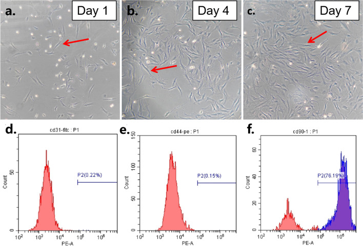
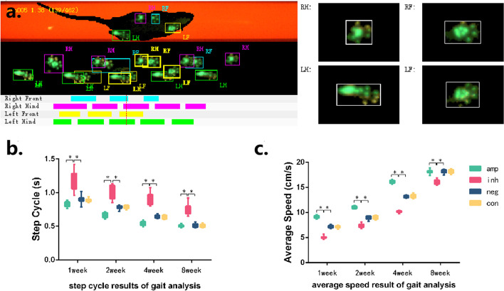
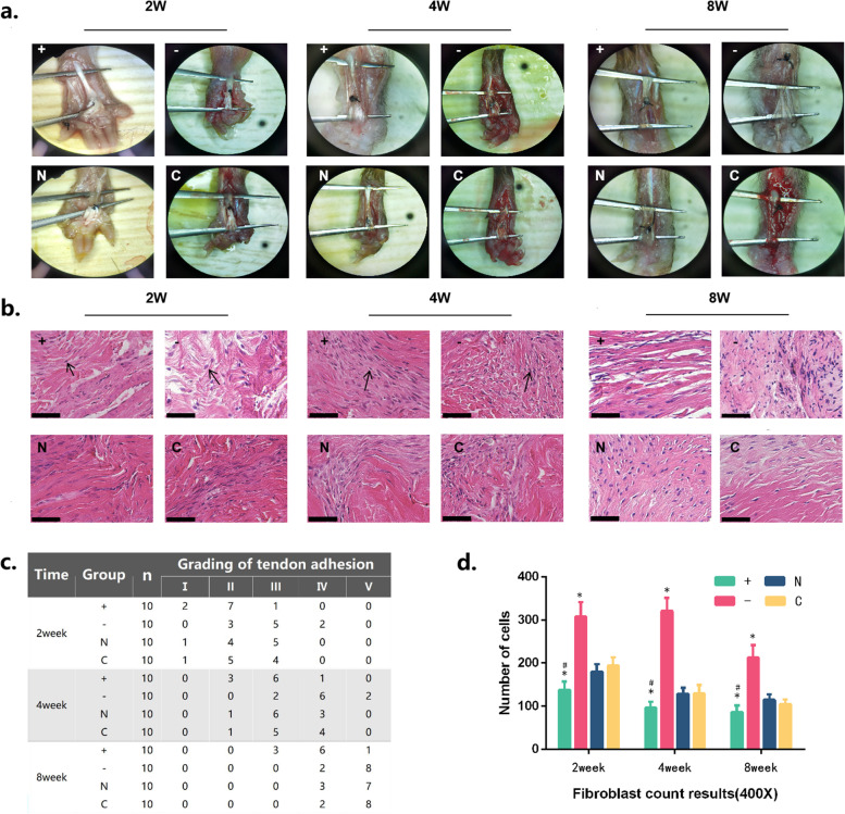
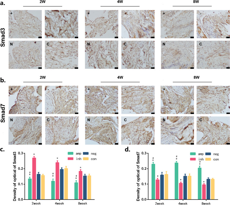
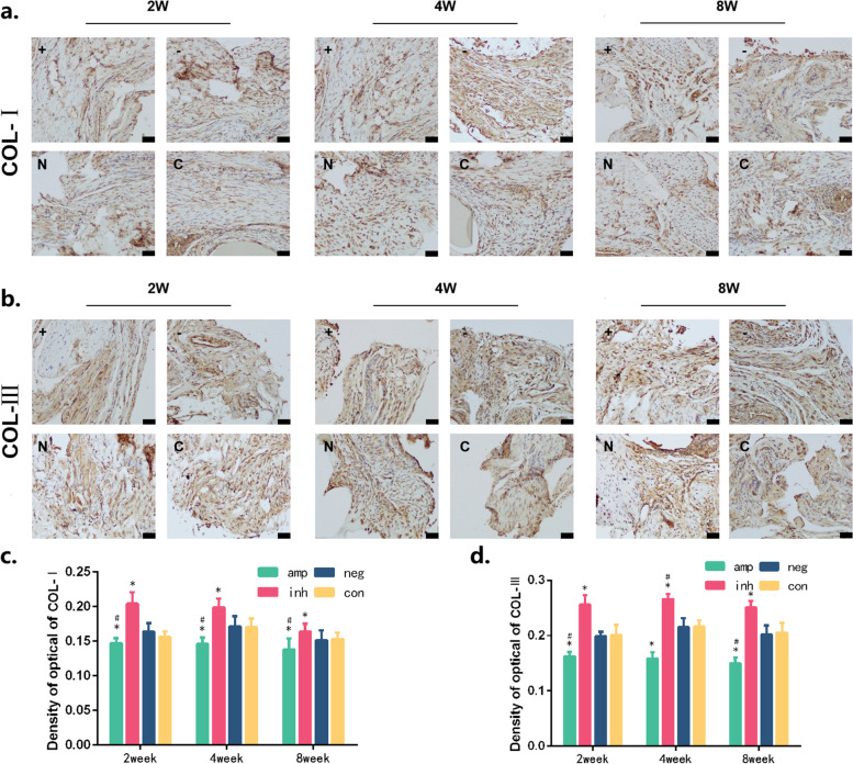
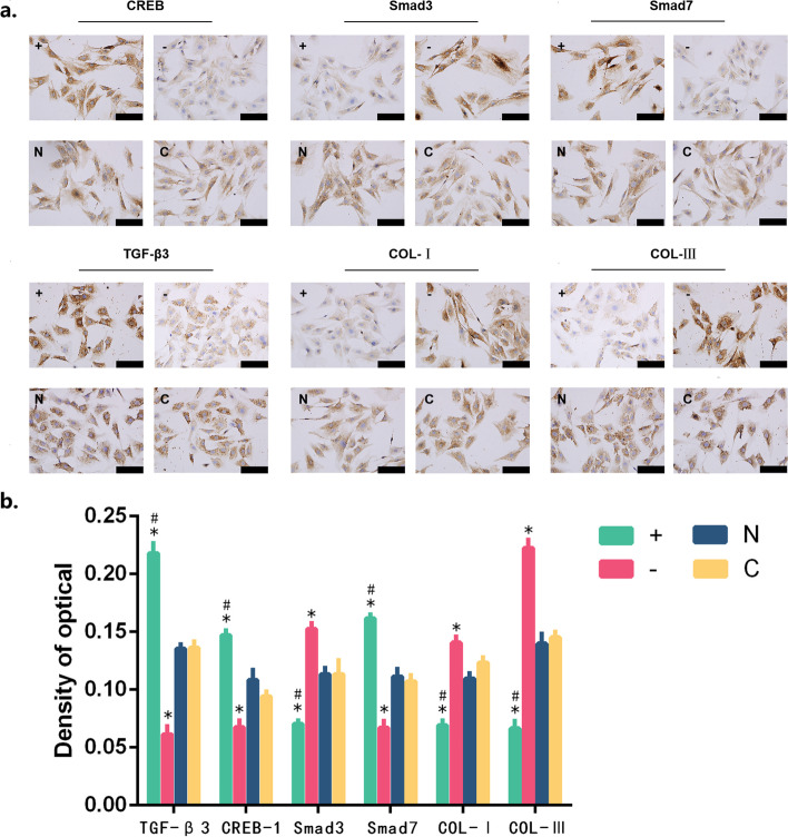
Method: All mice were divided into four groups of 1, 2, 4, and 8 weeks respectively. Each time group was divided into four treatment groups: the amplification group, the inhibition group, the negative group, and the control group. When the tendon injury model was established, the CREB-1 virus was injected into the tendon injury parts. A series of methods such as gait behaviourism, anatomy, histological examination, immunohistochemical examination and collagen staining were employed to assess the tendon healing and the protein expression of TGF-β3, CREB-1, Smad3/7 and type I/III collagen (COL-I/III). CREB-1 virus was sent to tendon stem cells to assess the protein expression of TGF-β1, TGF-β3, CREB-1, COL-I/III by methods such as immunohistochemistry and Western blot.
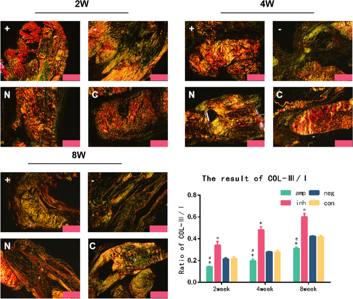
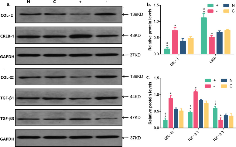
Results: The amplification group showed better gait behaviourism than the inhibition group in the healing process. The amplification group also had less adhesion than the negative group. Hematoxylin-eosin (HE) staining of tendon tissue sections showed that the number of fibroblasts in the amplification group was less than the inhibition group, and the immunohistochemical results indicated that the expression of TGF-β3, CREB-1, and Smad7 at each time point was higher than the inhibition group. The expression of COL-I/III and Smad3 in the amplification group was lower than the inhibition group at all time points. The collagen staining indicated that the ratio of type I/III collagen in the amplification group was higher than the negative group at 2,4,8 week. The CREB-1 amplification virus could promote the protein expression of TGF-β3, CREB-1 and inhibit the protein expression of TGF-β1 and COL-I/III in the tendon stem cells.
Conclusion: In the process of tendon injury healing, CREB-1 could promote the secretion of TGF-β3, so as to promote the tendon healing and have the effect of anti-adhesion in tendons. It might provide new intervention targets for anti-adhesion treatment of tendon injuries.
Keywords: Adhesion2; CREB-15; Healing3; TGF-β34; Tendon1.
© 2023. The Author(s).
Conflict of interest statement
The authors declare no competing interests.
Figures
Similar articles
-
Leptin Enhances M1 Macrophage Polarization and Impairs Tendon-Bone Healing in Rotator Cuff Repair: A Rat Model.Clin Orthop Relat Res. 2025 May 1;483(5):939-951. doi: 10.1097/CORR.0000000000003428. Epub 2025 Feb 19. Clin Orthop Relat Res. 2025. PMID: 39982019
-
[Influence and mechanism of extracellular vesicles derived from human dermal papilla cells on skin fibrosis in mice].Zhonghua Shao Shang Yu Chuang Mian Xiu Fu Za Zhi. 2025 Jun 20;41(6):559-568. doi: 10.3760/cma.j.cn501225-20240925-00348. Zhonghua Shao Shang Yu Chuang Mian Xiu Fu Za Zhi. 2025. PMID: 40588404 Free PMC article. Chinese.
-
[Experimental study on promotion of skin radiation damage repair by icarin via HIF-2α/VEGF/Notch pathway to enhance the paracrine function of adipose-derived stem cells].Zhongguo Xiu Fu Chong Jian Wai Ke Za Zhi. 2025 Jul 15;39(7):881-890. doi: 10.7507/1002-1892.202503089. Zhongguo Xiu Fu Chong Jian Wai Ke Za Zhi. 2025. PMID: 40659593 Free PMC article. Chinese.
-
The Black Book of Psychotropic Dosing and Monitoring.Psychopharmacol Bull. 2024 Jul 8;54(3):8-59. Psychopharmacol Bull. 2024. PMID: 38993656 Free PMC article. Review.
-
Platelet-rich therapies for musculoskeletal soft tissue injuries.Cochrane Database Syst Rev. 2014 Apr 29;2014(4):CD010071. doi: 10.1002/14651858.CD010071.pub3. Cochrane Database Syst Rev. 2014. PMID: 24782334 Free PMC article.
Cited by
-
Early activity after strong sutures helps to tendon healing in a rat tendon rupture model.Sci Rep. 2025 Jan 2;15(1):513. doi: 10.1038/s41598-024-84393-1. Sci Rep. 2025. PMID: 39747621 Free PMC article.
-
Ogerin induced activation of Gpr68 alters tendon healing.FASEB Bioadv. 2025 Apr 3;7(5):e70008. doi: 10.1096/fba.2024-00236. eCollection 2025 May. FASEB Bioadv. 2025. PMID: 40330429 Free PMC article.
References
-
- Childress MA, Beutler A. Management of chronic tendon injuries. Am Fam Physician. 2013;87(7):486–490. - PubMed
MeSH terms
Substances
LinkOut - more resources
Full Text Sources
Research Materials
