. 2023 Apr 25;18(1):52.
doi: 10.1186/s13000-023-01337-5.

Breast implant associated EBV-positive Diffuse Large B-cell lymphoma: an underrecognized entity?

Affiliations

Breast implant associated EBV-positive Diffuse Large B-cell lymphoma: an underrecognized entity?

Johanna Vets et al. Diagn Pathol. .

Abstract

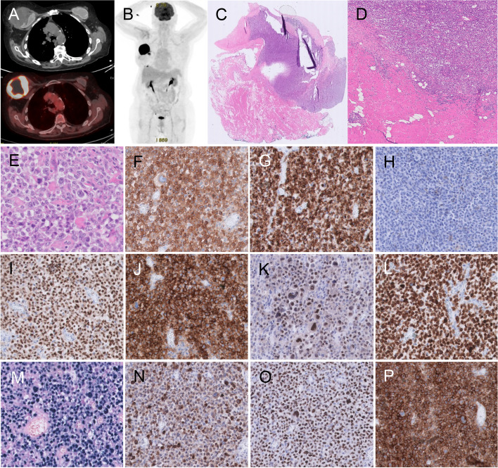
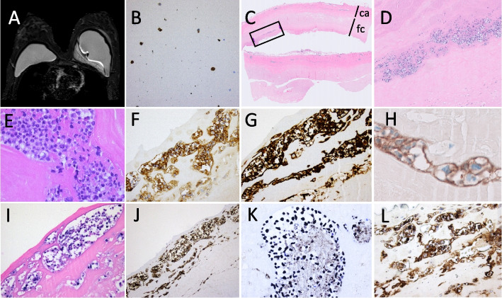
Breast-implant associated (BIA) lymphoma is an infrequent type of cancer occurring in the fluid and fibrous capsule around a textured breast implant. Recently, both the 2022 WHO 5th edition classification of Haematological tumours (WHO HAEM5) and 2022 International Consensus Classification of Mature Lymphoid Neoplasms (22ICC), recognized breast implant-associated Anaplastic Large Cell Lymphoma (BIA-ALCL) as a definitive entity, defined as a mature CD30-positive T-cell lymphoma, confined by a fibrous capsule, in a breast implant setting. Only few B-cell lymphomas have been reported in the literature to be associated with breast implants. Here we report two EBV-positive Diffuse Large B-cell lymphomas (EBV + DLBCL) in relation to a breast implant, both expressing CD30 as well as EBV latency type 3. Both lesions were considered as DLBCL associated with chronic inflammation (CI-DLBCL), but one presented as a 7 cm solid mass, while the other presented as a fibrin-associated DLBCL (FA-DLBCL) in an HIV patient. Clinically, both are in complete remission 6 months or longer after capsulectomy and graft removal, without additional chemotherapy.Such cases, characterized by large CD30-positive cells, can easily be misdiagnosed as BIA-ALCL if the cell of origin is not further established. Therefore, a diagnostic panel including lineage-specific B-and T-cell markers and EBER in situ hybridization is essential to recognize this rare entity, to understand lymphomagenesis, to predict outcome and to define clinical approach.

Keywords: BIA-DLBCL; Breast implant associated lymphoma; CI-DLBCL; EBV + DLBCL; FA-DLBCL.


Conflict of interest statement

The authors declare no competing interests.

Figures

Fig. 1
Fig. 1
Case 1. A-B. PET/CT shows a large tumoral mass at the upper-external quadrant of the right breast, in close contact with the implant with a layer of fluid surrounding the implant; There were no lymphadenopathies nor other suspicious PET-positive lesions. C-D. Low power view of the H&E of the tumor mass, composed of diffuse sheets of large atypical lymphoid cells. E. High power field view of HE of large malignant cells expressing CD30 (F), CD15 (G), but no T-cell markers, such as CD4 (H). The cells did express B-cell markers PAX5 (I), CD20 (J), OCT2 (K), CD79A (L) and were positive for EBER (M); The expression of LMP1 (N) and EBNA2 (O) indicate EBV viral latency type III; The tumor microenvironment is nearly absent, but the tumor cells diffusely express PD-L (P)
Fig. 2
Fig. 2
Case 2. A. MRI shows intact breast implants, and a thick-walled fluid collection with intralesional septations, but no contrast capturing, medially of the implant in the left breast; B. FNAC cytology shows large atypical cells (Papanicolaou stain), expressing CD30 and PAX5 (not shown); C. HE stain of the left capsulectomy specimen, showing a fibrous capsule (ca) with a fibrinous cover (fc), containing clusters of large atypical lymphoid cells (square in D, enlarged in D-E), expressing CD30 (F), and B-cell markers CD20 (G), OCT2, PAX5 and BOB1, and also the hypoxia marker CA-9 (H); Presence of EBV was shown by EBER ISH (I), displaying an EBV latency type III, with expression of LMP1 (J) and EBNA2 (K); There was no T-cell marker expression, but the cells were positive for PD-L1 (L)

References

    1. Campo E, Jaffe ES, Cook JR, Quintanilla-Martinez L, Swerdlow SH, Anderson KC, et al. The International Consensus Classification of Mature Lymphoid Neoplasms: a report from the Clinical Advisory Committee. Blood. 2022;140:1229–1253. doi: 10.1182/blood.2022015851. - DOI - PMC - PubMed
    1. Alaggio R, Amador C, Anagnostopoulos I, Attygalle AD, Araujo IB de O, Berti E, et al. The 5th edition of the World Health Organization Classification of Haematolymphoid Tumours: Lymphoid Neoplasms. Leukemia 36, 1720–48(2022) - PMC - PubMed
    1. Clemens MW, Jacobsen ED, Horwitz SM. 2019 NCCN Consensus Guidelines on the Diagnosis and Treatment of Breast Implant-Associated Anaplastic Large Cell Lymphoma (BIA-ALCL) Aesthetic Surg J. 2019;39:S3–13. doi: 10.1093/asj/sjy331. - DOI - PubMed
    1. Copie-Bergman C, Niedobitek G, Mangham DC, Selves J, Baloch K, Diss TC, et al. Epstein-Barr virus in B-cell lymphomas associated with chronic suppurative inflammation. J Pathol. 1997;183:287–292. doi: 10.1002/(SICI)1096-9896(199711)183:3<287::AID-PATH932>3.0.CO;2-Q. - DOI - PubMed
    1. Aozasa K, Takakuwa T, Nakatsuka S. Pyothorax-Associated Lymphoma. Adv Anat Pathol. 2005;12:324–331. doi: 10.1097/01.pap.0000194627.50878.02. - DOI - PubMed

MeSH terms