. 2023 Apr 25;16(1):36.
doi: 10.1186/s13041-023-01024-y.

Tibial fracture surgery in elderly mice caused postoperative neurocognitive disorder via SOX2OT lncRNA in the hippocampus

Affiliations

Tibial fracture surgery in elderly mice caused postoperative neurocognitive disorder via SOX2OT lncRNA in the hippocampus

Zhibin Xiao et al. Mol Brain. .

Abstract

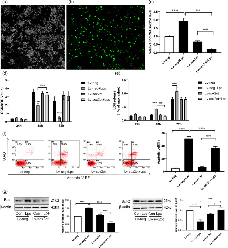
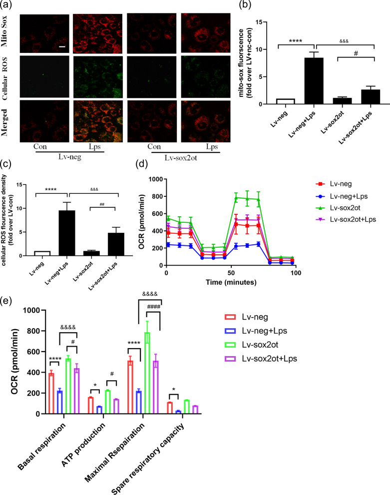
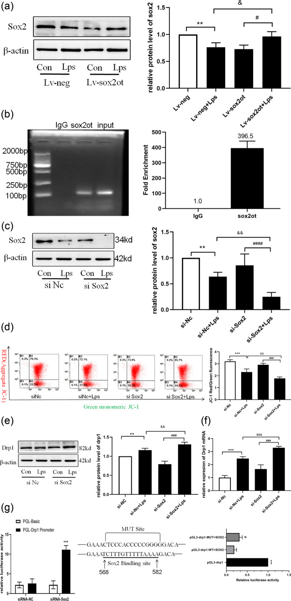
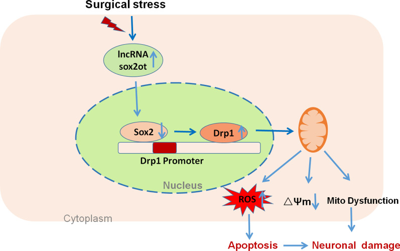
Increasing evidence indicates the major role of mitochondrial function in neurodegenerative disease. However, it is unclear whether mitochondrial dynamics directly affect postoperative neurocognitive disorder (PND). This study aimed to analyze the underlying mechanisms of mitochondrial dynamics in the pathogenesis of PND. Tibial fracture surgery was performed in elderly mice to generate a PND model in vivo. Cognitive behavior was evaluated 3 days post-surgery using novel object recognition and fear conditioning. A gradual increase in the SOX2OT mRNA level and decrease in the SOX2 mRNA level were noted, with impaired cognitive function, in the mice 3 days after tibial surgery compared with mice in the sham group. To evaluate the role of SOX2OT in PND, SOX2OT knockdown was performed in vitro and in vivo using lentivirus transfection in HT22 cells and via brain stereotactic injection of lentivirus, respectively. SOX2OT knockdown reduced apoptosis, inhibited oxidative stress, suppressed mitochondrial hyperdivision, attenuated surgery-induced cognitive dysfunction, and promoted downstream SOX2 expression in elderly mice. Furthermore, Sox2 alleviated mitochondrial functional damage by inhibiting the transcription of mitochondrial division protein Drp1. Our study findings indicate that SOX2OT knockout alleviates surgery-induced mitochondrial fission and cognitive function defects by upregulating the expression of Sox2 in mice, resulting in the inhibition of drp1 transcription. Therefore, regulation of the SOX2/Drp1 pathway may be a potential mechanism for the treatment of patients with PND.

Keywords: Drp1; Mitochondrial dynamics; Oxidative stress; Postoperative neurocognitive disorder; SOX2; SOX2OT.


Conflict of interest statement

The authors declare that they have no competing interests.

Figures

Fig. 1
Fig. 1
Experimental design and behavioral test results. a Mice were provided behavioral training 1 day before tibial surgery. The sham group mice were not subjected to tibial fracture. Behavioral tests were performed on day 3 post-surgery. b Comparison of the total activity, distance, and time spent in the central square. P < 0.05 vs. sham group; n = 4. c Comparison of total sniffing time and preference index for novel object. **P < 0.001 vs. sham group; n = 4. d Freezing time. **P < 0.005 vs. sham group; n = 4. Red arrows indicate electric shock stimuli, and green horn indicates sound stimuli
Fig. 2
Fig. 2
Upregulated lncRNA SOX2OT expression in the mouse model of tibial fracture. a GO term and KEGG pathway enrichment map of differential lncRNAs. b High expression of SOX2OT in lncRNA sequencing, verified using RT-PCR (P < 0.0001). c SOX2OT located on mouse chromosome 3 and Fxr1, Sox2, and Dnajc19 are upstream and downstream genes of SOX2OT in the Ensembl database. d Dnajc19, SOX2, and Fxr1 mRNA levels measured using RT-qPCR on days 1, 3, 7, and 14 after sham treatment or surgery. *P < 0.05, **P < 0.002 vs. sham group. e Western blot analysis showing SOX2 protein expression on days 1, 3, 7, and 14 day after sham treatment or surgery. *P < 0.05 vs. sham group; n = 6
Fig. 3
Fig. 3
SOX2OT knockdown suppresses apoptosis. a–c Fluorescent images showing virus-transfected HT22 cells and the stable knockdown of SOX2OT; SOX2OT expression levels detected using RT-PCR in the Con and LPS groups. *P < 0.05, ****P < 0.0001 vs. Lv-neg; ####P < 0.0001 vs. Lv-neg + LPS. d HT22 Lv-neg or Lv-SOX2OT cells were treated with LPS (100 ng/mL) for 24, 48, and 72 h, and then cell viability was determined. *P < 0.05 vs. Lv-neg; #P < 0.05 vs. Lv-neg + LPS. e Activity of lactate dehydrogenase in HT22 Lv-neg or Lv-SOX2OT cells cultured with LPS (100 ng/mL) for 24, 48, and 72 h using the LDH kit. *P < 0.05, ***P < 0.0003 vs. Lv-neg;####P < 0.0001 vs. Lv-neg + LPS. f Apoptosis detected using flow cytometry. ****P < 0.0001 vs. LV-neg; ####P < 0.0001 vs. Lv-SOX2OT + LPS; &&&&P < 0.0001 vs. Lv-neg. g Western blot analysis showing Bax and Bcl-2 protein expression after transfection with Lv-SOX2OT or Lv-neg lentiviral vectors. ****P < 0.0001 vs. Lv-neg; ####P < 0.0001 vs. Lv-SOX2OT + LPS; &&&&P < 0.0001 vs. Lv-neg; n = 4
Fig. 4
Fig. 4
SOX2OT knockdown prevents LPS-induced mitochondrial fission. a Mitochondrial morphology in the hippocampus determined using transmission electron microscopy. Scale bar = 0.5 μm. Analysis of mitochondrial damage score. P < 0.005 vs Sham. b Mitochondrial morphology in HT22 cells obtained using MitoTracker Red staining. Original magnification × 600; n = 4. c Percentage of cells with fragmented mitochondria. d: Mean mitochondrial volume (fold over Con + Nc); n = 4. e Expression of the Drp1, Fis1, Mfn, and Opa1 proteins determined using western blotting. *P < 0.05, ****P < 0.0001 vs. Lv-neg; ##P < 0.01 vs. Lv-neg + LPS; &&P < 0.009 vs. Lv-SOX2OT groups; n = 4
Fig. 5
Fig. 5
SOX2OT knockdown protected against LPS-induced mitochondrial dysfunction in HT22 cells. a Mitochondria-derived superoxide was stained with the MitoSox probe, and intracellular oxidants were stained with cellular ROS dye. Original magnification × 600; n = 4. b, c Statistical analysis of cellular ROS fluorescence density and MitoSox fluorescence in HT22 cells (fold over Lv + nc); n = 4. d–e Seahorse analysis showing OCR and quantitative statistical analysis of OCR. *P < 0.05 vs. Lv-neg; **P < 0.01, ****P < 0.01 vs. Lv-neg; ##P < 0.01 vs. Lv-neg; &&&P < 0.01 vs. Lv-neg + LPS; n = 5
Fig. 6
Fig. 6
SOX2OT knockdown attenuated surgery-induced cognitive dysfunction and promoted downstream SOX2 expression in elderly mice. a Experimental design and workflow. b The lentivirus Sox2ot was injected bilaterally into the CA1 region of the mouse hippocampus. c The GFP signal observed in CA1 neurons 7 days after injection of lentivirus confirmed the successful expression of Sox2ot or the control vector. Scale, 100 μm. d SOX2OT mRNA measured using RT-qPCR. **P < 0.001 vs Lv-neg. e Comparison of total activity, distance, and time spent in the central square; n = 4. f Comparison of total sniffing time and preference index for novel object. **P < 0.001 vs. Lv-neg group; n = 4. g Freezing time. **P < 0.005 vs. Lv-neg. e SOX2OT mRNA measured using RT-qPCR. **P < 0.001 vs Lv-neg. h SOX2 protein expression analyzed using western blotting. ***P < 0.0001, #P < 0.03, &&&P < 0.0001 vs Lv-neg. i Drp1 protein expression analyzed using western blotting. *P < 0.04, ###P < 0.0006, &&P < 0.003 vs Lv-neg; n = 4
Fig. 7
Fig. 7
SOX2OT binds to the downstream SOX2 target, which is related to mitochondrial fission protein Drp1. a Evaluating the transfection efficiency of Lv-SOX2OT lentivirus by detecting the expression of SOX2 using western blotting. *P < 0.05 vs. Lv-neg group, #P < 0.05 vs. Lv-SOX2OT group, && P < 0.004 vs. Lv-neg + LPS group; n = 4. b RIP-qPCR to verify the binding of downstream SOX2 target with lncRNA SOX2OT. *P < 0.05 vs. IgG group; n = 3. c Verifying the transfection effect of siRNA by detecting the expression of SOX2 using western blotting. **P < 0.007 vs. si-nc group, #### P < 0.0001 vs. si-SOX2 group; &&P < 0.004 vs. si-nc + LPS group; n = 4. d Flow cytometry analysis and JC-1 quantitative determination of mitochondrial membrane potential in HT22 cells. ****P < 0.0001 vs. si-nc group; ###P < 0.001 vs. si-SOX2 group; &&P < 0.004 vs. si-nc + LPS group; n = 4. e Drp1 expression detected using western blotting after transfection with si-NC or si-SOX2 vectors. **P < 0.006 vs. si-nc group; ###P < 0.0001 vs. si-SOX2 group; &&P < 0.009 vs. si-nc + LPS group; n = 4. f The drp1 mRNA level measured using RT-qPCR.***P<0.001 vs. si-NC; &&&P<0.001 vs. si-Nc+LPS; ###P<0.001 vs. si-SOX2. g Analysis of the binding reactivity between SOX2 and drp1 promoter region using Luciferase reporter assay. ***P < 0.0006 vs.pGL3-drp1 #P < 0.05 vs. pGL3-drp1-WT + SOX2; n = 3
Fig. 8
Fig. 8
Surgical stress leads to mitochondrial dynamics imbalance due to increased SOX2OT expression, promoting neuronal damage. SOX2OT negatively regulates SOX2 expression, which negatively regulates Drp1 expression. Surgical stress increases SOX2OT and Drp1 expression, leading to oxidative stress and mitochondrial-derived ROS production. Thus, the mitochondrial membrane potential is reduced and mitochondrial dysfunction occurs, leading to the development of postoperative cognitive dysfunction in elderly mice. SOX2OT knockdown can reduce oxidative stress and mitochondrial dysfunction, alleviate apoptosis, and protect against inflammation-induced cognitive dysfunction

Similar articles

Cited by

References

    1. Cortese GP, Burger C. Neuroinflammatory challenges compromise neuronal function in the aging brain: postoperative cognitive delirium and Alzheimer’s disease. Behav Brain Res. 2017;322:269–279. doi: 10.1016/j.bbr.2016.08.027. - DOI - PMC - PubMed
    1. Netto MB, de Oliveira Junior AN, Goldim M, Mathias K, Fileti ME, da Rosa N, et al. Oxidative stress and mitochondrial dysfunction contributes to postoperative cognitive dysfunction in elderly rats. Brain Behav Immun. 2018;73:661–669. doi: 10.1016/j.bbi.2018.07.016. - DOI - PubMed
    1. Moller JT, Cluitmans P, Rasmussen LS, Houx P, Rasmussen H, Canet J, et al. Long-term postoperative cognitive dysfunction in the elderly ISPOCD1 study. ISPOCD investigators. International study of post-operative cognitive dysfunction. Lancet. 1998;351:857–861. doi: 10.1016/S0140-6736(97)07382-0. - DOI - PubMed
    1. Monk TG, Weldon BC, Garvan CW, Dede DE, van der Aa MT, Heilman KM, et al. Predictors of cognitive dysfunction after major noncardiac surgery. Anesthesiology. 2008;108:18–30. doi: 10.1097/01.anes.0000296071.19434.1e. - DOI - PubMed
    1. Silbert BS, Evered LA, Scott DA. Incidence of postoperative cognitive dysfunction after general or spinal anesthesia for extracorporeal shock wave lithotripsy. Br J Anaesth. 2014;113:784–791. doi: 10.1093/bja/aeu163. - DOI - PubMed

Publication types

MeSH terms