. 2023 May 17;67(5):e0151922.
doi: 10.1128/aac.01519-22. Epub 2023 Apr 26.

PlyKp104, a Novel Phage Lysin for the Treatment of Klebsiella pneumoniae, Pseudomonas aeruginosa, and Other Gram-Negative ESKAPE Pathogens

Affiliations

PlyKp104, a Novel Phage Lysin for the Treatment of Klebsiella pneumoniae, Pseudomonas aeruginosa, and Other Gram-Negative ESKAPE Pathogens

Chad W Euler et al. Antimicrob Agents Chemother. .

Abstract

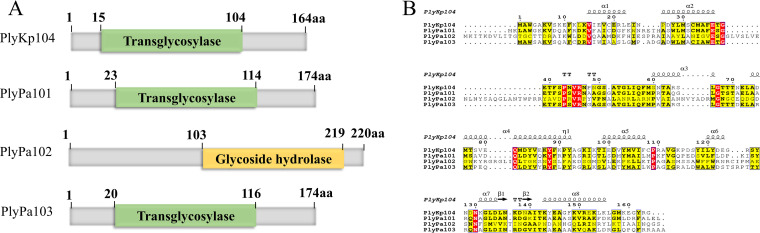
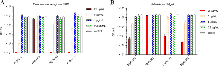
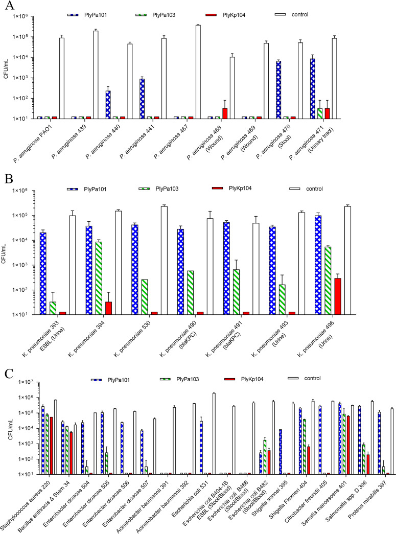
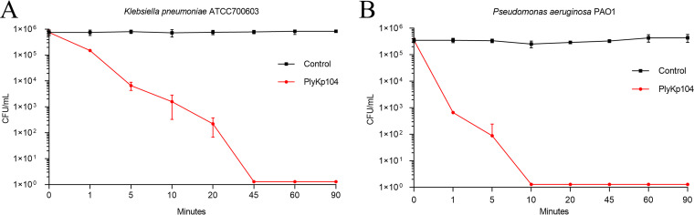
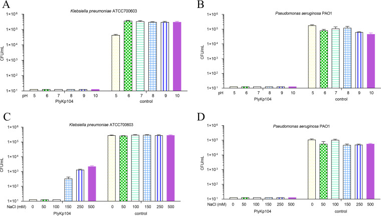
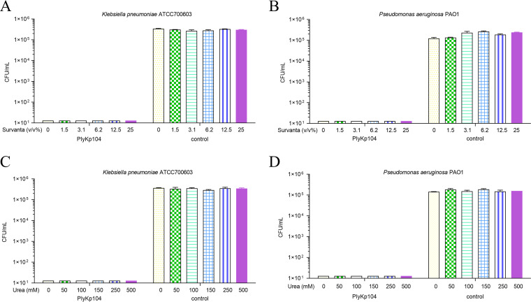
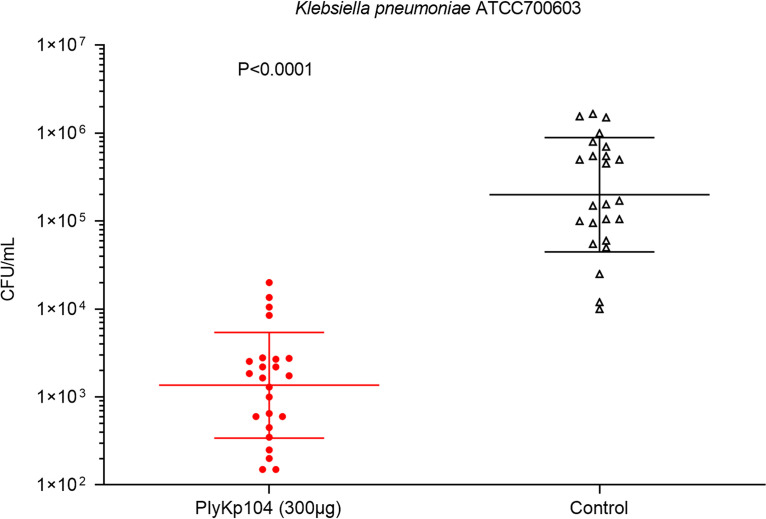
Klebsiella pneumoniae and Pseudomonas aeruginosa are two leading causes of burn and wound infections, pneumonia, urinary tract infections, and more severe invasive diseases, which are often multidrug resistant (MDR) or extensively drug resistant. Due to this, it is critical to discover alternative antimicrobials, such as bacteriophage lysins, against these pathogens. Unfortunately, most lysins that target Gram-negative bacteria require additional modifications or outer membrane permeabilizing agents to be bactericidal. We identified four putative lysins through bioinformatic analysis of Pseudomonas and Klebsiella phage genomes in the NCBI database and then expressed and tested their intrinsic lytic activity in vitro. The most active lysin, PlyKp104, exhibited >5-log killing against K. pneumoniae, P. aeruginosa, and other Gram-negative representatives of the multidrug-resistant ESKAPE pathogens (Enterococcus faecium, Staphylococcus aureus, K. pneumonia, Acinetobacter baumannii, P. aeruginosa, and Enterobacter species) without further modification. PlyKp104 displayed rapid killing and high activity over a wide pH range and in high concentrations of salt and urea. Additionally, pulmonary surfactants and low concentrations of human serum did not inhibit PlyKp104 activity in vitro. PlyKp104 also significantly reduced drug-resistant K. pneumoniae >2 logs in a murine skin infection model after one treatment of the wound, suggesting that this lysin could be used as a topical antimicrobial against K. pneumoniae and other MDR Gram-negative infections.

Keywords: ESKAPE; Gram negative; Klebsiella; Pseudomonas; antibiotic resistance; bacteriophage; endolysin; lysin; skin infection.


Conflict of interest statement

The authors declare no conflict of interest.

Figures

FIG 1
FIG 1
(A) Schematic representation of the four lysins identified and expressed. Domains and protein families were predicted by using Pfam (https://www.ebi.ac.uk/interpro/entry/pfam/#table). (B) Amino acid sequence alignment of the four lysins compared to PlyKp104. All alignments were generated by Clustal W (https://www.genome.jp/tools-bin/clustalw). The figure was generated by ESPript (https://espript.ibcp.fr/ESPript/ESPript/index.php). Strictly conserved residues are presented in red, highly conserved residues are in yellow, and dots represent gaps in the alignment. A schematic representation of the predicted secondary structure of PlyKp104 is shown above the sequences.
FIG 2
FIG 2
Bactericidal activity of lysins against P. aeruginosa PAO1 and Klebsiella sp. strain HM_4. Purified lysins were diluted to various concentrations and incubated with log-phase P. aeruginosa PAO1 or Klebsiella sp. strain HM_4 for 1 h at 37°C in 30 mM HEPES (pH 7.4). Values were established by serial dilution in 30 mM HEPES (pH 7.4) and plating to LB agar. (A) Bactericidal activity of lysins against P. aeruginosa PAO1. (B) Bactericidal activity of lysins against Klebsiella sp. strain HM_4. Experiments were conducted in triplicate. Error bars represent standard deviations.
FIG 3
FIG 3
Spectrum of lysin activity against various bacterial isolates. (A) P. aeruginosa; (B) K. pneumoniae; (C) other Gram-positive and Gram-negative species. Bacteria were incubated with 100 μg/mL of lysin in 30 mM HEPES buffer (pH 7.4) for 1 h at 37°C. Viable bacteria were enumerated by serial dilution in 30 mM HEPES buffer (pH 7.4) and plating to LB or BHI agar. Experiments were performed in triplicate; error bars represent standard deviations. Known drug resistance and/or clinical isolation site is given after the isolate name. ESBL, extended-spectrum β-lactamase; blaKPC, carbapenemase (carbapenem-hydrolyzing β-lactamase). See Table 1 for further information on bacterial strains.
FIG 4
FIG 4
Time-kill curve. Log-phase cells of (A) the ESBL-producing K. pneumoniae strain ATCC 700603 or (B) P. aeruginosa PAO1 were incubated for various times at 37°C with shaking in the presence of 100 μg/mL PlyKp104 in 30 mM HEPES buffer. Surviving bacteria were enumerated by serial dilution in 30 mM HEPES buffer and plating to LB agar. Experiments were performed in triplicate; error bars represent standard deviations.
FIG 5
FIG 5
(A and B) Effect of pH on the activity of PlyKp104. Log-phase cells of (A) the ESBL-producing K. pneumoniae strain ATCC 700603 or (B) P. aeruginosa PAO1 were incubated for 1 h at 37°C with 100 μg/mL lysin or buffer alone (control) in a 25 mM concentration of acetate buffer (pH 5.0), MES buffer (pH 6.0), HEPES buffer (pH 7.0 and 8.0), CHES buffer (pH 9.0), or CAPS buffer (pH 10.0). (C and D) Effect of NaCl on the activity of PlyKp104. Log-phase cells of (C) ESBL-producing K. pneumoniae strain ATCC 700603 or (D) P. aeruginosa PAO1 were incubated with 100 μg/mL PlyKp104 or buffer alone (control) for 1 h at 37°C in 30 mM HEPES and various concentrations of NaCl. Surviving bacteria were enumerated by serial dilution in 30 mM HEPES buffer and plating to LB agar. Experiments were performed in triplicate; error bars represent standard deviations.
FIG 6
FIG 6
Effects of Survanta and urea on the activity of PlyKp104. Log-phase cells of (A) the ESBL-producing K. pneumoniae strain ATCC 700603 or (B) P. aeruginosa PAO1 were incubated for 1 h at 37°C with 100 μg/mL of PlyKp104, or 30 mM HEPES buffer control, in the presence of the indicated concentrations of Survanta. Log-phase cells of (C) the ESBL-producing K. pneumoniae strain ATCC 700603 or (D) P. aeruginosa PAO1 were incubated with 100 μg/mL PlyKp104 for 1 h at 37°C in 30 mM HEPES and various concentrations of urea. Numbers of viable bacterial CFU were determined by serial dilution and plating. Experiments were performed in triplicate; error bars represent standard deviations.
FIG 7
FIG 7
The activity of PlyKp104 in the presence of human serum. Cells of (A) the ESBL-producing K. pneumoniae strain ATCC 700603 or (B) P. aeruginosa PAO1 were incubated for 1 h at 37°C with 100 μg/mL of the lysins in the presence of the indicated concentration of human serum in 30 mM HEPES buffer. Numbers of viable bacterial CFU are presented. Experiments were performed in triplicate; error bars represent standard deviations.
FIG 8
FIG 8
In vivo activity of PlyKp104 in a murine skin infection model of K. pneumoniae. Depilated CD1 mice were tape stripped and topically infected with 104 CFU of log-phase the ESBL-producing K. pneumoniae strain ATCC 700603. After 3 h, the infected areas were topically treated with 300 μg PlyKp104 in 50 μL CAPS-buffered saline, pH 6.0 (n = 12; 2 wounds per mouse) or 50 μL buffer control (n = 12; 2 wounds per mouse). Three hours after treatment, the animals were euthanized, and the infected skin was excised and homogenized in PBS. Samples were serially diluted in PBS and plated to LB agar for CFU enumeration. Horizontal lines indicate the geometric mean values of each group, error bars represent geometric standard deviations, and statistical significance was evaluated by Student’s t test.

Similar articles

Cited by

References

    1. Silby MW, Winstanley C, Godfrey SA, Levy SB, Jackson RW. 2011. Pseudomonas genomes: diverse and adaptable. FEMS Microbiol Rev 35:652–680. doi: 10.1111/j.1574-6976.2011.00269.x. - DOI - PubMed
    1. Shon AS, Bajwa RP, Russo TA. 2013. Hypervirulent (hypermucoviscous) Klebsiella pneumoniae: a new and dangerous breed. Virulence 4:107–118. doi: 10.4161/viru.22718. - DOI - PMC - PubMed
    1. Spencer RC. 1996. Predominant pathogens found in the European prevalence of infection in intensive care study. Eur J Clin Microbiol Infect Dis 15:281–285. doi: 10.1007/BF01695658. - DOI - PubMed
    1. Lyczak JB, Cannon CL, Pier GB. 2000. Establishment of Pseudomonas aeruginosa infection: lessons from a versatile opportunist. Microbes Infect 2:1051–1060. doi: 10.1016/s1286-4579(00)01259-4. - DOI - PubMed
    1. Winstanley C, O’Brien S, Brockhurst MA. 2016. Pseudomonas aeruginosa evolutionary adaptation and diversification in cystic fibrosis chronic lung infections. Trends Microbiol 24:327–337. doi: 10.1016/j.tim.2016.01.008. - DOI - PMC - PubMed

Publication types

Substances

LinkOut - more resources