. 2023 Jun 15;11(3):e0132623.
doi: 10.1128/spectrum.01326-23. Epub 2023 Apr 26.

Adaptive Laboratory Evolution Reveals the Selenium Efflux Process To Improve Selenium Tolerance Mediated by the Membrane Sulfite Pump in Saccharomyces cerevisiae

Affiliations

Adaptive Laboratory Evolution Reveals the Selenium Efflux Process To Improve Selenium Tolerance Mediated by the Membrane Sulfite Pump in Saccharomyces cerevisiae

Ao Gong et al. Microbiol Spectr. .

Abstract

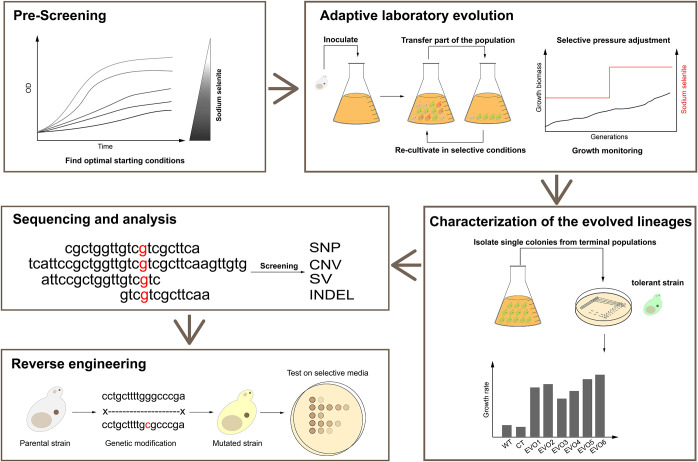
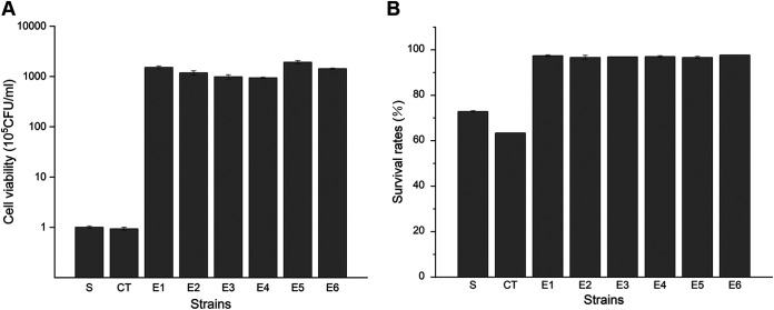
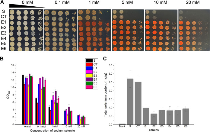
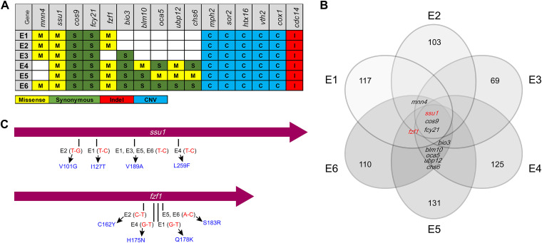
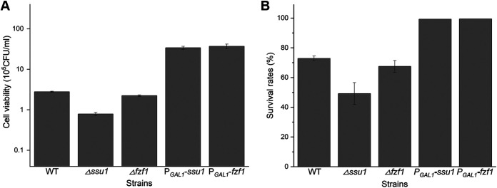
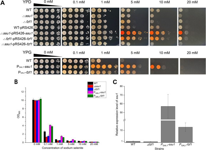
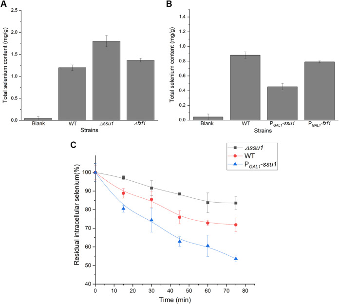
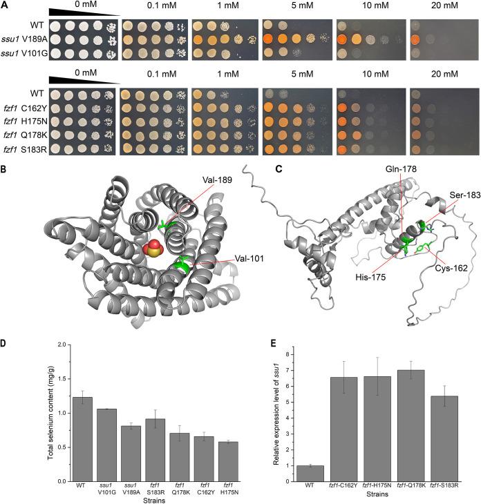
Selenium (Se) is a micronutrient in most eukaryotes, and Se-enriched yeast is the most common selenium supplement. However, selenium metabolism and transport in yeast have remained unclear, greatly hindering the application of this element. To explore the latent selenium transport and metabolism mechanisms, we performed adaptive laboratory evolution under the selective pressure of sodium selenite and successfully obtained selenium-tolerant yeast strains. Mutations in the sulfite transporter gene ssu1 and its transcription factor gene fzf1 were found to be responsible for the tolerance generated in the evolved strains, and the selenium efflux process mediated by ssu1 was identified in this study. Moreover, we found that selenite is a competitive substrate for sulfite during the efflux process mediated by ssu1, and the expression of ssu1 is induced by selenite rather than sulfite. Based on the deletion of ssu1, we increased the intracellular selenomethionine content in Se-enriched yeast. This work confirms the existence of the selenium efflux process, and our findings may benefit the optimization of Se-enriched yeast production in the future. IMPORTANCE Selenium is an essential micronutrient for mammals, and its deficiency severely threatens human health. Yeast is the model organism for studying the biological role of selenium, and Se-enriched yeast is the most popular selenium supplement to solve Se deficiency. The cognition of selenium accumulation in yeast always focuses on the reduction process. Little is known about selenium transport, especially selenium efflux, which may play a crucial part in selenium metabolism. The significance of our research is in determining the selenium efflux process in Saccharomyces cerevisiae, which will greatly enhance our knowledge of selenium tolerance and transport, facilitating the production of Se-enriched yeast. Moreover, our research further advances the understanding of the relationship between selenium and sulfur in transport.

Keywords: Se-enriched yeast; adaptive laboratory evolution; selenium; selenium efflux; selenium tolerance; yeasts.


Conflict of interest statement

The authors declare no conflict of interest.

Figures

FIG 1
FIG 1
Scheme of the experimental procedures to investigate selenium tolerance in S. cerevisiae. For prescreening, the growth inhibition of parental strains was measured in the presence of different concentrations of sodium selenite, and the optimal starting conditions were found and applied for subsequent serial cultivations. For adaptive laboratory evolution, strains were serially transferred into fresh YPD medium with the addition of increasing concentrations of sodium selenite for 100 days. For the characterization of the evolved yeast lineages, six clones were isolated from Ev1 to Ev6, and their selenium tolerance was characterized and evaluated. For sequencing and analysis, whole-genome sequencing of six single clones from E1 to E6 and one clone from the starting group was performed, and potentially functional mutations generated in the evolved strains were screened and identified. For reverse engineering, the determined mutations were introduced into the wild-type strain, and their contribution to the tolerance phenotype was tested.
FIG 2
FIG 2
Selenium adaptation of the unevolved and evolved strains. The starting strain is denoted S, the control strain (after serial transfer without sodium selenite) is denoted CT, and the six evolved strains are denoted E1 to E6. (A) Viability of various strains after selenium treatment. Cell samples from all tested strains were harvested after culture in YPD medium with 100 μM Na2SeO3 for 16 h, diluted, and plated. After incubation at 30°C for 3 days, the number of colonies on the plates was counted. (B) Survival rates of various strains after selenium treatment. Yeast cells were sampled during serial transfer in selenium-containing YPD medium, stained with propidium iodide, and analyzed by flow cytometry. The proportion of unstained cells was regarded as alive. The error bars represent the means and ranges from three independent experiments.
FIG 3
FIG 3
Selenium tolerance and intracellular selenium content in evolved strains. The starting strain is denoted S, the control strain (after serial transfer without sodium selenite) is denoted CT, and the six evolved strains are denoted E1 to E6. (A) Growth assays on agar plates. After incubation in liquid YPD medium overnight, cells were sampled and spotted onto 1.5% YPD agar plates supplemented with increasing concentrations of Na2SeO3. Pictures were taken 2 days later. (B) Growth assays in liquid medium. All strains were grown at 30°C overnight and then subcultured into fresh YPD liquid medium with increasing concentrations of sodium selenite for 16 h. The final OD600 of each strain was measured and recorded. (C) Intracellular selenium content of the evolved strains. After growth in YPD medium with 100 μM Na2SeO3, yeast cells were sampled and digested, and the selenium content was then analyzed using ICP-MS. The error bars represent the means and ranges from three independent experiments.
FIG 4
FIG 4
Summary of the mutated genes identified in evolution experiments. (A) Table of gene mutations present in more than three clones from each independently evolved population. (B) Venn diagram of genes with mutations that occurred and accumulated within E1 to E6. Each circle indicates the number of mutated genes found in the clone. ssu1, cos9, and fcy21 were found in all six evolved groups simultaneously, and fzf1 was found in five evolved groups simultaneously except for E3. mnn4 and bio3 were found in four evolved groups, while blm10, oca5, ubp12, and chs6 were found only in E4, E5, and E6. (C) Mutated positions of E1 to E6 in sequence diagrams of ssu1 and fzf1. The length of ssu1 is 1,377 bp, encoding a 458-aa membrane protein, and the length of fzf1 is 900 bp, encoding a 299-aa transcription factor. Red represents the base substitution, and blue represents the corresponding amino acid substitution.
FIG 5
FIG 5
ssu1 and fzf1 contribute to selenium adaptation in S. cerevisiae. (A) Viability of deletion and overexpression strains after selenium treatment. Cell samples from all tested strains were harvested after culture in YPG medium with 100 μM Na2SeO3 for 16 h, diluted, and plated. After incubation at 30°C for 3 days, the number of colonies on the plates was counted. (B) Survival rates of deletion and overexpression strains after selenium treatment. Yeast cells were sampled during serial transfer in selenium-containing YPG medium, stained with propidium iodide, and analyzed by flow cytometry. The proportion of unstained cells was regarded as alive. The error bars represent the means and ranges from three independent experiments.
FIG 6
FIG 6
Selenium tolerance and relative expression levels of ssu1 in the WT, Δfzf1, PGAL1-ssu1, and PGAL1-fzf1 strains. (A) Growth assays on agar plates. After incubation in YPD/YPG medium overnight, cells were sampled and spotted onto 1.5% YPD/YPG agar plates supplemented with increasing concentrations of Na2SeO3. Pictures were taken 2 days later. (B) Growth assays in liquid medium. All strains were grown at 30°C overnight and then subcultured into fresh YPG liquid medium with increasing concentrations of sodium selenite for 16 h. The final OD600 of each strain was measured and recorded. (C) The expression levels of ssu1 in the WT, Δfzf1, PGAL1-ssu1, and PGAL1-fzf1 strains were determined by qRT-PCR. The error bars represent the means and ranges from three independent experiments.
FIG 7
FIG 7
ssu1 and fzf1 affect intracellular selenium accumulation, and ssu1 is involved in selenium efflux. After growth in YPD/YPG medium with 100 μM Na2SeO3, yeast cells were sampled and digested, and the selenium content was then analyzed using ICP-MS. (A) The absence of ssu1 and fzf1 contributes to selenium accumulation in yeast cells. (B) The overexpression of ssu1 and fzf1 leads to a decrease in intracellular selenium. (C) Efflux of selenium from the WT, Δssu1, and PGAL1-ssu1 strains. Cells were loaded into YPG medium supplemented with 1 mM Na2SeO3 for 1 h to yield a final total intracellular concentration of 2 to 3 mg/g (dry weight). Selenium efflux was determined in Se-enriched cells suspended in fresh YPG medium by measuring the percent reduction of intracellular selenium. The error bars represent the means and ranges from three independent experiments.
FIG 8
FIG 8
Selenium tolerance of point mutation strains and structures of Ssu1p and Fzf1p. (A) Growth assays on agar plates. After incubation in YPD medium overnight, cells were sampled and spotted onto 1.5% YPD agar plates supplemented with increasing concentrations of Na2SeO3. Pictures were taken 2 days later. (B) AlphaFold-predicted structure and functional mutated sites of Ssu1p (https://alphafold.ebi.ac.uk/entry/P41930). The central molecule represents SeO32−. (C) AlphaFold-predicted structure and functional mutated sites of Fzf1p (https://alphafold.ebi.ac.uk/entry/P32805). (D) Measurement of the intracellular selenium contents of mutated strains. (E) Expression levels of ssu1 in mutated fzf1 strains.
FIG 9
FIG 9
The addition of sulfite impairs the selenite tolerance of yeast on YPG agar plates. (A) The growth inhibition of all tested strains was very limited on YPG agar plates with the addition of Na2SO3. (B) On YPG agar plates with the addition of Na2SeO3, the growth of the WT was severely inhibited, while the PGAL1-ssu1 strain could still tolerate up to 10 mM sodium selenite. (C) After incubation with 1 mM Na2SO3 for 6 h, yeast cells were washed, diluted, and spotted onto YPG agar plates with the addition of Na2SeO3, and the growth of all tested strains was inhibited further. The WT can only be sustained with 1 mM, and the PGAL1-ssu1 and PGAL1-fzf1 strains can only be maintained with 5 mM.
FIG 10
FIG 10
The expression level of ssu1 responds to selenite rather than sulfite in S. cerevisiae. (A) Expression levels of ssu1 in S. cerevisiae with different concentrations of sodium selenite or sodium sulfite. (B) Expression levels of fzf1 in S. cerevisiae with different concentrations of sodium selenite or sodium sulfite. (C) Expression levels of ssu1 in the Δfzf1 strain with different concentrations of sodium selenite or sodium sulfite. Yeast cells were incubated in YPD medium for 16 h at 30°C, different concentrations of selenite or sulfite were then added, and the cells were incubated for 2 h at 30°C. The same volume of water was added for the control groups. Finally, cells were harvested, and total RNA was extracted for qRT-PCR analysis.
FIG 11
FIG 11
Characterization of selenium species of Se-enriched yeasts by reversed-phase HPLC–ICP-MS. (A) Chromatograms of selenium species standards (150 ppb) of SeMet (selenomethionine), SeCys2 (selenocystine), and selenite. (B) Chromatograms of the tested yeast strains. All tested strains were inoculated into YPG medium containing 0.1 mM sodium selenite and cultivated for 16 h before being sampled and analyzed.

Similar articles

Cited by

References

    1. Hariharan S, Dharmaraj S. 2020. Selenium and selenoproteins: it’s [sic] role in regulation of inflammation. Inflammopharmacology 28:667–695. doi: 10.1007/s10787-020-00690-x. - DOI - PMC - PubMed
    1. Rayman MP. 2000. The importance of selenium to human health. Lancet 356:233–241. doi: 10.1016/S0140-6736(00)02490-9. - DOI - PubMed
    1. Yoshinaga M, How S, Blanco D, Murdoch IS, Grudny M, Powers SL, Molina N, Rosen BP, Welch AZ. 2018. Directed evolution of Saccharomyces cerevisiae for increased selenium accumulation. Microorganisms 6:81. doi: 10.3390/microorganisms6030081. - DOI - PMC - PubMed
    1. Kieliszek M, Błażejak S, Gientka I, Bzducha-Wróbel A. 2015. Accumulation and metabolism of selenium by yeast cells. Appl Microbiol Biotechnol 99:5373–5382. doi: 10.1007/s00253-015-6650-x. - DOI - PMC - PubMed
    1. Birringer M, Pilawa S, Flohé L. 2002. Trends in selenium biochemistry. Nat Prod Rep 19:693–718. doi: 10.1039/b205802m. - DOI - PubMed

Publication types

MeSH terms

LinkOut - more resources