Multicenter Study
. 2023 Apr 26;18(4):e0284748.
doi: 10.1371/journal.pone.0284748. eCollection 2023.

Point-of-care ultrasonography for risk stratification of non-critical suspected COVID-19 patients on admission (POCUSCO): A prospective binational study

Affiliations
Multicenter Study

Point-of-care ultrasonography for risk stratification of non-critical suspected COVID-19 patients on admission (POCUSCO): A prospective binational study

François Morin et al. PLoS One. .

Abstract

Background: Lung point-of-care ultrasonography (L-POCUS) is highly effective in detecting pulmonary peripheral patterns and may allow early identification of patients who are likely to develop an acute respiratory distress syndrome (ARDS). We hypothesized that L-POCUS performed within the first 48 hours of non-critical patients with suspected COVID-19 would identify those with a high-risk of worsening.

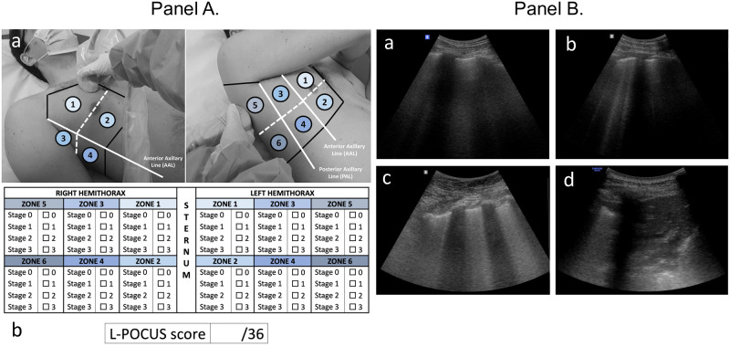
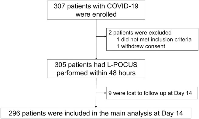
Methods: POCUSCO was a prospective, multicenter study. Non-critical adult patients who presented to the emergency department (ED) for suspected or confirmed COVID-19 were included and had L-POCUS performed within 48 hours following ED presentation. The lung damage severity was assessed using a previously developed score reflecting both the extension and the intensity of lung damage. The primary outcome was the rate of patients requiring intubation or who died within 14 days following inclusion.

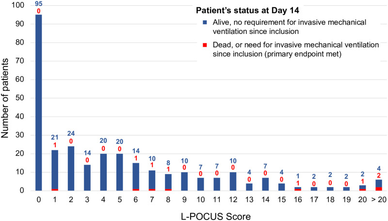
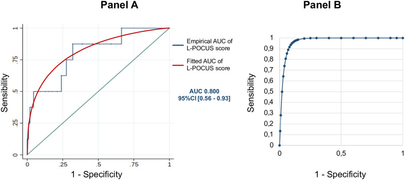
Results: Among 296 patients, 8 (2.7%) met the primary outcome. The area under the curve (AUC) of L-POCUS was 0.80 [95%CI:0.60-0.94]. The score values which achieved a sensibility >95% in defining low-risk patients and a specificity >95% in defining high-risk patients were <1 and ≥16, respectively. The rate of patients with an unfavorable outcome was 0/95 (0%[95%CI:0-3.9]) for low-risk patients (score = 0), 4/184 (2.17%[95%CI:0.8-5.5]) for intermediate-risk patients (score 1-15) and 4/17 (23.5%[95%CI:11.4-42.4]) for high-risk patients (score ≥16). In confirmed COVID-19 patients (n = 58), the AUC of L-POCUS was 0.97 [95%CI:0.92-1.00].

Conclusion: L-POCUS performed within the first 48 hours following ED presentation allows risk-stratification of patients with non-severe COVID-19.


Conflict of interest statement

Pr. Christophe Aubé declares personal scientific collaborations with Siemens Ultrasound, outside the submitted work. Pr. Francis Couturaud declares personal consulting fees and other from BMS, personal consulting fees and other from Bayer, personal consulting fees and other from MSD, outside the submitted work. Pr. Pierre-Marie Roy declares personal fees and other from Aspen, personal fees and other from Boehringer Ingelheim, personal fees and other from Bristol Myers Squibb, other from Bayer Health Care, outside the submitted work. Other authors declare no competing interests. This does not alter our adherence to PLOS ONE policies on sharing data and materials.

Figures

Fig 1
Fig 1. Lung point-of-care ultrasonography method (L-POCUS) and examples of four ultrasound aeration stages.
(Panel A). (a). Twelve chest areas of investigation following BLUE-PLUS Protocol: zone 1: upper anterior chest wall; zone 2: lower anterior chest wall; zone 3: upper lateral chest wall; zone 4: lower lateral chest wall; zone 5: upper posterolateral chest wall; zone 6: lower posterolateral chest wall. (b) L-POCUS score grid: Each zone was examined to establish which of four ultrasound parenchymal aeration stages it exhibited, and points are assigned to them according to their severity. Stage 0 or normal aeration (0 point): Lung sliding sign associated with respiratory movement of less than 3 B lines; Stage 1 or moderate loss of lung aeration (1 point): a clear number of multiple visible B-lines with horizontal spacing between adjacent B lines ≤ 7 mm (B1 lines); Stage 2 or severe loss of lung aeration (2 points): multiple B lines fused together that were difficult to count with horizontal spacing between adjacent B lines ≤ 3 mm, including “white lung”; and Stage 3 or pulmonary consolidation (3 points): hyperechoic lung tissue, accompanied by dynamic air bronchogram. (Panel B). (a) Stage 0 or normal aeration; (b) Stage 1 or moderate loss of lung aeration; (c) Stage 2 or severe loss of lung aeration; (d) Stage 3 or pulmonary consolidation.
Fig 2
Fig 2. Study flow chart.
COVID-19: Coronavirus disease 2019; L-POCUS: lung point of care ultrasonography; OSCI: ordinal scale for clinical improvement.
Fig 3
Fig 3. Distribution of L-POCUS score according to Ordinal Scale for Clinical Improvement (OSCI) at Day 14.
Fig 4
Fig 4. L-POCUS prognostic performance.
(Panel A) Receiver operating characteristic (ROC)) curve of prognostic performance of global L-POCUS with its area-under-the-curve (AUC) and its 95% confidence interval (95%CI). (Panel B) Receiver operating characteristic (ROC) curve of prognostic performance of L-POCUS with its area-under-the-curve (AUC) and its 95% confidence interval (95%CI) for positive SARS-CoV-2 RT-PCR patients.

Similar articles

Cited by

  • Advancing healthcare through thoracic ultrasound research in older patients.
    Scarlata S, Okoye C, Zotti S, Lauretani F, Nouvenne A, Cerundolo N, Bruni AA, Torrini M, Finazzi A, Mazzarone T, Lunian M, Zucchini I, Maccioni L, Guarino D, Fabbri Della Faggiola S, Capacci M, Bianco MG, Guarona G, Bellelli G, Monzani F, Virdis A, Antonelli Incalzi R, Ungar A, Ticinesi A; GRETA research group on thoracic ultrasound in the older patient, Italian Society of Geriatrics and Gerontology (SIGG). Scarlata S, et al. Aging Clin Exp Res. 2023 Dec;35(12):2887-2901. doi: 10.1007/s40520-023-02590-w. Epub 2023 Nov 11. Aging Clin Exp Res. 2023. PMID: 37950845 Free PMC article. Review.

References

    1. Salje H, Kiem CT, Lefrancq N, Courtejoie N, Bosetti P, Paireau J, et al.. Estimating the burden of SARS-CoV-2 in France. Science. 2020;369: 208–211. doi: 10.1126/science.abc3517 - DOI - PMC - PubMed
    1. Zhou F, Yu T, Du R, Fan G, Liu Y, Liu Z, et al.. Clinical course and risk factors for mortality of adult inpatients with COVID-19 in Wuhan, China: a retrospective cohort study. The Lancet. 2020;395: 1054–1062. doi: 10.1016/S0140-6736(20)30566-3 - DOI - PMC - PubMed
    1. Berlin DA, Gulick RM, Martinez FJ. Severe Covid-19. New England Journal of Medicine. 2020;0: null. doi: 10.1056/NEJMcp2009575 - DOI - PubMed
    1. Bernheim A, Mei X, Huang M, Yang Y, Fayad ZA, Zhang N, et al.. Chest CT Findings in Coronavirus Disease-19 (COVID-19): Relationship to Duration of Infection. Radiology. 2020;295: 200463. doi: 10.1148/radiol.2020200463 - DOI - PMC - PubMed
    1. Shi H, Han X, Jiang N, Cao Y, Alwalid O, Gu J, et al.. Radiological findings from 81 patients with COVID-19 pneumonia in Wuhan, China: a descriptive study. The Lancet Infectious Diseases. 2020;20: 425–434. doi: 10.1016/S1473-3099(20)30086-4 - DOI - PMC - PubMed

Publication types