. 2023 Apr 26;15(693):eabp9528.
doi: 10.1126/scitranslmed.abp9528. Epub 2023 Apr 26.

Cloning a profibrotic stem cell variant in idiopathic pulmonary fibrosis

Affiliations

Cloning a profibrotic stem cell variant in idiopathic pulmonary fibrosis

Shan Wang et al. Sci Transl Med. .

Abstract

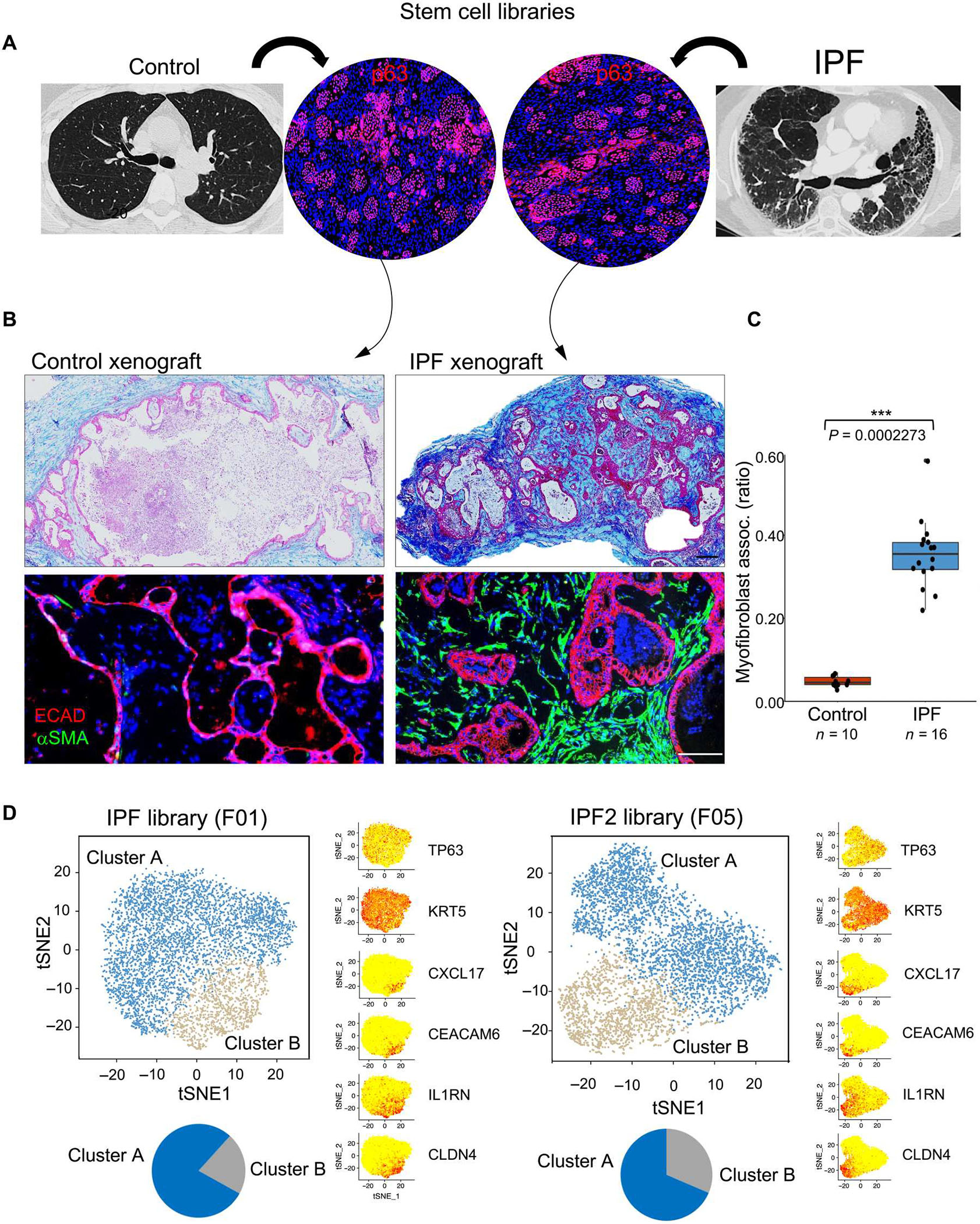
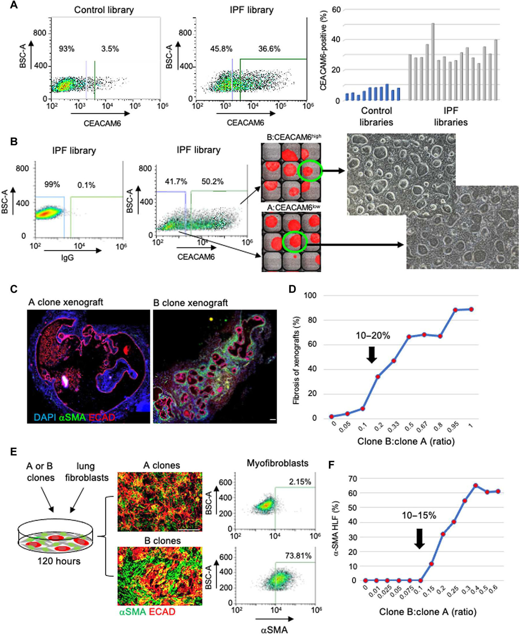
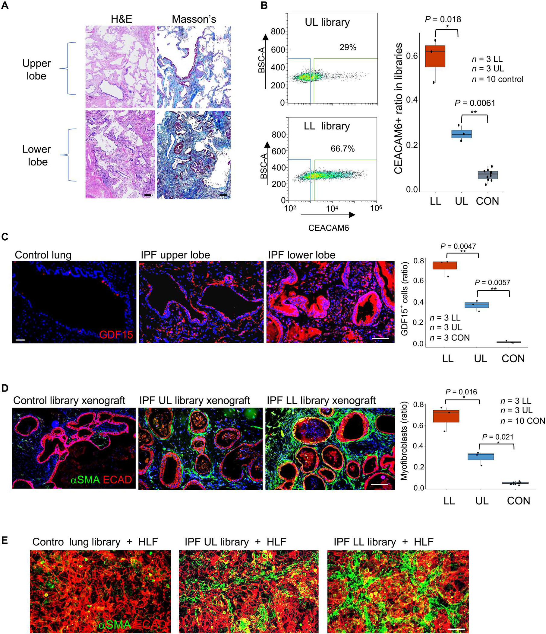
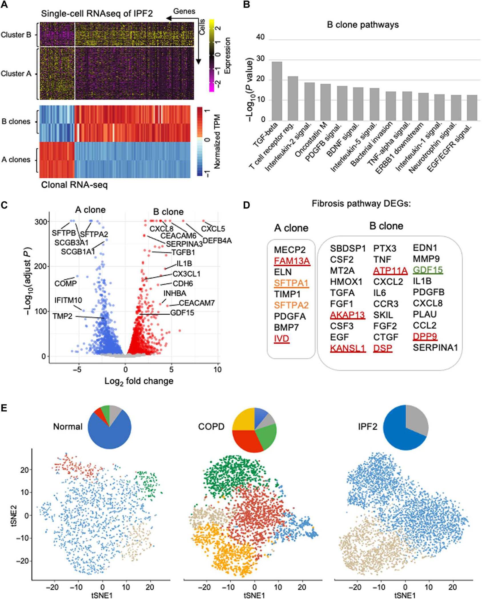
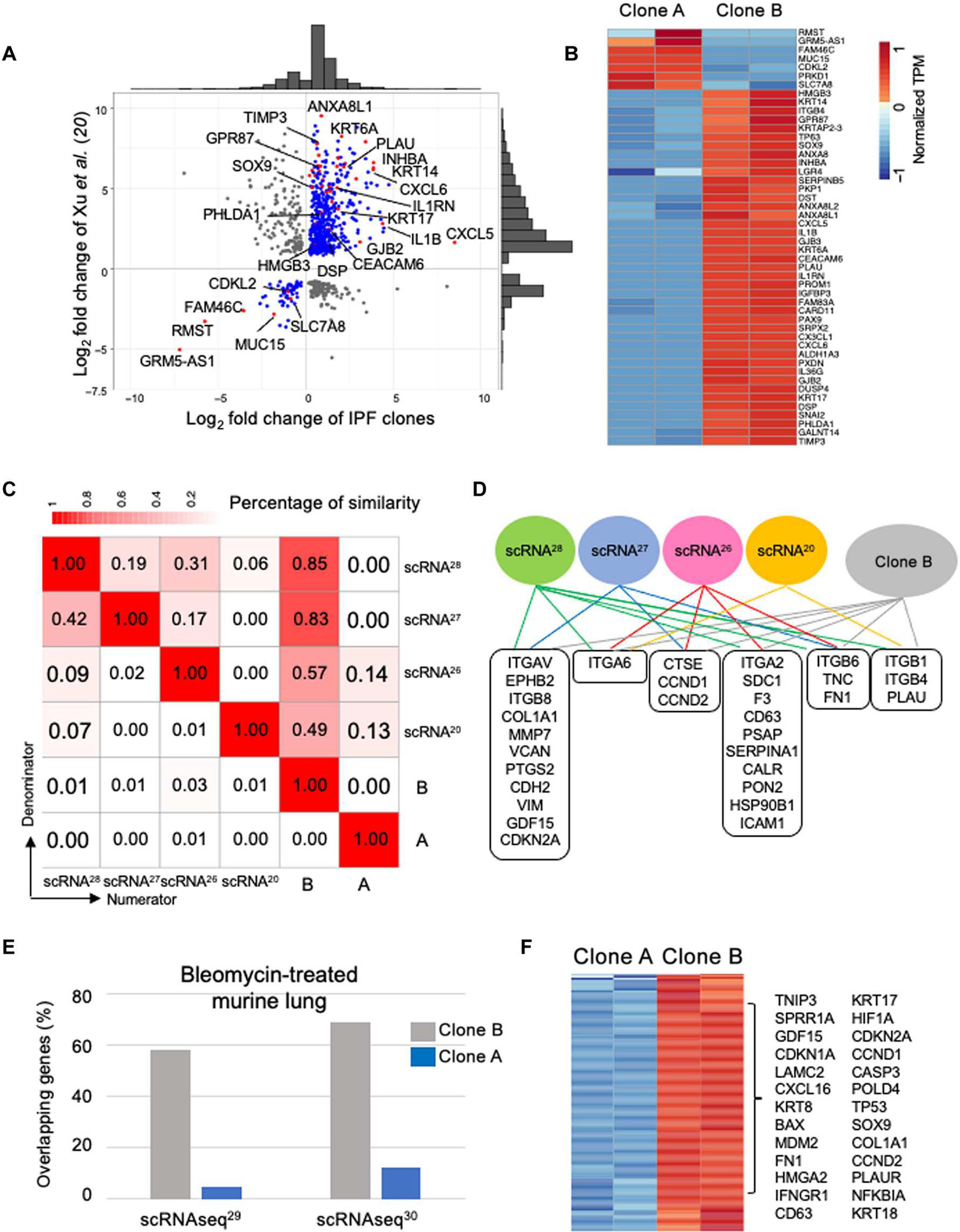
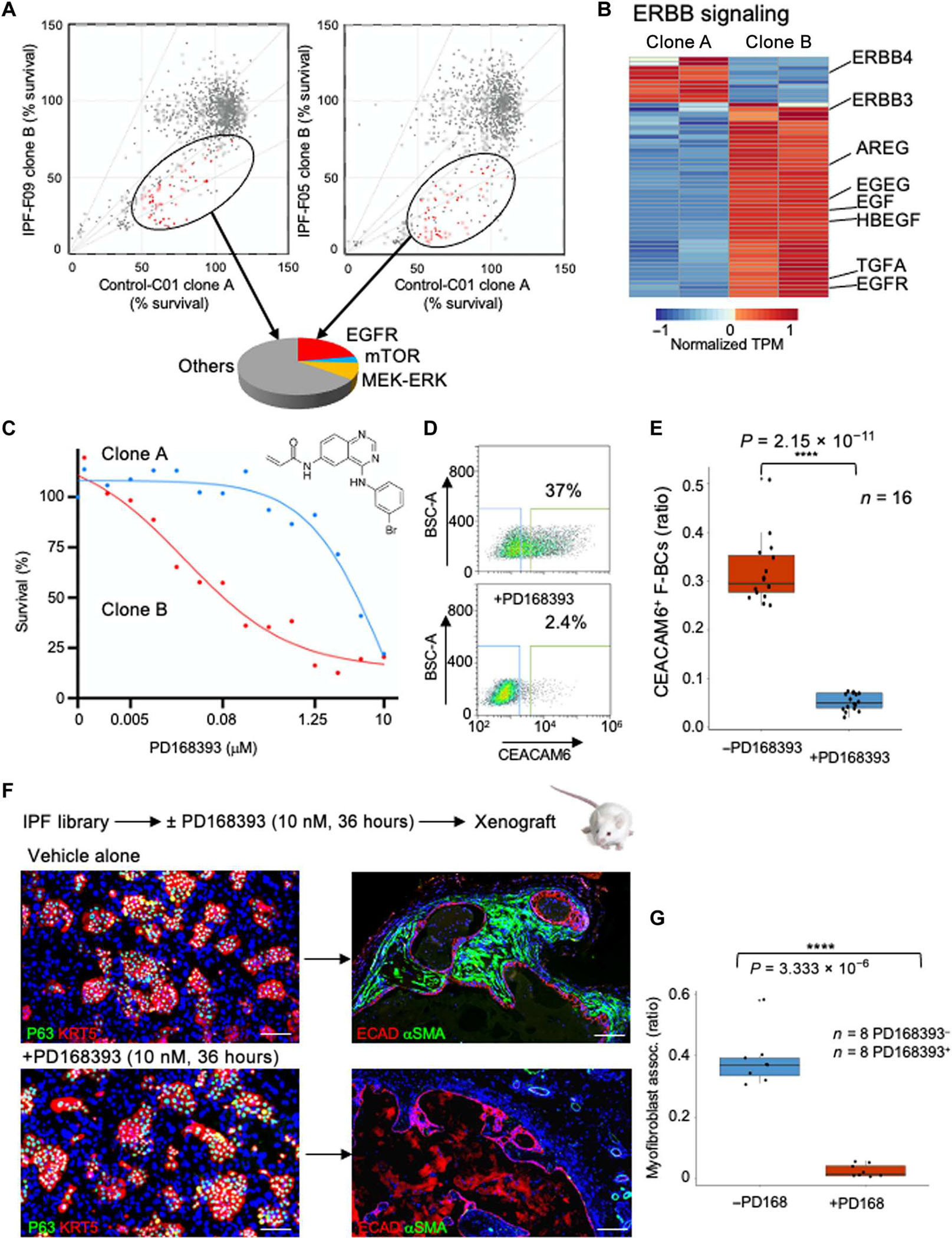
Idiopathic pulmonary fibrosis (IPF) is a progressive, irreversible, and rapidly fatal interstitial lung disease marked by the replacement of lung alveoli with dense fibrotic matrices. Although the mechanisms initiating IPF remain unclear, rare and common alleles of genes expressed in lung epithelia, combined with aging, contribute to the risk for this condition. Consistently, single-cell RNA sequencing (scRNA-seq) studies have identified lung basal cell heterogeneity in IPF that might be pathogenic. We used single-cell cloning technologies to generate "libraries" of basal stem cells from the distal lungs of 16 patients with IPF and 10 controls. We identified a major stem cell variant that was distinguished from normal stem cells by its ability to transform normal lung fibroblasts into pathogenic myofibroblasts in vitro and to activate and recruit myofibroblasts in clonal xenografts. This profibrotic stem cell variant, which was shown to preexist in low quantities in normal and even fetal lungs, expressed a broad network of genes implicated in organ fibrosis and showed overlap in gene expression with abnormal epithelial signatures identified in previously published scRNA-seq studies of IPF. Drug screens highlighted specific vulnerabilities of this profibrotic variant to inhibitors of epidermal growth factor and mammalian target of rapamycin signaling as prospective therapeutic targets. This profibrotic stem cell variant in IPF was distinct from recently identified profibrotic stem cell variants in chronic obstructive pulmonary disease and may extend the notion that inappropriate accrual of minor and preexisting stem cell variants contributes to chronic lung conditions.


Conflict of interest statement

Competing interests: W.X. and F.D.M. are CPRIT Scholars in Cancer Research. W.X., F.D.M., M.V., and C.P.C. have equity interest in Tract Pharmaceuticals, a biotechnology concern devoted to therapeutics for chronic inflammatory diseases and the cancers emerging from them. Tract Pharmaceuticals has licenses to patents related to the technology underlying this work. D.A.S. is a consultant for Vertex and the founder and chief scientific officer of Eleven P15 Inc.

Figures

Fig. 1.
Fig. 1.. IPF lung basal stem cell libraries promote fibrosis in immunodeficient mice.
(A) Generation of libraries of clonogenic TP63+ epithelial cells from control and IPF lungs. (B) Histological sections of nodules formed from subcutaneous xenografting of cells from control and IPF basal cell libraries stained with Masson’s trichrome (collagen; blue, top) and immunolabeling with antibodies to ECAD (red) and αSMA (green, bottom). Scale bars, 200 μm. (C) Whisker box plot depicts the morphometric quantification of the percentage of epithelial cystic surfaces occupied by submucosal myofibroblasts in xenografts of 10 control and 16 IPF libraries. (D) tSNE profiles of scRNA-seq data of two IPF patient–derived basal cell libraries showing both normal basal cells (cluster A; blue) and a variant denoted as “cluster B” (gray) that is marked by the differential expression of CXCL17, CEACAM6, IL1RN, and CLDN4. Distal airway basal cell markers TP63 and KRT5 were broadly expressed across clusters A and B.
Fig. 2.
Fig. 2.. Cloning a major profibrotic variant from IPF lung.
(A) FACS profiling (left) and a histogram showing the quantification of CEACAM6+ cells in 10 control and 16 IPF stem cell libraries. BSC-A, back scatter area. (B) FACS-aided isolation and single-cell cloning of CEACAM6+ and CEACAM6 cells from IPF basal cell libraries in 384-well plates for clonal expansion. Fluorescein isothiocyanate–mouse immunoglobulin G (IgG) was used as the isotype control. Scale bar, 100 μm. (C) ECAD (red) and αSMA (green) IF on sections of nodules formed from xenografts of A (left) and B clones. Scale bar, 100 μm. (D) Graphical presentation of induction of αSMA+ myofibroblasts by xenograft nodules formed by defined ratios of cells of A and B clones. (E) From left: Schematic of coculture of A or B clones with human normal lung fibroblasts (HLFs), IF images of 72-hour cocultures of clone A + HLF (top) or clone B + HLF (bottom) stained with antibodies to ECAD (red) and αSMA (green), and FACS quantification of the αSMA+ cells in the respective cocultures. Scale bar, 100 μm. (F) Graphical representation of αSMA+ cells quantified by FACS profiling induced by cocultures of human lung fibroblasts with defined ratios of cells from A and B clones.
Fig. 3.
Fig. 3.. Cluster B cells in regions of low and high UIP histopathology.
(A) From left: Drawing portraying biased UIP histopathology in lower lobes of IPF lung, patient-matched histological sections of upper (top) and lower (bottom) lung stained with hematoxylin and eosin (H&E) and Masson’s trichrome (collagen; blue). Scale bars, 100 μm. (B) From left: FACS profiling of CEACAM6+ cells from basal cell libraries generated from corresponding regions of upper and lower lung as well as whisker box plot of CEACAM6+ cells in libraries derived from upper and lower lungs from three IPF cases and from 10 control libraries. (C) From left: GDF15 IF in histological sections of control and upper (UL) and lower lobes (LB) of IPF lung as well as whisker box plot quantification of GDF15 fluorescence in sections from 10 control and three IPF patient–matched lower and upper lobes. Scale bars, 100 μm. (D) ECAD (red) and αSMA+ (green) IF of nodule sections from xenografts of basal cell libraries from control and IPF lower and upper lobes as well as whisker box plot presentation of morphometric data across 10 control and three IPF cases. Scale bar, 100 μm. (E) ECAD (red) and αSMA+ (green) IF of 72-hour cocultures of control, upper lobe, and lower lobe libraries with HLFs. Scale bar, 100 μm.
Fig. 4.
Fig. 4.. Cluster B cells display profibrotic signatures distinct from COPD variants.
(A) Differential gene expression profiles from IPF library scRNA-seq and clonal RNA-seq data of IPF clones A and B. TPM, transcripts per million. (B) Histogram depicting most significant (P < 1.0 × 10⁻¹³) pathways determined by NCATS (National Center for Advancing Translational Sciences) BioPlanet pathway analysis of RNA-seq differentially expressed genes of IPF clone B versus clone A. (C) Volcano plot of RNA-seq differential gene expression in in vitro–differentiated cluster B cells relative to cluster A cells. (D) Fibrosis pathway–associated differentially expressed genes (DEGs) in IPF A and B clones. (E) tSNE profiles of scRNA-seq basal cell libraries from control, COPD, and IPF lungs showing the respective clusters of normal distal airway basal cells (cluster A; blue), IPF cluster B (gray), squamous cell metaplasia (orange), inflammatory squamous cell metaplasia (red), and goblet cell metaplasia (green).
Fig. 5.
Fig. 5.. Cluster B clone profiles encompass IPF scRNA-seq populations.
(A) Scatter plot with marginal histograms comparing fold change of differentially expressed genes from RNA-seq data of sorted epithelial cells from IPF lung (20) with cluster A and cluster B clones from IPF library. (B) Expression heatmap of consistent genes identified in sorted IPF epithelial cells (20) with the cluster A and cluster B IPF clones. (C) Similarity matrix between expression signatures of populations identified by four scRNA-seq studies of IPF lungs (, –28) and IPF cluster A and cluster B clones. (D) Common genes identified by at least two scRNA-seq studies and IPF cluster B clone. (E) Histogram of percentage of overlap in gene expression between epithelial populations emerging in murine lung after bleomycin and IPF cluster A and cluster B clones. (F) Consistent differentially expressed genes from both murine bleomycin studies and IPF cluster A and cluster B clones.
Fig. 6.
Fig. 6.. Vulnerability of cluster B cells to bioactive small molecules.
(A) Two-dimensional survival plots comparing cluster A and cluster B clones from two IPF cases after exposure to a bioactive small-molecule library. Ovals encompass molecules that selectively disfavor cluster B cells in both IPF cases, and the pie chart categorizes the pathways affected by these drugs. mTOR, mammalian target of rapamycin; MEK-ERK, mitogen-activated protein kinase kinase–extracellular signal–regulated kinase. (B) Expression heatmap of genes in the ERBB pathway in IPF clones A and B. (C) Dose response curves of EGFR inhibitor PD168393 for cluster A and cluster B cells. Error bars, SD. (D) CEACAM6 FACS profile of IPF basal cell library before and after exposure to PD168393. (E) Whisker box plot of CEACAM6+ cells in all 16 IPF libraries as a function of exposure to the EGFR inhibitor. (F) Schematic of in vitro exposure of IPF basal cell library to PD168393 followed by xenografting into immunodeficient mice. IF images of libraries (left) and xenograft nodules using the indicated markers. Scale bars, 100 μm. (G) Whisker box plot of morphometric analysis of myofibroblast association in xenografts of eight IPF libraries with or without in vitro exposure to PD168393.

References

    1. Lederer DJ, Martinez FJ, Idiopathic pulmonary fibrosis. N. Engl. J. Med. 379, 797–798 (2018). - PubMed
    1. Wolters PJ, Blackwell TS, Eickelberg O, Loyd JE, Kaminski N, Jenkins G, Maher TM, Molina-Molina M, Noble PW, Raghu G, Richeldi L, Schwarz MI, Selman M, Wuyts WA, Schwartz DA, Time for a change: Is idiopathic pulmonary fibrosis still idiopathic and only fibrotic? Lancet Respir. Med. 6, 154–160 (2018). - PMC - PubMed
    1. Selman M, Pardo A, The leading role of epithelial cells in the pathogenesis of idiopathic pulmonary fibrosis. Cell. Signal. 66, 109482 (2020). - PubMed
    1. Nogee LM, Dunbar III AE, Wert SE, Askin F, Hamvas A, Whitsett JA, A mutation in the surfactant protein C gene associated with familial interstitial lung disease. N. Engl. J. Med. 344, 573–579 (2001). - PubMed
    1. Selman M, Lin H-M, Montano M, Jenkins AL, Estrada A, Lin Z, Wang G, DiAngelo SL, Guo X, Umstead TM, Lang CM, Pardo A, Phelps DS, Floros J, Surfactant protein A and B genetic variants predispose to idiopathic pulmonary fibrosis. Hum. Genet. 113, 542–550 (2003). - PubMed

Publication types

LinkOut - more resources