. 2023 Apr 26;15(693):eade6285.
doi: 10.1126/scitranslmed.ade6285. Epub 2023 Apr 26.

Sleep deprivation exacerbates microglial reactivity and Aβ deposition in a TREM2-dependent manner in mice

Affiliations

Sleep deprivation exacerbates microglial reactivity and Aβ deposition in a TREM2-dependent manner in mice

Samira Parhizkar et al. Sci Transl Med. .

Abstract

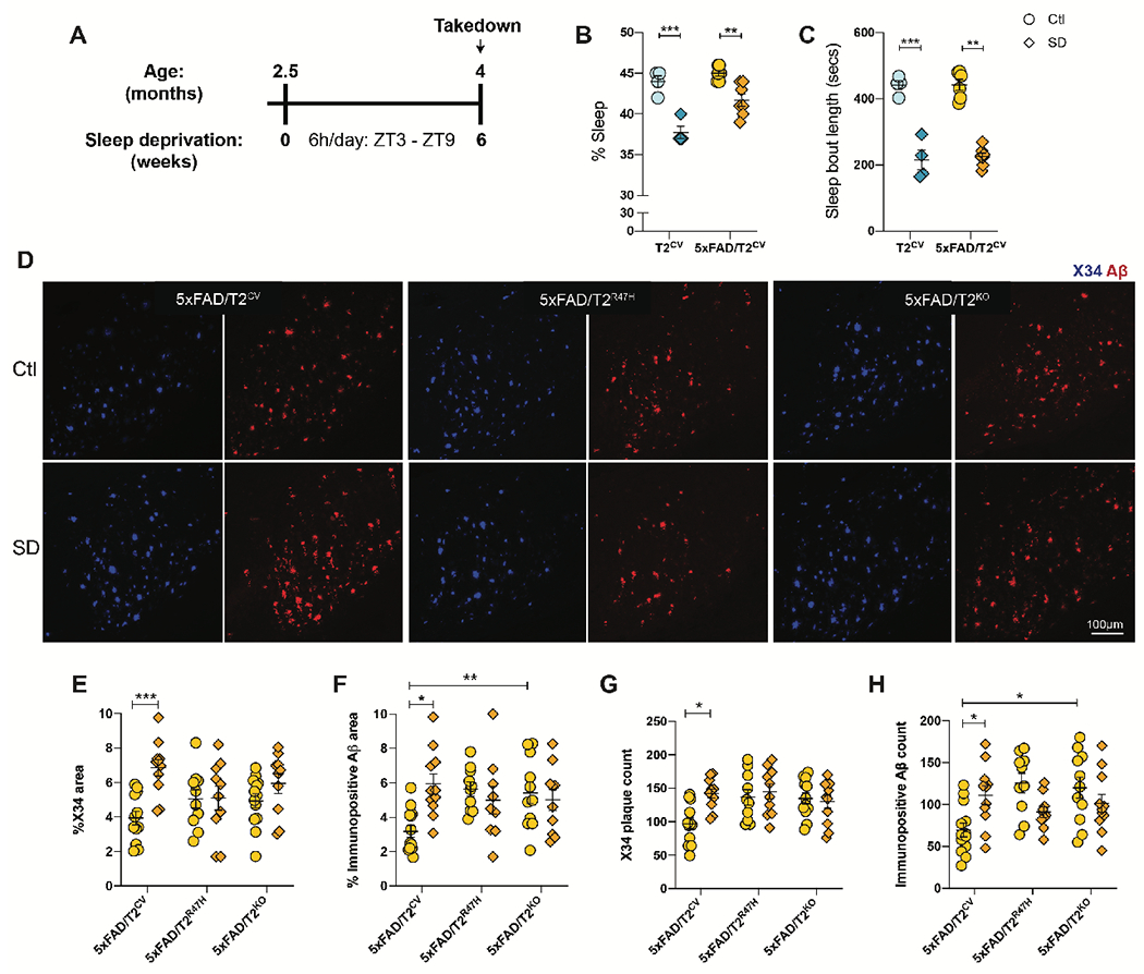
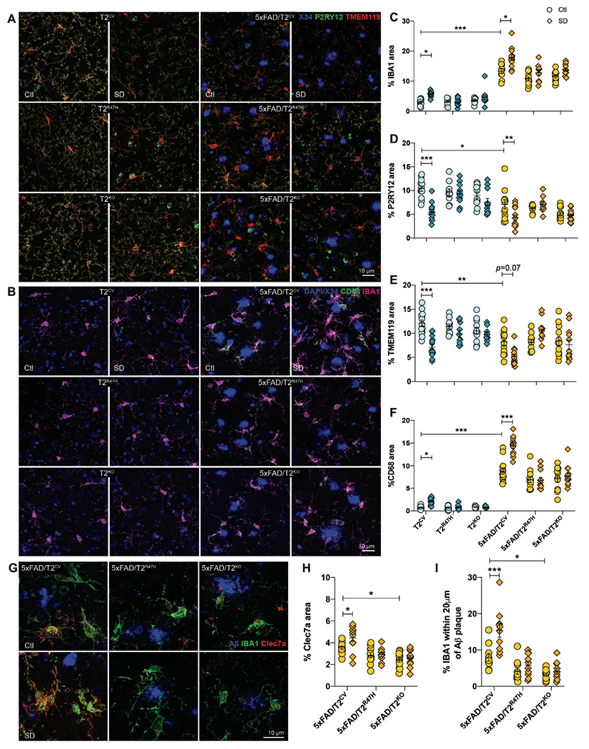
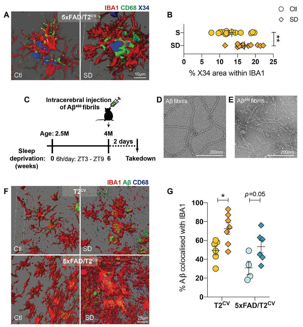
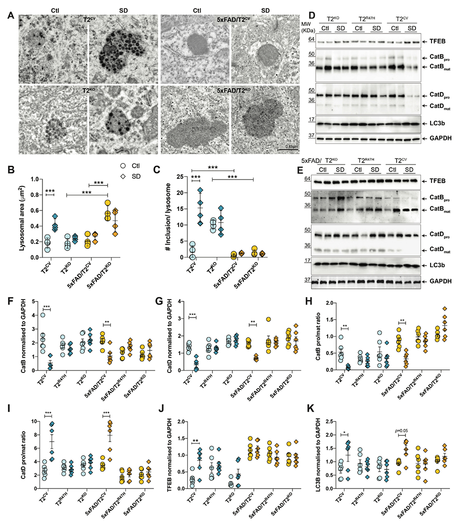
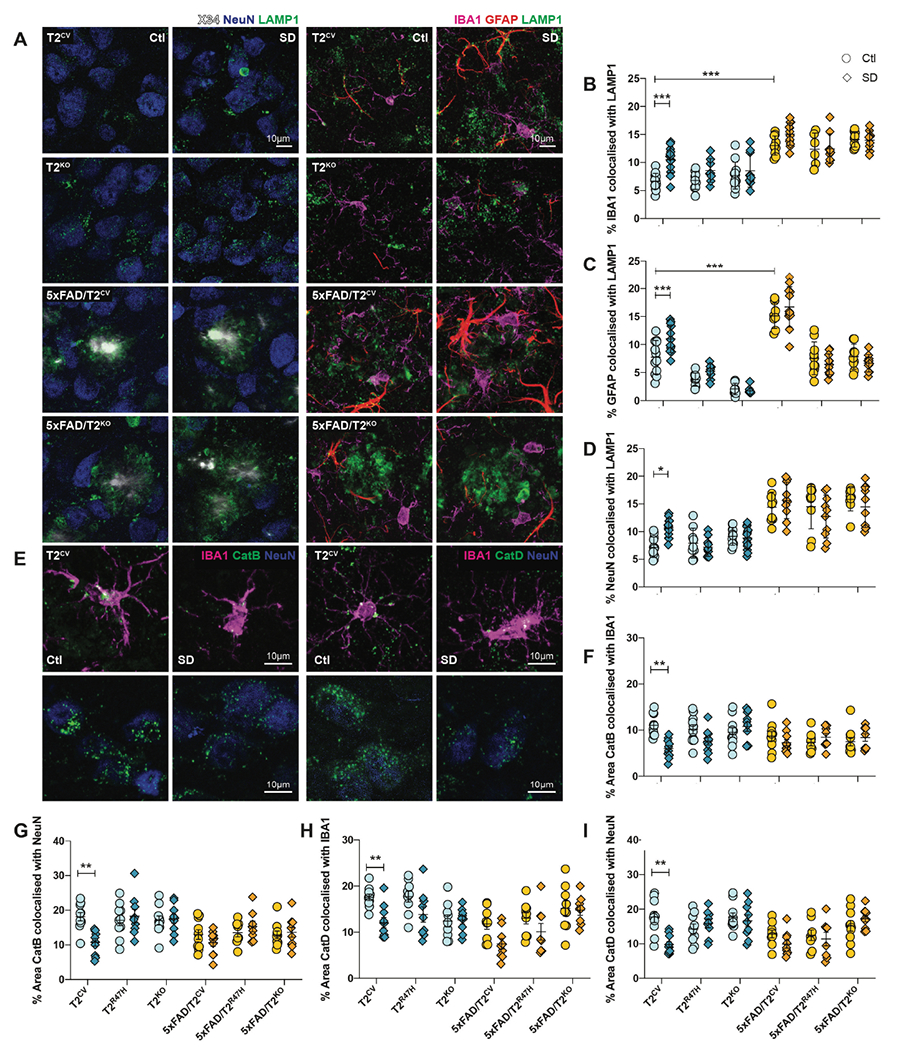
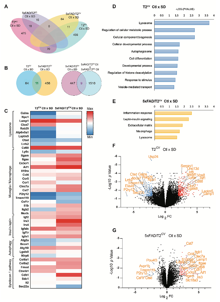
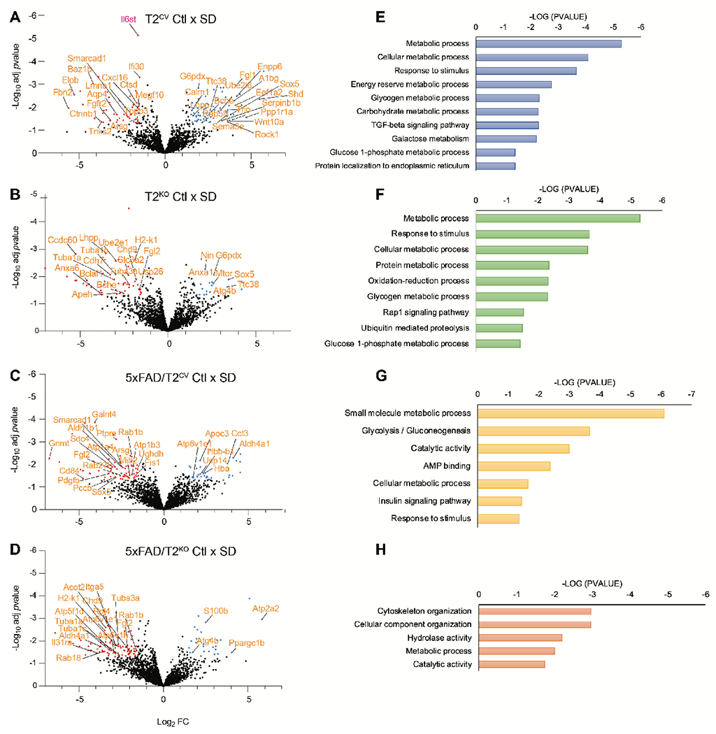
Sleep loss is associated with cognitive decline in the aging population and is a risk factor for Alzheimer's disease (AD). Considering the crucial role of immunomodulating genes such as that encoding the triggering receptor expressed on myeloid cells type 2 (TREM2) in removing pathogenic amyloid-β (Aβ) plaques and regulating neurodegeneration in the brain, our aim was to investigate whether and how sleep loss influences microglial function in mice. We chronically sleep-deprived wild-type mice and the 5xFAD mouse model of cerebral amyloidosis, expressing either the humanized TREM2 common variant, the loss-of-function R47H AD-associated risk variant, or without TREM2 expression. Sleep deprivation not only enhanced TREM2-dependent Aβ plaque deposition compared with 5xFAD mice with normal sleeping patterns but also induced microglial reactivity that was independent of the presence of parenchymal Aβ plaques. We investigated lysosomal morphology using transmission electron microscopy and found abnormalities particularly in mice without Aβ plaques and also observed lysosomal maturation impairments in a TREM2-dependent manner in both microglia and neurons, suggesting that changes in sleep modified neuro-immune cross-talk. Unbiased transcriptome and proteome profiling provided mechanistic insights into functional pathways triggered by sleep deprivation that were unique to TREM2 and Aβ pathology and that converged on metabolic dyshomeostasis. Our findings highlight that sleep deprivation directly affects microglial reactivity, for which TREM2 is required, by altering the metabolic ability to cope with the energy demands of prolonged wakefulness, leading to further Aβ deposition, and underlines the importance of sleep modulation as a promising future therapeutic approach.


Conflict of interest statement

Competing interests

DMH is as an inventor on a patent licensed by Washington University to C2N Diagnostics on the therapeutic use of anti-tau antibodies, US patent 9,834,596. DMH co-founded and is on the scientific advisory board of C2N Diagnostics. DMH is on the scientific advisory board of Denali, Genentech, and Cajal Neuroscience and consults for Alector and Asteroid

Figures

Figure 1.
Figure 1.. Sleep deprivation exacerbates amyloid plaque deposition in 5xFAD mice in a TREM2-dependent manner.
(A) Design of sleep deprivation study. (B, C) Shown is the percentage sleep (B) and sleep bout length recorded during sleep deprivation (SD) compared to normally sleeping control (Ctl) mice expressing TREM2 common variant (T2CV). Data were recorded over a 24h time period. (D) Representative confocal images of immunohistochemically stained fibrillar Aβ plaques (X34) and immunopositive plaques (6E10) in the subiculum of 5xFAD/T2CV, 5xFAD/T2R47H and 5xFAD/T2KO mice that were either sleep deprived or allowed normal sleep as control (Ctl). (E,F,G,H) Quantifications of immunohistochemical images shown in panel D; n=10-14 mice/genotype. Data represent mean ± S.E.M. Two-way ANOVA, Tukey’s multiple comparison test; *P<0.05; **P<0.01; ***P<0.001. ZT, Zeitgeber time (where ZT 0 is the start of the light phase and ZT 12 is the start of the dark phase).
Figure 2.
Figure 2.. Sleep deprivation alters microglial reactivity according to TREM2 expression.
(A) Shown are representative confocal images of the subiculum immunostained with X34, P2RY12 or TMEM119, which detect fibrillar Aβ plaques and homeostatic microglia, respectively. (B) Shown are representative confocal images of the subiculum immunostained with X34 and costained with CD68+ phagolysosomes within microglia (green) and IBA1+ microglia (magenta), with DAPI nuclear stain (blue). (C) The percentage of subiculum area stained positive for IBA1 is shown (including microglia close to or far away from amyloid plaques) quantified from panel B. (D) The percentage of subiculum area stained positive for P2RY12 quantified from panel A. (E) The percentage of subiculum area stained positive for TMEM119 is shown. (F) The percentage of subiculum area stained positive for CD68 quantified from panel A is shown. (G) Representative confocal images of subiculum immunopositive for Aβ (6E10 antibody, blue), IBA1 (green) and Clec7a (red). (H) The percentage of the subiculum area stained positive for Clec7a. (I) The percentage of IBA1+ microglia within 20μm of Aβ plaques in the subiculum were quantified. n=10-14 mice/genotype. SD, Sleep deprived; Ctl, normally sleeping control mice. Data represent mean ± S.E.M. Two-way ANOVA, Sidak’s multiple comparison test; *P<0.05; **P<0.01; ***P<0.001.
Figure 3.
Figure 3.. Sleep deprivation results in an increase in Aβ within microglia.
(A) 3D reconstructed images of X34, CD68 and IBA1 immunostained subiculum from normally sleeping control (Ctl) 5xFAD/T2CV mice compared to sleep deprived (SD) 5xFAD/T2CV mice. (B) Quantification of the percentage of X34-positive (blue) area within CD68+ stained microglia (green). n=10-14 mice/genotype. Unpaired t-test. (C) Study design for intracerebral inoculation of 488-labelled Aβ fibrils into the subiculum of sleep deprived T2CV and 5xFAD/T2CV mice versus normally sleeping controls. (D,E) Representative transmission electron microscopy images of unlabeled (D) and 488-labelled (E) Aβ fibrils. (F) Shown are 3D reconstructed images of 488-labelled Aβ (green), CD68 (blue) and IBA1 (red) immunostaining of subiculum from T2CV control mice (n=6-7 mice/group) and 5xFAD/T2CV mice (n=4-6/group). (G) Shown is the percentage of 488-labelled Aβ fibrils within IBA1+ microglia quantified from panel F. Data represent mean ± S.E.M. Two-way ANOVA, Sidak’s multiple comparison test; *P<0.05; **P<0.01; ***P<0.001. ZT, zeitgeiber time.
Figure 4.
Figure 4.. Lysosomal impairments after sleep deprivation are dependent on TREM2 expression.
(A) Representative transmission electron microscopy images of lysosomes in subiculum from T2CV, 5xFAD/T2CV, T2KO and 5xFAD/T2KO mice that were sleep deprived (SD) or normally sleeping controls (Ctrl). (B, C) Quantification of lysosomal area (B) and number of electron dense inclusions per lysosome (C) from transmission electron microscopy images in panel A. Two-way ANOVA, Tukey’s multiple comparison test; ***P<0.001. (D) Immunoblotting of hippocampal lysates for TFEB, cathepsins B and D, LC3b, with GAPDH as loading control, for T2CV, T2R47H and T2KO groups that were sleep deprived (SD) or normally sleeping (Ctl). (E) Immunoblotting of hippocampal lysates for TFEB, cathepsins B and D, LC3b, with GAPDH as loading control, for 5xFAD/T2CV, 5xFAD/T2R47H and 5xFAD/T2KO groups that were sleep deprived (SD) or normally sleeping (Ctl). (F,G) Quantification of immunoblots for (F) cathepsin B and (G) cathepsin D normalized to GAPDH. (H,I) Quantification of immunoblots for the pro form to mature form ratio of (H) cathepsin B and (I) cathepsin D. (J, K) Quantification of immunoblots for TFEB (J) and LC3b (K) normalized to GAPDH. (n=6/genotype). All data represent mean ± S.E.M. Two-way ANOVA, Sidak’s multiple comparison test; *P<0.05; **P<0.01; ***P<0.001.
Figure 5.
Figure 5.. Cell-specific lysosomal protein changes after sleep deprivation.
(A) Left panels show representative confocal images of X34, NeuN and LAMP1 immunostaining of subiculum from T2CV, 5xFAD/T2CV, T2KO, and 5xFAD/T2KO mice that were sleep deprived (SD) or normally sleeping controls (Ctl). Right panels show representative confocal images of IBA1, GFAP and LAMP1 immunostaining of subiculum from T2CV, 5xFAD/T2CV, T2KO, and 5xFAD/T2KO mice that were sleep deprived (SD) or normally sleeping controls (Ctl). (B) Quantification of the percentage of subiculum stained for colocalized IBA1 and LAMP1. (C) Quantification of the percentage of subiculum stained for colocalized GFAP and LAMP1. (D) Quantification of the percentage of subiculum stained for colocalized NeuN and LAMP1. (E) Left panels show representative confocal images of subiculum from T2CV mice costained with IBA1 (magenta), cathepsin B (CatB, green), and NeuN (blue). Right panels show representative confocal images of subiculum from sleep deprived or normally sleeping T2CV mice costained with IBA1 (magenta), cathepsin D (CatD, green), and NeuN (blue). (F, G) Quantification of the percentage of subiculum in which cathepsin B colocalizes with IBA1 (F) or NeuN (G). (H,I) Quantification of the percentage of subiculum in which cathepsin D colocalizes with IBA1 (H) or NeuN (I). n=9-11 mice/genotype. Data represent mean ± S.E.M. Two-way ANOVA, Tukey’s multiple comparison test; *P<0.05; **P<0.01; ***P<0.001.
Figure 6.
Figure 6.. Sleep deprivation triggers transcriptional pathways depending on TREM2 and Aβ plaques .
(A) Venn diagram illustrates the number of genes mutually inclusive or exclusive in the four different mouse genotypes: T2CV, 5xFAD/T2CV, T2KO, and 5xFAD/T2KO. (B) Venn diagrams compare the number of genes mutually inclusive or exclusive for sleep deprived (SD) T2CV mice and 5xFAD/T2CV mice, and their normally sleeping controls (Ctl), as well as for sleep deprived 5xFAD/T2CV mice compared to normally sleeping 5xFAD mice as control. (C) Heat map of RNA sequences that were upregulated or downregulated in bulk hippocampal tissue from T2CV and 5xFAD/T2CV mice, either sleep deprived (SD) or normally sleeping controls (Ctl), grouped according to gene ontology pathways. (D, E) Shown are gene ontology pathways affected by sleep deprivation in T2CV mice (D) and 5xFAD/T2CV mice (E). (F, G) Volcano plot of gene expression changes in T2CV mice (F) and 5xFAD/T2CV mice (G) that were sleep deprived (SD) or allowed sleep normally (Ctl). Significantly downregulated genes are indicated by blue dots and upregulated genes by red dots. n=10/genotype. Significance for volcano plots was marked at p<0.05 with a 2-fold change in Log2.
Figure 7.
Figure 7.. Sleep deprivation induces metabolic dysregulation in the mouse CSF proteome.
(A-D) Shown are volcano plots of the CSF proteomes for T2CV mice (A), 5xFAD/T2CV mice (B), T2KO mice (C) and 5xFAD/T2KO mice (D), that were either sleep deprived (SD) or allowed to sleep normally as a control (Ctl). Significantly downregulated proteins are indicated by blue dots and upregulated proteins are indicated by red dots. (E-H) Gene ontology functional pathways are shown for CSF proteomes for T2CV mice (E), 5xFAD/T2CV mice (F), T2KO mice (G) and 5xFAD/T2KO mice (H). n=9-11 mice/genotype. Significance for volcano plots was marked at p<0.05 with a 1.5-fold change in Log2.

References

    1. Sabia S, Fayosse A, Dumurgier J, van Hees VT, Paquet C, Sommerlad A, Kivimäki M, Dugravot A, Singh-Manoux A, Association of sleep duration in middle and old age with incidence of dementia. Nat. Commun 12, 2289 (2021). - PMC - PubMed
    1. Prince T-M, Abel T, The impact of sleep loss on hippocampal function. Learn. Mem 20, 558–69 (2013). - PMC - PubMed
    1. Xie L, Kang H, Xu Q, Chen MJ, Liao Y, Thiyagarajan M, O’Donnell J, Christensen DJ, Nicholson C, Iliff JJ, Takano T, Deane R, Nedergaard M, Sleep Drives Metabolite Clearance from the Adult Brain. Science. 342, 373–377 (2013). - PMC - PubMed
    1. Achariyar TM, Li B, Peng W, Verghese PB, Shi Y, McConnell E, Benraiss A, Kasper T, Song W, Takano T, Holtzman DM, Nedergaard M, Deane R, Glymphatic distribution of CSF-derived apoE into brain is isoform specific and suppressed during sleep deprivation. Mol. Neurodegener 11, 74 (2016). - PMC - PubMed
    1. Bellesi M, de Vivo L, Chini M, Gilli F, Tononi G, Cirelli C, Sleep Loss Promotes Astrocytic Phagocytosis and Microglial Activation in Mouse Cerebral Cortex. J. Neurosci 37, 5263–5273 (2017). - PMC - PubMed

Publication types