. 2023 Jun:62:102707.
doi: 10.1016/j.redox.2023.102707. Epub 2023 Apr 20.

The deubiquitinase USP11 ameliorates intervertebral disc degeneration by regulating oxidative stress-induced ferroptosis via deubiquitinating and stabilizing Sirt3

Affiliations

The deubiquitinase USP11 ameliorates intervertebral disc degeneration by regulating oxidative stress-induced ferroptosis via deubiquitinating and stabilizing Sirt3

Jian Zhu et al. Redox Biol. 2023 Jun.

Abstract

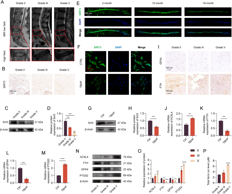
Increasing studies have reported that intervertebral disc degeneration (IVDD) is the main contributor and independent risk factor for low back pain (LBP), it would be, therefore, enlightening that investigating the exact pathogenesis of IVDD and developing target-specific molecular drugs in the future. Ferroptosis is a new form of programmed cell death characterized by glutathione (GSH) depletion, and inactivation of the regulatory core of the antioxidant system (glutathione system) GPX4. The close relationship of oxidative stress and ferroptosis has been studied in various of diseases, but the crosstalk between of oxidative stress and ferroptosis has not been explored in IVDD. At the beginning of the current study, we proved that Sirt3 decreases and ferroptosis occurs after IVDD. Next, we found that knockout of Sirt3 (Sirt3-/-) promoted IVDD and poor pain-related behavioral scores via increasing oxidative stress-induced ferroptosis. The (immunoprecipitation coupled with mass spectrometry) IP/MS and co-IP demonstrated that USP11 was identified to stabilize Sirt3 via directly binding to Sirt3 and deubiquitinating Sirt3. Overexpression of USP11 significantly ameliorate oxidative stress-induced ferroptosis, thus relieving IVDD by increasing Sirt3. Moreover, knockout of USP11 in vivo (USP11-/-) resulted in exacerbated IVDD and poor pain-related behavioral scores, which could be reversed by overexpression of Sirt3 in intervertebral disc. In conclusion, the current study emphasized the importance of the interaction of USP11 and Sirt3 in the pathological process of IVDD via regulating oxidative stress-induced ferroptosis, and USP11-mediated oxidative stress-induced ferroptosis is identified as a promising target for treating IVDD.

Keywords: De-ubiquitination; IVDD; Oxidative stress-induced ferroptosis; Sirt3; USP11.


Conflict of interest statement

Declaration of competing interest The authors have declared that no competing interest exists.

Figures

Fig. 1
Fig. 1
(A) Magnetic resonance imaging of patients with different levels of IVDD according to Pfirrmann grades. (B) The results of IHC for human NP tissue sections demonstrated that the expression of Sirt3 was decreased with the progression of degeneration. (C, D) The expression change of Sirt3 in IVDD was further confirmed by western blotting assay, which was accordance with the result of IHC. (E). The IF for mouse intervertebral disc tissue sections proved the down-regulated Sirt3 after IVDD. (F) The expression of Sirt3 was lower in HNP cells treated with TBHP. (G, H) The administration of TBHP for HNP cells made the Sirt3 expression dropped significantly compared with control group. (I) IHC results demonstrated the lower expression of GPX4 and FTH after IVDD. (J–M) The results of qPCR proved that the expression of ACSL4 and PTGS2 was enhanced while GPX4 and FTH was decreased after IVDD. (N, O) The result of WB further confirmed the above results. (P) ELISA proved that the ferric ion increased with the progression of IVDD. ∗ for p < 0:05, ∗∗ for p < 0:01, ∗∗∗for p < 0:001, ∗∗∗∗ for p < 0:0001.
Fig. 2
Fig. 2
(A) HE staining showed the morphology of mice discs, more severe rupture between the annulus fibrosus and nucleus pulposus was observed in Sirt3−/− group. (B) SF staining proved that KO of Sirt3 was associated with more severe IVDD with internuclear fibrosis. (C) Histological grades were calculated based on HE staining and SF staining results. (D) IHC results revealed that higher expression of ADAMTS5 and MMP3 in Sirt3−/− group after 6 months from the construction of IVDD model was confirmed. (E) The co-immunofluorescence of ACAN and MMP3 for HNP cells indicated that KO of Sirt3 promoted higher expression of ACAN and lower expression of MMP3. (F) Enhanced expression of ADAMTS5 and MMP3 and down-regulated expression of ACAN and Col2 was observed in WB results. (G) Reduced signal intensity was observed in Sirt3−/− group, which proved that KO of Sirt3 leaded to more severe IVDD. (H–K) The qPCR results accorded with the results above. (L–O) Pain-related behavioral scores, including pressure tolerance, distance walked, total active time and maximum speed, were significantly compromised in Sirt3−/− group with increasing IVDD duration. ∗ for p < 0:05, ∗∗ for p < 0:01, ∗∗∗for p < 0:001, ∗∗∗∗ for p < 0:0001, ns for no significance.
Fig. 3
Fig. 3
(A) Detailed information of the experimental group in the part of the experiment involving oxidative stress. (B, C) KO of Sirt3 could significantly decrease the expression of anti-oxidative stress genes (HO-1, NQO1, SOD2 and SCC7A11). (D) The superoxide was increased with increasing time of TBHP treatment, and higher superoxide was observed in Sirt3-KO group at each time point. (E–H) The above result was further confirmed by qPCR. (I) Mitochondrial superoxide was up-regulated after the treatment of TBHP, which was exacerbated by KO of Sirt3. (J) Detailed information of the experimental group in investigating the effect of KO of Sirt3 on oxidative stress-induced ferroptosis. (K, L) The WB result revealed that the expression of anti-ferroptosis genes (GPX4 and FTH) were decreased while pro-ferroptosis genes (PTGS2 and ACSL4) was increased in NP cells after treated with TBHP, the deletion of Sirt3 exacerbated ferroptosis induced by oxidative stress, which could be reversed by specific oxidative stress inhibitor NAC. (M) As shown in the result of ELISA, the content of MDA and ferric ion was increased significantly in Sirt3−/−, while GSH was dropped. (N, O) Non-peroxidative state shows red fluorescence, while peroxidative state shows green fluorescence in BODIPY assay. The result suggested that the level of lipid peroxidation was the most up-regulated in Sirt3 KO group after exposed to TBHP. (P) Mitochondrial morphology changed the most in Sirt3 KO accompanied with administration of TBHP, which was reversed partly by NAC. (Q–T) The result of qPCR was in accordance with the above results. ∗ for p < 0:05, ∗∗ for p < 0:01, ∗∗∗for p < 0:001, ∗∗∗∗ for p < 0:0001. (For interpretation of the references to colour in this figure legend, the reader is referred to the Web version of this article.)
Fig. 4
Fig. 4
(A) The result of IP/MS demonstrated that USP11 was identified to be a putative protein binding to Sirt3. To further confirm the result of IP/MS, Co-IP analysis was conducted subsequently. (B) USP11 precipitated Sirt3 in NP cells, confirming the interaction of USP11 and Sirt3. (C) Reverse Co-IP revealed that USP11 was precipitated by Sirt3 in HP cells. The Co-IP analysis using epitope-tagged proteins was further performed. (D, E) the Co-IP test with exogenous Flag-tagged USP11, or Myc-tagged Sirt3 in HEK 293 T cells could precipitate endogenous Sirt3 or USP11, respectively. (F, G) Exogenous Flag-tagged USP11 and Myc-tagged Sirt3 could co-precipitate each other efficiently in HEK 293 T cells. (H) The data above proved the interactions of USP11 and Sirt3, which could be enhanced after IVDD. (I) The schematic diagram showed that USP11 and Sirt3 contains several domains. (J, K) Full length or truncated segments of Flag-USP11 and Myc-Sirt3 containing different domains were transfected in HEK 293 T cells to investigate the binding region. (L) The Co-IP revealed that the fragment containing DUSP domain of USP11 was able to bind Sirt3. (M) Reverse Co-IP proved that USP11 interacted with M2 domain of Sirt3.
Fig. 5
Fig. 5
(A) The expression of Sirt3 declined spontaneously with time in the presence of protein synthesis inhibitor cycloheximide (CHX, 10 μg/ml), which could be reversed by proteasome specific inhibitor MG132. Next, siRNA-USP11 or siRNA-Ctrl was transfected into HNP cells and the expression of Sirt3 was analyzed using immunoblotting. (B) The expression of Sirt3 dropped significantly after knockdown of USP11. (C) On the contrary, overexpression of USP11 stabilized the expression of Sirt3. (D) The results above were confirmed again in HEK 293 T cells. (E, F) Overexpression of normal USP11 could upregulate the Sirt3 expression, while overexpression of USP11 with C318S mutant did not. (G) The de-ubiquitination of Sirt3 was abolished effectively by knockdown of USP11. (H) HEK 293 T cells were co-transfected with plasmid of Flag-USP11, Myc-Sirt3 and HA-Ub. The results demonstrated that the deubiquitinate effect of USP11 on Sirt3 was proved. (I) The de-ubiquitination of Sirt3 was abolished in IVD in USP11−/− mice compared with wild mice. (J) Higher de-ubiquitination of Sirt3 and higher expression of Sirt3 was observed after IVDD in mice injected with AAV-USP11. (K) USP11 could cleave the Lys48-polyubiquitin chain instead of Lys63-polyubiquitin chain. (L). Lys48-linked poly-ubiquitination was necessary for de-ubiquitination of Sirt3 regulated by USP11.
Fig. 6
Fig. 6
(A) Detailed information of the experimental group in the part of the experiment. (B, C) Ferroptosis events were exacerbated by knockdown of Sirt3, including increased expression of ACSL4 and PTGS2, and decreased expression of GPX4 and FTH, which could be ameliorated by overexpression of USP11. (D) ELISA revealed that contents of MDA and ferric ion in supernatant were surged significantly, but GSH dropped with statistical significance in siRNA-Sirt3 group, which could be reversed by co-transfected with plasmid-USP11. (E–H) The results of qPCR for ferroptosis-related genes showed the same trend. (I) After plasmid-USP11 treatment, the morphology of mitochondria maintained well, whereas pretreatment with siRNA-Sirt3resulted in a significant decrease in mitochondrial cristae. (J) The feature of BODIPY assay is that the higher the green fluorescence intensity, the higher the lipid peroxidation level. Thus, as presented, the level of lipid peroxidation was much higher after the treatment of siRNA-Sirt3, which was reversed partly by plasmid-USP11. (K, L) WB proved that siRNA-Sirt3 resulted in enhanced expressions of ADAMTS5 and MMP3 and down-regulated expressions of ACAN and Col2, while plasmid-USP11 could mitigate IVDD. (M − P) The results above were further confirmed using qPCR. ∗ for p < 0:05, ∗∗ for p < 0:01, ∗∗∗for p < 0:001, ∗∗∗∗ for p < 0:0001. (For interpretation of the references to colour in this figure legend, the reader is referred to the Web version of this article.)
Fig. 7
Fig. 7
(A) Detailed information of the experimental group in the part of the experiment. (B, C) KO of USP11 resulted in more severe IVDD, indicated by disrupted boundary between NP and AF tissue and decreased NP cells. (D) IHC results revealed that ADAMTS5 and MMP3 were increased significantly after KO of USP11, while AAV-Sirt3 could improve IVDD both in WT and USP11−/− mice. (E) KO of USP11 resulted in increased expression of MMP3 (green fluorescence) and decreased expression of ACAN (red fluorescence), which was reversed partly by AAV-Sirt3. (F, G) WB further confirmed the protective effect of AAV-Sirt3 on IVDD induced by KO of USP11. (H–K) The results above were further confirmed by qPCR. (L–O) KO of USP11 resulting in poor pain-related behavioral scores was proved, which, however, could be improved by AAV-Sirt3. ∗ for p < 0:05, ∗∗ for p < 0:01, ∗∗∗for p < 0:001, ∗∗∗∗ for p < 0:0001. (For interpretation of the references to colour in this figure legend, the reader is referred to the Web version of this article.)
Fig. 8
Fig. 8
USP11 regulates oxidative stress-induced ferroptosis after IVDD by deubiquitinating Sirt3. Ferroptosis is associated closely with the process of IVDD. The direct interaction of USP11 and Sirt3 is a crucial molecular event that inhibit oxidative stress-induced ferroptosis, thus improves IVDD and relives pain reaction.
figs1
figs1

References

    1. Morgan T., Wu J., Ovchinikova L., Lindner R., Blogg S., Moorin R. A national intervention to reduce imaging for low back pain by general practitioners: a retrospective economic program evaluation using Medicare Benefits Schedule data. BMC Health Serv. Res. 2019;19:983. doi: 10.1186/s12913-019-4773-y. - DOI - PMC - PubMed
    1. Yadav R.I., Long L., Yanming C. Comparison of the effectiveness and outcome of microendoscopic and open discectomy in patients suffering from lumbar disc herniation. Medicine (Baltim.) 2019;98 doi: 10.1097/MD.0000000000016627. - DOI - PMC - PubMed
    1. Hoy D., Brooks P., Blyth F., Buchbinder R. The Epidemiology of low back pain. Best Pract. Res. Clin. Rheumatol. 2010;24:769–781. doi: 10.1016/j.berh.2010.10.002. - DOI - PubMed
    1. Francisco V., Pino J., González-Gay M.Á., Lago F., Karppinen J., Tervonen O., Mobasheri A., Gualillo O. A new immunometabolic perspective of intervertebral disc degeneration. Nat. Rev. Rheumatol. 2022;18:47–60. doi: 10.1038/s41584-021-00713-z. - DOI - PubMed
    1. Guo W., Mu K., Zhang B., Sun C., Zhao L., Li H.-R., Dong Z.-Y., Cui Q. The circular RNA circ-GRB10 participates in the molecular circuitry inhibiting human intervertebral disc degeneration. Cell Death Dis. 2020;11:612. doi: 10.1038/s41419-020-02882-3. - DOI - PMC - PubMed

Publication types

MeSH terms

LinkOut - more resources