. 2023 Jul 1;435(13):168128.
doi: 10.1016/j.jmb.2023.168128. Epub 2023 Apr 25.

Omicron Spike Protein is Vulnerable to Reduction

Affiliations

Omicron Spike Protein is Vulnerable to Reduction

Zhong Yao et al. J Mol Biol. .

Abstract

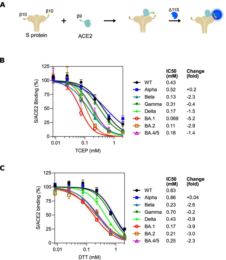
SARS-CoV-2 virus spike (S) protein is an envelope protein responsible for binding to the ACE2 receptor, driving subsequent entry into host cells. The existence of multiple disulfide bonds in the S protein makes it potentially susceptible to reductive cleavage. Using a tri-part split luciferase-based binding assay, we evaluated the impacts of chemical reduction on S proteins from different virus variants and found that those from the Omicron family are highly vulnerable to reduction. Through manipulation of different Omicron mutations, we found that alterations in the receptor binding module (RBM) are the major determinants of this vulnerability. Specifically we discovered that Omicron mutations facilitate the cleavage of C480-C488 and C379-C432 disulfides, which consequently impairs binding activity and protein stability. The vulnerability of Omicron S proteins suggests a mechanism that can be harnessed to treat specific SARS-CoV-2 strains.

Keywords: ACE2; SARS-CoV-2; binding; disulfide; receptor binding motif (RBM).


Conflict of interest statement

Declaration of Competing Interest The authors declare that they have no known competing financial interests or personal relationships that could have appeared to influence the work reported in this paper.

Figures

None
Graphical abstract
Figure 1
Figure 1
Different SARS-CoV-2 variant S proteins demonstrate varied vulnerabilities to chemical reduction. (A) Schematic diagram of the tNLuc-based S/ACE2 binding assay. (B-C) S proteins derived from early VOCs (Alpha, Beta, Gamma and Delta) or Omicron variants (BA.1, BA.2 and BA4/5) were treated with TCEP (B) or DTT (C) in various concentrations as indicated, followed by tNLuc-based S/ACE2 binding assay. The readings were normalized using corresponding S protein samples without compound treatment. Results are summary of four independent experiments with two technical replicates in each experiment. Data were fitted into the inhibition kinetics model with normalized response and variable slope. Obtained kinetics are presented in average curves. Fold changes in IC50 were calculated by comparing the WT S protein.
Figure 2
Figure 2
Omicron mutations in RBM are the major determinant in vulnerability to chemical reduction. (A) The impacts of omicron mutations were examined in WT/Omicron S protein chimeras. WT (S: WT), Omicron (S: Omicron), chimera of Omicron S1 and WT S2 (S: S1-Omi), chimera with Omicron NTD and WT backbone (S: NTD-Omi) or chimera with Omicron RBM and WT backbone (S: RBM-Omi) S proteins were pretreated with the indicated concentrations of TCEP followed by ACE2 binding assay. (B) Binding effects were further examined in RBD proteins. WT S protein (S: WT), Omicron S protein (S: Omicron), WT RBD (RBD: WT), Omicron RBD (RBD: Omi) or chimeric RBD with Omicron RBM on WT backbone (RBD: RBM-Omi) were pretreated with the indicated concentrations of TCEP followed by ACE2 binding assay. Readings were normalized using corresponding protein constructs without compound treatment. Results are summary of four (A) or three (B) independent experiments with two technical replicates in each experiment. Data were fitted into the inhibition kinetics model with normalized response and variable slope. Obtained kinetics are presented in average curves. Fold changes in IC50 were calculated by comparing the WT S protein (A, B*) or WT RBD protein (B**).
Figure 3
Figure 3
Detailed analysis of the importance of Omicron RBM mutations in the vulnerability to chemical reduction. (A) 3D and linear structure of the Omicron RBD. Cystines are highlighted in red and represented as red spheres. Mutations are also represented as spheres and shown in different colors based on their grouping into three clusters. (B-C) Analysis of loss-of-function mutants. In B, WT and Omicron S proteins and S proteins of the Omicron backbone with cluster A or B mutations reverted to WT sequence (S: Omi_A or S: Omi_B) were analysed. In C, WT RBD protein, WT RBD carrying Omicron RBM (RBD: RBM-Omi), and WT RBD protein carrying Omicron RBM with clusters A, B or C reverted back to WT sequence (RBD: RBM-Omi_A, RBD: RBM-Omi_B or RBD: RBM-Omi_C) were analysed. Protein constructs are listed in Figure S4. (D-E) Analysis of gain-of-function mutants. In D, WT and Omicron S proteins, and S proteins of WT backbone with an RBM cluster mutated to its Omicron sequence (S: A-Omi, S: B-Omi or S: C-Omi) were analyzed. In E, WT RBD protein and Omicron RBD protein (RBD: Omi), alongside RDB proteins with WT backbone and an RBM cluster mutated to its Omicron sequence (RBD: A-Omi, RBD: B-Omi or RBD: C-Omi) were analyzed. Proteins in B-E were pretreated with the indicated concentrations of TCEP followed by ACE2 binding assay. Protein constructs are listed in Figure S4. Readings were normalized with corresponding S or RBD proteins without compound treatment. Results are summary of three independent experiments with two technical replicates in each experiment. Data were fitted into the inhibition kinetics model with normalized response and variable slope. Obtained kinetics are presented in average curves. Fold changes in IC50 were calculated by comparing the WT S or RBD protein.
Figure 4
Figure 4
Mass spectrometry analysis of reduced cysteines in RBD. (A) Strategy of the MS analysis. (B) WT or Omicron RBD proteins were treated with 0.1 mM TCEP or mock treated and analyzed as described in A. The intensities of peptides containing alkylated cysteines derived from RBD proteins are presented as a dot plot. The data are normalized to the intensities of nontreated Omicron RBD sample. Black dots stand for common peptides appearing in both WT and Omicron RBD; blue dots stand for WT-specific peptides; green dots stand for Omicron specific peptides.
Figure 5
Figure 5
Impacts of disulfide cleavage in Omicron RBD. (A) Disulfide cleavage was mimicked by paired cysteine-to-serine mutation. Protein stability of the mutants was measured as the abundance of each protein in culture medium of the cells transiently expressing the protein. The results are normalized using co-expressed Cypridina luciferase. (B) The binding activity of each cysteine-to-serine mutant was measured using a tNLuc-based assay with different concentrations of ACE2. The results are presented as mean ± SEM of three independent experiments with two technical replicates in each experiment.

References

    1. Baker J.M., Nakayama J.Y., O’Hegarty M., McGowan A., Teran R.A., Bart S.M., Mosack K., Roberts N., et al. SARS-CoV-2 B.1.1.529 (Omicron) Variant Transmission Within Households - Four U.S. Jurisdictions, November 2021-February 2022. MMWR Morb. Mortal. Wkly. Rep. 2022;71:341–346. doi: 10.15585/mmwr.mm7109e1. - DOI - PMC - PubMed
    1. Lewnard J.A., Hong V.X., Patel M.M., Kahn R., Lipsitch M., Tartof S.Y. Clinical outcomes associated with SARS-CoV-2 Omicron (B.1.1.529) variant and BA.1/BA.1.1 or BA.2 subvariant infection in Southern California. Nat. Med. 2022;28:1933–1943. doi: 10.1038/s41591-022-01887-z. - DOI - PMC - PubMed
    1. Viana R., Moyo S., Amoako D.G., Tegally H., Scheepers C., Althaus C.L., Anyaneji U.J., Bester P.A., et al. Rapid epidemic expansion of the SARS-CoV-2 Omicron variant in southern Africa. Nature. 2022;603:679–686. doi: 10.1038/s41586-022-04411-y. - DOI - PMC - PubMed
    1. Cai Y., Zhang J., Xiao T., Lavine C.L., Rawson S., Peng H., Zhu H., Anand K., et al. Structural basis for enhanced infectivity and immune evasion of SARS-CoV-2 variants. Science. 2021;373:642–648. doi: 10.1126/science.abi9745. - DOI - PMC - PubMed
    1. Alter G., Yu J., Liu J., Chandrashekar A., Borducchi E.N., Tostanoski L.H., McMahan K., Jacob-Dolan C., et al. Immunogenicity of Ad26.COV2.S vaccine against SARS-CoV-2 variants in humans. Nature. 2021;596:268–272. doi: 10.1038/s41586-021-03681-2. - DOI - PMC - PubMed

Publication types

Substances

Supplementary concepts

LinkOut - more resources