. 2023 Jun;299(6):104742.
doi: 10.1016/j.jbc.2023.104742. Epub 2023 Apr 24.

A heme pocket aromatic quadrupole modulates gas binding to cytochrome c'-β: Implications for NO sensors

Affiliations

A heme pocket aromatic quadrupole modulates gas binding to cytochrome c'-β: Implications for NO sensors

Hannah R Adams et al. J Biol Chem. 2023 Jun.

Abstract

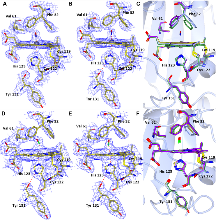
The structural basis by which gas-binding heme proteins control their interactions with NO, CO, and O2 is fundamental to enzymology, biotechnology, and human health. Cytochromes c' (cyts c') are a group of putative NO-binding heme proteins that fall into two families: the well-characterized four alpha helix bundle fold (cyts c'-α) and an unrelated family with a large beta-sheet fold (cyts c'-β) resembling that of cytochromes P460. A recent structure of cyt c'-β from Methylococcus capsulatus Bath revealed two heme pocket phenylalanine residues (Phe 32 and Phe 61) positioned near the distal gas-binding site. This feature, dubbed the "Phe cap," is highly conserved within the sequences of other cyts c'-β but is absent in their close homologs, the hydroxylamine-oxidizing cytochromes P460, although some do contain a single Phe residue. Here, we report an integrated structural, spectroscopic, and kinetic characterization of cyt c'-β from Methylococcus capsulatus Bath complexes with diatomic gases, focusing on the interaction of the Phe cap with NO and CO. Significantly, crystallographic and resonance Raman data show that orientation of the electron-rich aromatic ring face of Phe 32 toward distally bound NO or CO is associated with weakened backbonding and higher off rates. Moreover, we propose that an aromatic quadrupole also contributes to the unusually weak backbonding reported for some heme-based gas sensors, including the mammalian NO sensor, soluble guanylate cyclase. Collectively, this study sheds light on the influence of highly conserved distal Phe residues on heme-gas complexes of cytochrome c'-β, including the potential for aromatic quadrupoles to modulate NO and CO binding in other heme proteins.

Keywords: aromatic quadrupole; carbon monoxide; cytochrome; nitric oxide.


Conflict of interest statement

Conflict of interest The authors declare that they have no conflicts of interest with the contents of this article.

Figures

Figure 1
Figure 1
Hemes A and B of McCP-β. With either CO (A and B) or NO bound (D and E). Only one orientation of CO is seen with an angle of 174/173°. Two orientations of NO are seen in monomer A and one in monomer B. The aromatic ring of Phe 32 can be seen to be rotated away from the CO molecule in the second heme. Comparison of Fe(II) McCP-β (heme A in blue and heme B in green), and both hemes A (purple) and B (gold) with CO (C) or NO (F) bound, Phe 32 can be seen to move upon introduction of a ligand to the distal side of the heme. McCP-β, cyt c′-β from Methylococcus capsulatus (Bath).
Figure 2
Figure 2
Room temperature resonance Raman spectra of 6cCO McCP-β complexes prepared with12CO and13CO recorded in the low frequency (left panel) and high frequency (right panel) regions, together with12CO to13CO difference spectra. McCP-β, cyt c′-β from Methylococcus capsulatus (Bath);
Figure 3
Figure 3
Relationship between ν(FeCO) and ν(CO) frequencies in 6cCO heme complexes, including data for McCP-β. Vibrational data are taken from Table 3. The solid line is the reported empirical relationship between the ν(FeCO) and ν(CO) frequencies of model porphyrins (arising from variations in Fe(II)→CO(π∗) backbonding) (22). Heme protein 6cCO complexes exhibit a vibrational trend similar to that of model complexes, which allows ν(FeCO) and ν(CO) frequencies to report on the heme pocket polarity. McCP-β, cyt c′-β from Methylococcus capsulatus (Bath).
Figure 4
Figure 4
Room-temperature resonance Raman spectra of Fe(II)NO McCP-β prepared with14NO (black) and15NO (red), together with14NO—15NO difference spectra (blue) in the high frequency (left panel) and low frequency (right panel) regions. Spectra at pH 4.0 correspond to a 5cNO complex, whereas spectra at pH 10 correspond to a 6cNO complex (asterisks denote minor contributions of 5cNO species). A mixture of 5cNO and 6cNO species is observed at pH 7.0. All RR spectra were recorded with 407 nm excitation, except for the low-frequency region of the Fe(II)NO complex at pH 10, which was recorded using both 407 and 442 nm, the latter revealing a 6cNO ν(FeNO) mode at 545 cm−1. McCP-β, cyt c′-β from Methylococcus capsulatus (Bath).
Figure 5
Figure 5
Stopped-flow spectroscopy data for the reaction of ferrous McCP-β with NO at pH 7.5 showing the spectra obtained from global fitting of diode array data to a simple a → b model where a represents the initial 6cNO complex and b the final spectrum at the end of the reaction. The inset shows representative time courses collected at 415 and 395 nm, together with fits to single exponential functions. The observed rate constant for 6cNO→5cNO conversion, measured at pH 7.5, remains effectively unchanged (kobs ∼0.6 ± 0.05 s−1) when the NO concentration is varied from 0.01 to 0.05 mM. McCP-β, cyt c′-β from Methylococcus capsulatus (Bath).
Figure 6
Figure 6
Stopped-flow UV–visible characterization of the transient Fe(II)O2complex of wt McCP-β (25 °C, pH 8.9).Top panel, upon reaction with 650 μM O2, the Fe(II) state (red dashed trace) converts to an Fe(II)O2 complex (blue trace) within the stopped-flow mixing time, followed by biphasic autoxidation to the Fe(III) state (magenta trace) with rate constants, kox(1) = 4.8 s−1 (20% ΔAbs) and kox(2) = 0.37 s−1 (80% ΔAbs). Bottom panel, UV–visible spectra of Fe(II) protein (red) and the initial species formed (1 ms after mixing) with various final O2 concentrations (32–650 μM). The inset shows an O2 binding saturation curve with the absorption increase at 414 nm plotted versus O2 concentration and fit to a hyperbolic function, ΔA414 = (ΔAmax × [O2])/Kd + [O2] (solid line). An O2 binding curve was also prepared using the change in absorption at 430 nm (data not shown). The average Kd value obtained from these binding curves is 74 ± 13 μM. McCP-β, cyt c′-β from Methylococcus capsulatus (Bath).
Figure 7
Figure 7
Hemes A and B of F32V McCP-β. With either NO (A and B) or CO bound (D and E). Comparison of Fe(II) F32V hemes A (blue) and B (green) and both hemes A (purple) and B (gold) with NO (C) or CO (F) bound. Only one orientation of CO can be seen with a slightly more bent geometry than native McCP-β at angles of 164/165°. Two orientations of NO can be seen: one points toward Leu 28 (heme A) and the other toward Gly 82 (heme B). These have Fe–N distances of 1.90 and 1.97 Å and Fe–N–O angles of 110° and 92° in hemes A and B, respectively. McCP-β, cyt c′-β from Methylococcus capsulatus (Bath).
Figure 8
Figure 8
Hemes A and B of F61V McCP-β. With either NO (A and B) or CO bound (D and E). Comparison of Fe(II) F61V hemes A (blue) and B (green) and both hemes A (purple) and B (gold) with NO (C) or CO (F) bound. Only one orientation of both NO and CO can be seen with angles of 166° and 177° (CO) and 130° and 135° (NO). McCP-β, cyt c′-β from Methylococcus capsulatus (Bath).

References

    1. Caranto J.D., Vilbert A.C., Lancaster K.M. Nitrosomonas europaea cytochrome P460 is a direct link between nitrification and nitrous oxide emission. Proc. Natl. Acad. Sci. U. S. A. 2016;113:14704–14709. - PMC - PubMed
    1. Zahn J.A., Arciero D.M., Hooper A.B., Dispirito A.A. Cytochrome c′ of Methylococcus Capsulatus Bath. Eur. J. Biochem. 1996;240:684–691. - PubMed
    1. Poret-Peterson A.T., Graham J.E., Gulledge J., Klotz M.G. Transcription of nitrification genes by the methane-oxidizing bacterium, Methylococcus capsulatus strain Bath. ISME J. 2008;2:1213–1220. - PubMed
    1. Adams H.R., Krewson C., Vardanega J.E., Fujii S., Moreno-Chicano T., Sambongi Y., et al. One fold, two functions: Cytochrome P460 and cytochrome c′-β from the methanotroph Methylococcus capsulatus (Bath) Chem. Sci. 2019;10:3031–3041. - PMC - PubMed
    1. Smith M.A., Majer S.H., Vilbert A.C., Lancaster K.M. Controlling a burn: outer-sphere gating of hydroxylamine oxidation by a distal base in cytochrome P460. Chem. Sci. 2019;10:3756–3764. - PMC - PubMed

Publication types

LinkOut - more resources