. 2023 Aug 2;35(8):3053-3072.
doi: 10.1093/plcell/koad116.

An unexpected hydratase synthesizes the green light-absorbing pigment fucoxanthin

Affiliations

An unexpected hydratase synthesizes the green light-absorbing pigment fucoxanthin

Tianjun Cao et al. Plant Cell. .

Abstract

The ketocarotenoid fucoxanthin and its derivatives can absorb blue-green light enriched in marine environments. Fucoxanthin is widely adopted by phytoplankton species as a main light-harvesting pigment, in contrast to land plants that primarily employ chlorophylls. Despite its supreme abundance in the oceans, the last steps of fucoxanthin biosynthesis have remained elusive. Here, we identified the carotenoid isomerase-like protein CRTISO5 as the diatom fucoxanthin synthase that is related to the carotenoid cis-trans isomerase CRTISO from land plants but harbors unexpected enzymatic activity. A crtiso5 knockout mutant in the model diatom Phaeodactylum tricornutum completely lacked fucoxanthin and accumulated the acetylenic carotenoid phaneroxanthin. Recombinant CRTISO5 converted phaneroxanthin into fucoxanthin in vitro by hydrating its carbon-carbon triple bond, instead of functioning as an isomerase. Molecular docking and mutational analyses revealed residues essential for this activity. Furthermore, a photophysiological characterization of the crtiso5 mutant revealed a major structural and functional role of fucoxanthin in photosynthetic pigment-protein complexes of diatoms. As CRTISO5 hydrates an internal alkyne physiologically, the enzyme has unique potential for biocatalytic applications. The discovery of CRTISO5 illustrates how neofunctionalization leads to major diversification events in evolution of photosynthetic mechanisms and the prominent brown coloration of most marine photosynthetic eukaryotes.


Conflict of interest statement

Conflict of interest statement. T.C., H.Z., and X.L. have filed a patent application related to the work described in this manuscript. The other authors declare that they have no competing interests.

Figures
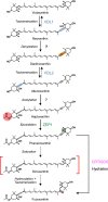
Figure 1.
Figure 1.
Previous and updated models of the fucoxanthin biosynthesis pathway in diatoms and haptophytes. The pathway from phaneroxanthin to fucoxanthin is updated, with the previously proposed scheme shown with dashed arrows. VDL, violaxanthin de-epoxidase-like; ZEP, zeaxanthin epoxidase. The enzymes that generate diadinoxanthin from neoxanthin or haptoxanthin from allenoxanthin have not been identified and are thus indicated with question marks. The functional groups newly generated by each enzymatic step are highlighted by differently colored backgrounds. Among the carotenoids shown, only violaxanthin and neoxanthin are produced in green plants. In addition, the diatom/haptophyte neoxanthin synthase, VDL1, is not present in green plants. Note that this study indicates that dinoxanthin is not an intermediate in the biosynthetic pathway of fucoxanthin in diatoms and haptophytes.
Figure 2.
Figure 2.
Molecular characterization and pigment phenotype of the CRTISO5-knockout mutant from the diatom P. tricornutum. A) Genotypes and appearances of WT, the crtiso5-1 mutant, and a complemented line (crtiso5-1-c1). A scheme showing the insertion site of the 1.4-kb zeocin resistance cassette (Ble) in the CRTISO5 gene is shown on the top. Binding sites for primers CRTISO5forward (Cf), CRTISO5reverse (Cr), and Bleforward (Bf) are labeled with triangles. Photographs in the middle show cuvettes with liquid cultures of the 3 strains. Electrophoretic analysis of PCR products using the indicated primer pair (Cf + Cr pair; Bf + Cr pair; 23s_fwd; and 23s_rev pair; Supplemental Table S1) are shown below. In the complemented line, amplification of the fragment of the reintroduced native gene was strongly favored over the longer fragment of the gene with Ble insertion. The plastome-encoded 23S rRNA gene was amplified as a control. B) HPLC traces of pigment extracts from WT, the knockout, and the complemented mutant. Pigment abbreviations are as follows: Car, carotene; Chl a, chlorophyll a; Chl c1/c2, chlorophyll c1/c2; Ddx, diadinoxanthin; Fx, fuxoxanthin; Phx, phaneroxanthin; unk., unknown pigment.
Figure 3.
Figure 3.
In vitro characterization of recombinant CRTISO5 from P. tricornutum. A) Reaction scheme of the last 2 steps in fucoxanthin biosynthesis catalyzed by ZEP1 and CRTISO5, showing only the part of the molecules that is modified by the reactions. B, C) In the in vitro assays, the substrate Phx was incubated in reaction buffer for 2 h at room temperature with different additives, followed by pigment extraction and HPLC analysis. In B), the resulting chromatograms showed no enzymatic activity when only FADred or only CRTISO5 were added. Addition of CRTISO5 and FAD or CRTISO5, FAD and NADPH also yielded no activity, while addition of CRTISO5 and FADred led to almost complete conversion of Phx to Fx. Lowering dissolved oxygen in the assay by addition of CGG even resulted in a complete conversion, indicating that the keto oxygen in Fx is not derived from molecular oxygen. In C), the addition of CRTISO5 and FADred to Hpx did not yield detectable pigment conversion, while recombinant ZEP1 from P. tricornutum with cofactors converted Hpx to Phx; addition of both ZEP1 and CRTISO5 together with cofactors resulted in the formation of Fx from Hpx via Phx. Each of the substrate–enzyme–cofactor mixture compositions in panels B) and C) were tested 3 times, and the effects of each enzyme or cofactor were consistent each time. Pigment or cofactor abbreviations are as follows: FAD, flavin adenine dinucleotide; Fx, fuxoxanthin; Hpx, haptoxanthin; NADPH, reduced nicotinamide adenine dinucleotide phosphate; Phx, phaneroxanthin.
Figure 4.
Figure 4.
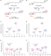
CRTISO5 from P. tricornutum synthesizes fucoxanthin by addition of water to phaneroxanthin. A, B) Proposed conversion of phaneroxanthin to fucoxanthin catalyzed by CRTISO5, and expected MS decay products of fucoxanthin when using either normal water (A) or heavy-oxygen water (B) as cosubstrate; C, D) APCI-MS scans (positive ion mode) of fucoxanthin generated by recombinant CRTISO5 in vitro using buffer containing normal water (C, 23 spectra averaged; calculated mass of 659.4306 for [M + H]+) or 90% heavy-oxygen water (D, 23 spectra; calculated mass of 661.4349 for [M + H]+); E, F) APCI-MS-MS scans (positive ion mode) of the peaks in C) at m/z of 659.4318 (E; 29 spectra) and in D) at 661.4352 (F, 22 spectra); for major fragment ion peaks, the eliminated molecules, m/z values and relative abundancies are indicated; peak labels for unknown fragments in panels C) and D) italicized. Masses of fucoxanthin and its fragment ions were consistently increased by 2 units when the reaction was performed with heavy-oxygen water. Difference between calculated and measured mass was below 2 ppm (below 0.0015 u) for all peaks analyzed in MS mode.
Figure 5.
Figure 5.
Identification of residues important for the enzymatic activity of CRTISO5. A) A snapshot of the docking of FADred and phaneroxanthin (Phx) into CRTISO5. Amino acids in proximity to the active site that were chosen for mutational analyses and also a nearby water molecule are labeled. Distances from Y306 to the triple bond (1), the water molecule to the triple bond (2), and H303 to the water molecule (3) are indicated. B) Relative CRTISO5 activities of the point mutants compared to the WT CRTISO5 protein (see Materials and Methods for details of quantification). Results from 3 replicate assays (performed on 3 independent mixtures of substrates, enzymes, and cofactors) were shown, with error bars indicating standard deviations, and each dot representing the result from an independent assay. C) Partially collapsed phylogenetic tree including CRTISO and CRTISO-like proteins from plants, algae, and cyanobacteria used for reconstruction of ancestral sequences at the nodes labeled N0 to N5 (see Supplemental Fig. S22 for detailed tree). Based on the ML tree shown in Supplemental Fig. S1, the proteins of the cyanobacterial CrtD family were used as outgroup; branch lengths are proportional to the number of substitutions per site (see scale bar); the cluster L5 contains the CRTISO5 sequences; Chrom., chromalveolate algae; Cyanos, cyanobacteria; Prasinos, prasinophyte algae. D) Ancestral sequence reconstruction for the substrate-interacting part in CRTISO5 at the nodes labeled in the phylogenetic tree in C); except for the sequence at N0, the majority of positions in the reconstructed sequences had a probability of >0.95 (see Supplemental Fig. S23) as being correctly identified. Amino acids differing from the sequence of the preceding node are printed in white on background colors corresponding to the node colors in C). Amino acid positions in the ruler below the sequences correspond to those in the CRTISO5 sequence from P. tricornutum, while asterisks denote the amino acids that were mutated for experimental validation of the active site in CRTISO5.
Figure 6.
Figure 6.
Photophysiological characterizations of the crtiso5-1 mutant. A) Relative protein accumulation of photosynthetic complexes calculated from quantitative immunoblots. The FCPs and subunits of photosystem I (PsaB), photosystem II (PsbD), and ATPase (ATPB) were quantified. B) Functional absorption cross section of PSII was obtained by Fluorescence Induction and Relaxation kinetics (FIRe fluorometer) for WT, the crtiso5-1 knockout mutant, and the complemented line (crtiso5-1-c1) in blue light. C) Fluorescence emission of whole cells at 687 nm with different excitation wavelengths, measured at 77 K. Spectra were normalized to the peak at 439 nm. D) The light-limited slope of the photosynthesis–irradiance curve (α). Photosynthesis was measured as oxygen evolution in red light. E) Fluorescence spectra of whole cells at 77 K during excitation at 438 nm. Spectra were normalized to the peak at 688 nm. All bar graphs show the average, and the error bars indicate standard deviations. Panel A) shows statistical results following a Student's t-test (*P < 0.05, **P < 0.015, ***P < 0.005, and ***P < 0.0001; ns, not significant, defined as P > 0.05). Panels B) and D) show statistical results from an ANOVA test followed by a Tukey post hoc. The replicates for WT or the crtiso5-1 mutant were independent cultures. The ANOVA and t-test results are provided in Supplemental Data Set 3.

Comment in

Similar articles

Cited by

References

    1. Ahn JW, Kim KJ. Crystal structure of 1′-OH-carotenoid 3,4-desaturase from Nonlabens dokdonensis DSW-6. Enzyme Microb Technol. 2015:77:29–37. 10.1016/j.enzmictec.2015.05.005 - DOI - PubMed
    1. Alipui OD, Zhang D, Schulz H. Direct hydration of 3-octynoyl-CoA by crotonase: a missing link in Konrad Bloch's enzymatic studies with 3-alkynoyl thioesters. Biochem Biophys Res Commun. 2002:292(5):1171–1174. 10.1006/bbrc.2001.2024 - DOI - PubMed
    1. Altschul SF, Madden TL, Schaffer AA, Zhang J, Zhang Z, Miller W, Lipman DJ. Gapped BLAST and PSI-BLAST: a new generation of protein database search programs. Nucleic Acids Res. 1997:25(17):3389–3402. 10.1093/nar/25.17.3389 - DOI - PMC - PubMed
    1. Armstrong GA, Alberti M, Leach F, Hearst JE. Nucleotide sequence, organization, and nature of the protein products of the carotenoid biosynthesis gene cluster of Rhodobacter capsulatus. Mol Gen Genet. 1989:216(2–3):254–268. 10.1007/BF00334364 - DOI - PubMed
    1. Bai Y, Cao T, Dautermann O, Buschbeck P, Cantrell MB, Chen Y, Lein CD, Shi X, Ware MA, Yang F, et al. . Green diatom mutants reveal an intricate biosynthetic pathway of fucoxanthin. Proc Natl Acad Sci U S A. 2022:119(38):e2203708119. 10.1073/pnas.2203708119 - DOI - PMC - PubMed

Publication types