. 2023 Apr 26;14(1):2400.
doi: 10.1038/s41467-023-38038-y.

Mini-PCDH15 gene therapy rescues hearing in a mouse model of Usher syndrome type 1F

Affiliations

Mini-PCDH15 gene therapy rescues hearing in a mouse model of Usher syndrome type 1F

Maryna V Ivanchenko et al. Nat Commun. .

Abstract

Usher syndrome type 1 F (USH1F), caused by mutations in the protocadherin-15 gene (PCDH15), is characterized by congenital deafness, lack of balance, and progressive blindness. In hair cells, the receptor cells of the inner ear, PCDH15 is a component of tip links, fine filaments which pull open mechanosensory transduction channels. A simple gene addition therapy for USH1F is challenging because the PCDH15 coding sequence is too large for adeno-associated virus (AAV) vectors. We use rational, structure-based design to engineer mini-PCDH15s in which 3-5 of the 11 extracellular cadherin repeats are deleted, but which still bind a partner protein. Some mini-PCDH15s can fit in an AAV. An AAV encoding one of these, injected into the inner ears of mouse models of USH1F, produces a mini-PCDH15 which properly forms tip links, prevents the degeneration of hair cell bundles, and rescues hearing. Mini-PCDH15s may be a useful therapy for the deafness of USH1F.


Conflict of interest statement

D.P.C. is a cofounder of Skylark Bio. M.V.I. is a consultant for Skylark Bio. President and Fellows of Harvard College, Ohio State Innovation Foundation, Massachusetts Eye and Ear Infirmary have filed patent application “AAV vectors encoding mini-PCDH15 and uses thereof”, inventors D.P.C., A.A.I., M.S., M.V.I. and C.W.P.; US (PCT/US2020/029968). The present disclosure provides isolated nucleic acids, vectors, and rAAV.9.PHP.B comprising a transgene encoding a mini-PCDH15, and methods of treating hearing loss using the same. The other authors declare no competing interests.

Figures
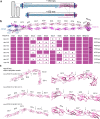
Fig. 1
Fig. 1. Knowledge of atomic structures allows rational design of mini-PCDH15 constructs.
a Arrangement of the tip-link proteins PCDH15 and CDH23 at stereocilia tips. Each protein has multiple extracellular cadherin (EC) repeats forming a chain ~170 nm long. Mini-PCDH15s that lack 3–5 EC repeats are about 25–40% shorter, while the resulting tip links are 10–15% shorter. b upper panel, Composite atomic structure of wild-type PCDH15 (magenta) bound to EC1 + EC2 of CDH-23 (blue; adapted from Choudhary et al., 2020). lower panel, The EC repeats deleted from each mini-PCDH15 version. c AlphaFold2 monomeric structure models for the three shortest mini-PCDH15s (V4, V7, and V8), calculated without utilizing a structural template. The predictions with or without a structural template are nearly identical. Right panels are higher magnification images of the junctions between EC repeats (arrows).
Fig. 2
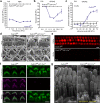
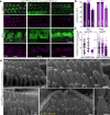
Fig. 2. Gfi1-Cre conditional Pcdh15 knockout mouse cochleas show severe functional and morphological changes.
a Average auditory brainstem response (ABR) thresholds as a function of frequency in P35 mice. Pcdh15fl/fl,Gfi1-Cre+/− (n = 6) mice showed a profound hearing loss at P35 relative to Pcdh15fl/fl control mice (n = 9). b Knockout mice had no distortion-product otoacoustic emission (DPOAE) responses (n = 6) compared to Pcdh15fl/fl hearing control mice (n = 9). c DPOAE emission amplitude at 16 kHz in P35 Pcdh15fl/fl,Gfi1-Cre+/− (n = 5) was significantly reduced compared to the control mice (n = 4). d Representative scanning electron micrographs taken from P6 mice show disorganized inner hair cells (IHCs) (upper row) and outer hair cells (OHCs) (lower row) stereocilia morphology in Pcdh15fl/fl,Gfi1-Cre+/− hair cells (right) as compared to Pcdh15fl/fl controls (left). e Representative confocal microscopy images of FM1-43 dye loading by IHCs and OHCs from the apical region of the cochlea at P10. FM1-43 loading was completely abolished, indicating no functional mechanotransduction. f Anti-PCDH15 labeling (magenta) and phalloidin co-staining (green) in Pcdh15fl/fl control mice (left) demonstrated normal PCDH15 trafficking to stereocilia tips at P6, which was absent in Pcdh15fl/fl, Gfi1-Cre+/− knockouts (right). g Immunogold scanning electron microscopy labeling of a P6 IHC immunostained with anti-PCDH15 primary antibody and 12 nm gold-conjugated secondary antibody. PCDH15 trafficked to the tips of stereocilia in Pcdh15fl/fl control mice and was abolished in Pcdh15fl/fl,Gfi1-Cre+/− mice. Scale bars: (d) 1 µm; (e, f) 5 µm; (g) 0.5 µm. All data are presented as mean values ± SEM. Source data are provided as a Source Data file.
Fig. 3
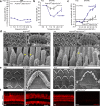
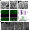
Fig. 3. Myo15-Cre conditional knockout mice show delayed functional and morphological cochlear pathology.
a P35, Pcdh15fl/fl,Myo15-Cre+/− knockout mice (n = 10) show lack of auditory brainstem response (ABR) thresholds at all frequencies and sound levels tested, compared to Pcdh15fl/fl hearing control mice (n = 9). b At P35, knockout mice (n = 6) have residual distortion-product otoacoustic emission (DPOAE) responses to high sound level stimuli in the mid-range frequencies, compared to Pcdh15fl/fl control mice (n = 9). c Despite residual responses at high sound levels, DPOAE emission amplitude at 11.3 kHz in P35 Pcdh15fl/fl,Myo15-Cre+/− mice (n = 8) was significantly reduced compared to Pcdh15fl/fl control mice (n = 9). d Scanning electron micrographs of P6 stereocilia in Pcdh15fl/fl hearing control mice (left) and Myo15-Cre conditional knockout mice (right) demonstrate healthy bundle morphology in neonatal knockout mice. High magnification images (lower panel) show extensive links between adjacent stereocilia (yellow arrows), including intact tip links in control mice as well as in knockout mice; however most middle row stereocilia in knockout mice had lost their normal shape, forming elongated tips. IHCs inner hair cells, OHCs outer hair cells. e Scanning electron micrographs of mature P35 stereocilia in Pcdh15fl/fl control mice (left) and Pcdh15fl/fl,Myo15-Cre+/− knockout mice (right) show that hair cells survive and the architecture of the organ of Corti is preserved at this age. However high magnification micrographs demonstrate shortened and missing middle- and short-row stereocilia in knockout mice. f In P6 mice, uptake of FM1-43 dye reveals a similar proportion of open transduction channels in Pcdh15fl/fl mice and Pcdh15fl/fl,Myo15-Cre+/− mice. At P35, however, uptake was abolished in Pcdh15fl/fl,Myo15-Cre+/− mice, indicating no open transduction channels. Scale bars: (d) (upper panel) 1 µm; (d) (lower panel) 500 nm; (e) 1 µm; (f) 20 µm. All data are presented as mean values ± SEM. Source data are provided as a Source Data file.
Fig. 4
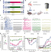
Fig. 4. AAV-mini-PCDH15s rescue hearing in the Myo15-Cre mouse model of USH1F.
a Schematic of the three mini-PCDH15 constructs that are short enough to be packaged into AAV capsids. The lengths of the DNA sequences are shown. Vectors used a CMV promoter and BGH polyadenylation sequence but not a WPRE. b AAV9-PHP.B was used to deliver the different mini PCDH15 versions at P1 via round-window membrane injection. IF immunofluorescence, EM electron microscopy. c Auditory brainstem response (ABR) testing showed differential rescue with different AAV-mini-PCDH15 versions. Representative click ABR traces from P35 Pcdh15fl/fl hearing control mice, from uninjected Pcdh15fl/fl,Myo15-Cre+/− deaf knockout mice, and from knockout mice injected with AAV-mini-PCDH15-V4, -V7, or -V8. SP, Summating potential. d Average click ABR thresholds for P35 uninjected Pcdh15fl/fl control mice (n = 4), uninjected Pcdh15fl/fl,Myo15-Cre+/− conditional knockout mice (n = 6), and conditional knockout mice injected with AAV-mini-PCDH15-V4 (n = 7), AAV- HA-mini-PCDH15-V4 (n = 14), AAV-mini-PCDH15-V7 (n = 12), or AAV-mini-PCDH15-V8 (n = 5). Significance was determined by a two-tailed unpaired t-test. P values: uninjected Pcdh15fl/fl,Myo15-Cre+/− vs. AAV-mini-PCDH15-V7, p = 0.0008 (***); Pcdh15fl/fl,Myo15-Cre+/− vs. AAV-mini-PCDH15-V4, p < 0.0001 (****); Pcdh15fl/fl vs. AAV-mini-PCDH15-V4, p = 0.08 (ns); AAV-HA-mini-PCDH15-V4 vs. AAV-mini-PCDH15-V4, p = 0.84 (ns). e Average tone ABR thresholds as a function of frequency for P35 uninjected Pcdh15fl/fl control mice (n = 9), uninjected Pcdh15fl/fl,Myo15-Cre+/− conditional knockout mice (n = 10), and conditional knockout mice injected with AAV-mini-PCDH15-V4 (n = 7), HA-tagged mini-PCDH15-V4 (n = 14), mini-PCDH15-V7 (n = 12), or mini-PCDH15-V8 (n = 5). f, g Average distortion-product otoacoustic emission (DPOAE) thresholds and average DPOAE emission amplitudes at 11.3 kHz, for P35 hearing control mice (n = 9) and uninjected Pcdh15fl/fl,Myo15-Cre+/− mice (n = 6), and for those injected with AAV-mini-PCDH15-V4 (n = 7), -V7 (n = 12), or -V8 (n = 5). All data are presented as mean values ± SEM. Source data are provided as a Source Data file.
Fig. 5
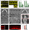
Fig. 5. Delivery of AAV-mini-PCDH15-V4 rescues stereocilia bundle morphology and mechanotransduction.
a Representative confocal microscopy images taken from the middle turn of the cochlea of P35 outer hair cells (OHCs) stained with phalloidin. Panels show Pcdh15fl/fl hearing control mice, uninjected Myo15-Cre conditional knockout mice or knockout mice injected with AAV-mini-PCDH15-V4. Phalloidin staining (yellow) shows rescued hair bundles in injected cochleas. Asterisk indicates a typical partially rescued hair bundle with >55% intact stereocilia per bundle. b Quantification of rescue rate of inner hair cells (IHCs) and OHCs in mice injected with AAV-mini-PCDH15-V4 in the apical, middle, and basal regions of the cochlea (n = 12 cochleas). Data are presented as mean values ± SEM. c Scanning electron micrographs of single hair bundles. Panels show an OHC of a hearing control mouse and OHCs and an IHC of an untreated conditional knockout. Knockout bundles are severely disrupted. d OHCs of a conditional knockout treated with AAV-mini-PCDH15-V4 showing normal bundle morphology. e IHCs of a conditional knockout treated with AAV-mini-PCDH15-V4 showing normal bundle morphology. f High magnification images of boxed areas in (e). Tip links were observed (yellow arrows) in rescued bundles. g Representative confocal microscopy images, from the apical/mid-apical region of the cochlea, of FM1-43 dye loaded IHCs and OHCs at P35. Pcdh15fl/fl mice (left), uninjected knockout mice (middle), mice injected with AAV-mini-PCDH15-V4 (right). h Average percentage of IHCs and OHCs loaded with FM1-43 in conditional knockout mice injected with AAV-mini-PCDH15-V4 (n = 6), and in Pcdh15fl/fl control mice (n = 5). Data are presented as mean values ± SEM. Scale bars: (a) 5 µm; (c, d, e) 1 µm; (f) 100 nm; (g) 15 µm. Source data are provided as a Source Data file.
Fig. 6
Fig. 6. Anti-HA staining of HA-tagged mini-PCDH15-V4 demonstrates localization at the lower end of the tip link.
a Representative confocal microscopy images at P35 from apical, middle and basal turns of the cochlea show anti-HA staining of HA-mini-PCDH15-V4 (magenta) at the tips of stereocilia (co-stained for phalloidin, green) in knockout cochleas treated at P1 with AAV-HA-mini-PCDH15-V4. IHCs inner hair cells, OHCs outer hair cells. b Transduction efficiency in IHCs and OHCs at P35 in treated Pcdh15fl/fl,Myo15-Cre+/− conditional knockout mice (n = 12). Data are presented as mean values ± SEM. c Quantification of fluorescence intensity of anti-HA labeling in IHCs and OHCs from apical, middle and basal turns of the cochleas in mice treated with AAV-HA-mini-PCDH15-V4. Symbols represent integrated fluorescence intensity in arbitrary units (arb. units) in individual cells (apex: n = 56 IHCs, n = 144 OHCs; middle: n = 49 IHCs, n = 198 OHCs; base: n = 42 IHCs, n = 138 OHCs). Cells were measured from four separate cochleas. Data are presented as mean values ± SD. Significance was determined by a two-tailed unpaired t-test. P values: apex IHCs vs. middle IHCs, p = 0.45 (ns); apex IHCs vs. base IHCs, p < 0.0001 (****); middle IHCs vs. base IHCs, p < 0.0001 (****); apex OHCs vs. middle OHCs, p = 0.40 (ns); apex OHCs vs. base OHCs, p < 0.0001 (****); apex OHCs vs. base OHCs, p < 0.0001 (****). d Immunogold scanning electron microscopy demonstrated localization of HA-tagged mini-PCDH15-V4 at the tips of stereocilia (yellow arrow), similar to the localization of wild-type PCDH15 detected with anti-PCDH15, confirming that mini-PCDH15-V4 targets properly. Scale bars: (a) 5 μm, (d) 100 nm. Source data are provided as a Source Data file.
Fig. 7
Fig. 7. Delivery of AAV-HA-mini-PCDH15-V4 rescues stereocilia tip links in the R245X knockout mouse model.
a Scanning electron micrographs of the organ of Corti from Pcdh15R245X/+ hearing control mice at P6 (upper panel). Hair bundles have normal morphology and tip links connect the tips of adjacent stereocilia (yellow arrows). Organ of Corti from Pcdh15R245X/R245X homozygous null mice at P6 (lower panel). Bundles are severely disrupted, stereocilia are shortened, and no tip links are detected (yellow asterisk) although the lateral links are present. b Hair cells in Pcdh15R245X/R245X homozygous knockout mice expressing HA-mini-PCDH15-V4. Bundle morphology was partially or fully restored. High magnification images demonstrate the presence of tip links (yellow arrows) in rescued bundles at P6. c Representative confocal microscopy images of anti-PCDH15 labeling (magenta) of organ of Corti along with actin staining (phalloidin, green) demonstrated normal bundle morphology and PCDH15 localization to the stereocilia tips in Pcdh15R245X/+ control mice (left). In Pcdh15R245X/R245X knockout mice the hair bundles were severely disorganized and no anti-PCDH15 labeling was detected (right). d Representative confocal microscopy images of the cochlea showing anti-HA staining (magenta). No staining was observed at the tips of stereocilia in the untreated Pcdh15R245X/R245X mice (left). Anti-HA staining was observed at stereocilia tips in knockout cochleas treated with AAV-HA-mini-PCDH15-V4 (right). e Transduction efficiency in inner hair cells (IHCs) and outer hair cells (OHCs), measured with anti-HA labeling at P6, in Pcdh15R245X/R245X mice treated at P1 (n = 13). Almost all hair cells in all cochlear regions were transduced. Data are presented as mean values ± SEM. f Morphology rescue rate of IHCs and OHCs, assessed by fluorescent actin label, in mice injected with AAV-HA-mini-PCDH15-V4 in the apical, middle, and basal regions of the cochlea (n = 13). Most bundles were partially or fully rescued. Data are presented as mean values ± SEM. g Immunogold scanning electron microscopy of P9 IHCs expressing HA-mini-PCDH15-V4. Gold beads demonstrate localization of HA-tagged mini-PCDH15-V4 at the tips of stereocilia (yellow arrows) and along the surface of stereocilia, confirming that mini-PCDH15-V4 targets properly in Pcdh15R245X/R245X mice. Scale bars: (a, b) 500 nm, (c, d) 10 µm, (f) 200 nm. Source data are provided as a Source Data file.
Fig. 8
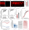
Fig. 8. Delivery of AAV-mini-PCDH15-V4 rescues mechanotransduction and the ABR in Pcdh15R245X/R245X mice.
a Representative confocal microscopy images of FM1-43 dye loading in inner hair cells (IHCs) and outer hair cells (OHCs) from the middle region of the cochlea at P6. Pcdh15R245X/+ mice (left), uninjected Pcdh15R245X/R245X (right). b FM1-43 dye loading in Pcdh15R245X/R245X knockout mice injected with AAV-mini-PCDH15-V4. Loading was restored in most hair cells. c Average percentage of IHCs and OHCs loaded with FM1-43; knockout mice injected with AAV-mini-PCDH15-V4 (red, n = 8), uninjected Pcdh15R245X/+ normal control mice (gray, n = 6). About 80% of knockout cells loaded after treatment. d Representative transduction currents measured at P10-P17 (top) in response to −175 to 1135-nm bundle deflections (bottom). A Pcdh15R245X/+ hearing control IHC (left); a Pcdh15R245X/R245X knockout IHC expressing mini-PCDH15-V4 (right). e Peak transduction currents at P10-P17 from Pcdh15R245X/+ controls (n = 6 cells) and Pcdh15R245X/R245X knockouts treated with AAV-mini-PCDH15-V4 (n = 9 cells). Cells from five separate Pcdh15R245X/+ cochleas and six separate Pcdh15R245X/R245X treated knockout cochleas were studied. f Normalized open probability as a function of stimulus probe displacement for Pcdh15R245X/+ controls (black, n = 6 cells) and treated Pcdh15R245X/R245X knockouts (red, n = 9 cells). g Average auditory brainstem response (ABR) thresholds as a function of frequency in P35 Pcdh15R245X/+ controls (blue, n = 10), uninjected knockout (black, n = 4) and knockout mice injected with AAV-mini-PCDH15-V4 mice (red, n = 5). h Representative ABR traces at P35 from controls, uninjected knockout mice, and treated knockout mice. SP summating potential. Scale bars: (a, b) 10 µm. All data are presented as mean values ± SEM. Source data are provided as a Source Data file.

References

    1. Maguire CA, Corey DP. Viral vectors for gene delivery to the inner ear. Hear Res. 2020;394:107927. doi: 10.1016/j.heares.2020.107927. - DOI - PubMed
    1. Peters CW, Maguire CA, Hanlon KS. Delivering AAV to the central nervous and sensory systems. Trends Pharmacol. Sci. 2021;42:461–474. doi: 10.1016/j.tips.2021.03.004. - DOI - PMC - PubMed
    1. Delmaghani, S. & El-Amraoui, A. Inner ear gene therapies take off: current promises and future challenges. J. Clin. Med.9, 2309 (2020). - PMC - PubMed
    1. Bulcha JT, Wang Y, Ma H, Tai PWL, Gao G. Viral vector platforms within the gene therapy landscape. Signal Transduct. Target Ther. 2021;6:53. doi: 10.1038/s41392-021-00487-6. - DOI - PMC - PubMed
    1. Gyorgy B, et al. Gene transfer with AAV9-PHP.B rescues hearing in a mouse model of Usher syndrome 3A and transduces hair cells in a non-human primate. Mol. Ther. Methods Clin. Dev. 2019;13:1–13. doi: 10.1016/j.omtm.2018.11.003. - DOI - PMC - PubMed

Publication types

Supplementary concepts