. 2023 Apr 26;13(1):6829.
doi: 10.1038/s41598-023-32631-3.

Never in mitosis gene A-related kinase-8 promotes proliferation, migration, invasion, and stemness of breast cancer cells via β-catenin signalling activation

Affiliations

Never in mitosis gene A-related kinase-8 promotes proliferation, migration, invasion, and stemness of breast cancer cells via β-catenin signalling activation

Eunji Kang et al. Sci Rep. .

Abstract

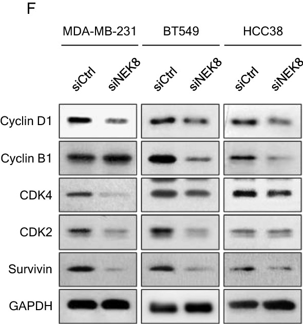
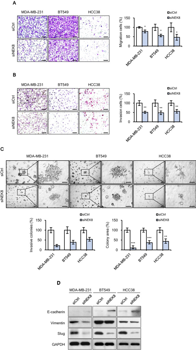
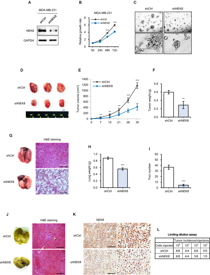
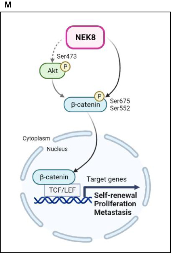
Never in mitosis gene A (NIMA)-related kinase-8 (NEK8) is involved in cell cycle progression, cytoskeleton development, and DNA damage repair. However, its role in breast cancer has not yet been explored. To investigate this, NEK8 was knocked down in MDA-MB-231, BT549, and HCC38 breast cancer cell lines. We observed a decrease in cell proliferation and colony formation owing to regulation of the G1/S and G2/M transitions. Furthermore, the expression of several cell cycle regulatory proteins was altered, including that of cyclin D1, cyclin B1, CDK4, CDK2, and surviving. NEK8 knockdown impaired cell migration and invasion as well as reduced the expression of epithelial-mesenchymal transition markers. Regarding stem-cell characteristics, NEK8 knockdown decreased the tumour sphere formation, aldehyde dehydrogenase activity, and stem-cell marker expression, including that of CD44, Sox2, Oct4a, and Nanog. Further analysis revealed that NEK8 interacts with β-catenin. Also, NEK8 knockdown promoted β-catenin degradation. NEK8-silenced MDA-MB-231 cells inhibited xenograft tumour growth, metastasis, and tumour initiation in vivo. Using the Oncomine and TNMplot public databases, we found a significant correlation between NEK8 overexpression and poor clinical outcomes in breast cancer patients. Thus, NEK8 may be a crucial regulator of breast cancer progression and a potential therapeutic target.


Conflict of interest statement

The authors declare no competing interests.

Figures

Figure 1
Figure 1
Increased never in mitosis gene A-related kinase-8 (NEK8) expression is associated with poor survival in patients with breast cancer. (A) NEK8 mRNA expression in normal breast tissues and invasive breast carcinomas from the Oncomine database. (B) NEK8 mRNA expression in normal breast tissues and invasive ductal breast carcinoma from the Oncomine database. (C) NEK8 mRNA expression in normal breast tissues and mucinous breast carcinoma from the Oncomine database. Error bars, standard deviation; ***P < 0.001, **P < 0.01, *P < 0.05. (D) NEK8 mRNA expression determined using RNA-sequencing in normal and tumour tissues from TNMplot.com. (E) NEK8 expression in breast invasive carcinoma and normal tissues from the Gene Expression Profiling Interactive Analysis (GEPIA) database. Kaplan–Meier curves indicating (F) overall survival (OS) and (G) distant metastasis-free survival (DMFS) of patients with breast cancer were produced using the Kaplan–Meier plotter. (H) NEK8 mRNA level in breast cancer and normal cell lines. GAPDH was used as a loading control. (I) NEK8 protein expression in breast cancer and normal cell lines. HSC70 was used as a loading control. The blots were cut prior to incubation with antibodies.
Figure 2
Figure 2
Knockdown of never in mitosis gene A-related kinase-8 (NEK8) attenuates proliferation and induces cell cycle arrest in breast cancer cells. MDA-MB-231, BT549, and HCC38 cells were transfected with control (siCtrl) or NEK8-silencing (siNEK8) small interfering RNA. (A) On day 3 after transfection, western blotting was used to confirm the knockdown of NEK8 in the siNEK8 group compared with that in the siCtrl group. (B) NEK8 knockdown inhibited the proliferation of breast cancer cells after 24, 48, and 72 h. The graph is representative of at least three independent experiments (n ≥ 3), where each experiment was performed in triplicate. Error bars, standard deviation (SD); ***P < 0.001, **P < 0.01, *P < 0.05. (C) NEK8 knockdown decreased colony formation. Representative images at least three independent experiments that showed similar results of colony growth (n ≥ 3). (D) In the 3D Matrigel matrix, cells with NEK8 knockdown showed reduced proliferation after 5 days. Error bars, SD; ***P < 0.001, **P < 0.01, *P < 0.05. (E) Distribution of cell cycle phases measured using flow cytometry. (F) Expression of cell cycle-related genes was confirmed using western blotting 72 h after NEK8 knockdown. GAPDH was used as a loading control. The figure is representative of at least three independent experiments that showed similar results (n ≥ 3). The blots were cut prior to incubation with antibodies.
Figure 2
Figure 2
Knockdown of never in mitosis gene A-related kinase-8 (NEK8) attenuates proliferation and induces cell cycle arrest in breast cancer cells. MDA-MB-231, BT549, and HCC38 cells were transfected with control (siCtrl) or NEK8-silencing (siNEK8) small interfering RNA. (A) On day 3 after transfection, western blotting was used to confirm the knockdown of NEK8 in the siNEK8 group compared with that in the siCtrl group. (B) NEK8 knockdown inhibited the proliferation of breast cancer cells after 24, 48, and 72 h. The graph is representative of at least three independent experiments (n ≥ 3), where each experiment was performed in triplicate. Error bars, standard deviation (SD); ***P < 0.001, **P < 0.01, *P < 0.05. (C) NEK8 knockdown decreased colony formation. Representative images at least three independent experiments that showed similar results of colony growth (n ≥ 3). (D) In the 3D Matrigel matrix, cells with NEK8 knockdown showed reduced proliferation after 5 days. Error bars, SD; ***P < 0.001, **P < 0.01, *P < 0.05. (E) Distribution of cell cycle phases measured using flow cytometry. (F) Expression of cell cycle-related genes was confirmed using western blotting 72 h after NEK8 knockdown. GAPDH was used as a loading control. The figure is representative of at least three independent experiments that showed similar results (n ≥ 3). The blots were cut prior to incubation with antibodies.
Figure 3
Figure 3
Knockdown of never in mitosis gene A-related kinase-8 (NEK8) inhibits the migration and invasion of breast cancer cells. (A) Migration and (B) invasion of MDA-MB-231, BT549, and HCC38 cells determined using Transwell assays (×200 magnification; scale bar, 50 μm). (C) Cells were grown on 3D Matrigel for 5 days; then, branching structures were counted using phase-contrast microscopy (×40 magnification; scale bar, 100 μm). The graph is representative of at least three independent experiments (n ≥ 3), where each experiment was performed in triplicate. Error bars, standard deviation; ***P < 0.001, **P < 0.01, *P < 0.05. (D) Expression of the epithelial-mesenchymal transition‑related proteins E‑cadherin, vimentin and Slug using western blotting. GAPDH was used as a loading control. The figure is representative of at least two independent experiments (n ≥ 2). The blots were cut prior to incubation with antibodies.
Figure 4
Figure 4
Knockdown of never in mitosis gene A-related kinase-8 (NEK8) inhibits tumour sphere formation, aldehyde dehydrogenase 1 (ALDH1) positive sub-populations, and drug resistance. (A) Representative images of spheres induced by culturing MDA-MB-231, BT549, and HCC38 cells treated with siCtrl (control) or NEK8-silencing (siNEK8) in mammosphere medium. NEK8 knockdown inhibited sphere formation in MDA-MB-231, BT549, and HCC38 cells (×40 magnification; scale bar, 100 μm). n = 3; Error bars, standard deviation (SD); ***P < 0.001, **P < 0.01, *P < 0.05. (B) Stemness in MDA-MB-231, BT549, and HCC38 cells was assessed with a ALDEFLUOR assay. Cell populations that disappeared in the presence of the specific ALDH inhibitor diethylaminobenzaldehyde (DEAB) were gated as ALDHhigh cells. (C) Expression of cancer stem-cell-related proteins CD44, SOX2, Oct4a, and Nanog upon the knockdown of NEK8 in breast cancer cells. GAPDH was used as a loading control. The figure is representative of at least two independent experiments (n ≥ 2). The blots were cut prior to incubation with antibodies. (D) NEK8 knockdown decreased resistance to cisplatin treatment. The graph is representative of at least two independent experiments (n ≥ 2), where each experiment was performed in triplicate. Error bars indicate SD; ***P < 0.001, **P < 0.01, **P < 0.05.
Figure 5
Figure 5
Knockdown of never in mitosis gene A-related kinase-8 (NEK8) inhibits β-catenin signalling in breast cancer cells. (A) The expression of total β-catenin, phospho-β-cateninS675, phospho-β-cateninS552, and phospho-AKTS473 in breast cancer cells transfected with NEK8-silencing (siNEK8) was detected using western blotting. (B) Representative western blot images demonstrating subcellular localisation of β-catenin in the nucleus and cytosol. (C) Confocal micrographs showing decreased nuclear β-catenin levels in MDA-MD-231, BT549, and HCC38 cells treated with siCtrl or siNEK8 for 72 h. GAPDH and β-actin were used as loading controls for whole cell lysates, and Lamin B was used as a loading control for nuclear fractions. The figure is representative of at least three independent experiments that showed similar results (n ≥ 3). (D) Stabilisation of β-catenin in NEK8 knockdown cells. The cells were treated with cycloheximide (CHX; 20 μg/mL) and collected at the indicated times for western blotting. (E) The cells were untreated or treated with MG132 (5 μM) for 6 h, and then subjected to western blotting. (F) Co-immunoprecipitation analysis of the interaction between NEK8 and β-catenin in MDA-MB-231 cells. The blots were cut prior to incubation with antibodies.
Figure 6
Figure 6
Never in mitosis gene A-related kinase-8 (NEK8) knockdown significantly inhibits tumour initiation and metastasis in MDA-MB-231 xenografts. (A) NEK8 expression in stable transfectants confirmed using western blotting. (B) Short hairpin RNA (shRNA) silencing of NEK8 attenuated MDA-MB-231 proliferation. (C) Phase-contrast images of 3D Matrigel growth of MDA-MB-231 cells with or without NEK8 knockdown (×40 magnification; scale bar, 100 μm). The results are representative of three independent experiments (n = 3). (DF) Representative images and tumour sizes at 35 days after implantation showing that NEK8 knockdown decreased tumour volume and weight (n = 3). Images are representative of at least two experiments (n ≥ 2). (G) Representative haematoxylin and eosin (H&E) images of lung metastases. Scale bar = 50 μm. (H) Lung weights in the lung metastasis model. Values are presented as mean ± standard deviation (SD) (n = 3). ***P < 0.001. (I) Numbers of metastatic foci in the lungs. Values are presented as mean ± SD (n = 3). ***P < 0.001. (J) Representative H&E images of liver metastases. Scale bar, 50 μm. (K) Immunohistochemistry analysis to confirm the expression of NEK8 in tumours from either shCtrl or shNEK8 group. Scale bar, 10 μm. (L) Numbers of tumours generated and the results of the limiting dilution assay (106, 104, 103, and 102 cells). Numbers of tumours generated are shown in the table. (M) Schematic representation of the mechanism underlying the effect of NEK8 silencing on breast cancer cell proliferation, migration, invasion, and stemness mediated through the β-catenin signalling pathway. Created using BioRender (BioRender.com). NEK8 causes increased translocation of β-catenin to the nucleus through the phosphorylation of β-catenin. NEK8 also regulates the phosphorylation of AKT, which may affect the phosphorylation of β-catenin, as well as its activation and nuclear translocation, contributing to the enhancement of β-catenin transcriptional activity, which then results in increased self-renewal, proliferation, and metastasis of breast cancer cells.
Figure 6
Figure 6
Never in mitosis gene A-related kinase-8 (NEK8) knockdown significantly inhibits tumour initiation and metastasis in MDA-MB-231 xenografts. (A) NEK8 expression in stable transfectants confirmed using western blotting. (B) Short hairpin RNA (shRNA) silencing of NEK8 attenuated MDA-MB-231 proliferation. (C) Phase-contrast images of 3D Matrigel growth of MDA-MB-231 cells with or without NEK8 knockdown (×40 magnification; scale bar, 100 μm). The results are representative of three independent experiments (n = 3). (DF) Representative images and tumour sizes at 35 days after implantation showing that NEK8 knockdown decreased tumour volume and weight (n = 3). Images are representative of at least two experiments (n ≥ 2). (G) Representative haematoxylin and eosin (H&E) images of lung metastases. Scale bar = 50 μm. (H) Lung weights in the lung metastasis model. Values are presented as mean ± standard deviation (SD) (n = 3). ***P < 0.001. (I) Numbers of metastatic foci in the lungs. Values are presented as mean ± SD (n = 3). ***P < 0.001. (J) Representative H&E images of liver metastases. Scale bar, 50 μm. (K) Immunohistochemistry analysis to confirm the expression of NEK8 in tumours from either shCtrl or shNEK8 group. Scale bar, 10 μm. (L) Numbers of tumours generated and the results of the limiting dilution assay (106, 104, 103, and 102 cells). Numbers of tumours generated are shown in the table. (M) Schematic representation of the mechanism underlying the effect of NEK8 silencing on breast cancer cell proliferation, migration, invasion, and stemness mediated through the β-catenin signalling pathway. Created using BioRender (BioRender.com). NEK8 causes increased translocation of β-catenin to the nucleus through the phosphorylation of β-catenin. NEK8 also regulates the phosphorylation of AKT, which may affect the phosphorylation of β-catenin, as well as its activation and nuclear translocation, contributing to the enhancement of β-catenin transcriptional activity, which then results in increased self-renewal, proliferation, and metastasis of breast cancer cells.

Similar articles

Cited by

References

    1. Siegel RL, Miller KD, Fuchs HE, Jemal A. Cancer statistics, 2021. CA Cancer J. Clin. 2021;71:7–33. doi: 10.3322/caac.21654. - DOI - PubMed
    1. Riggio AI, Varley KE, Welm AL. The lingering mysteries of metastatic recurrence in breast cancer. Br. J. Cancer. 2021;124:13–26. doi: 10.1038/s41416-020-01161-4. - DOI - PMC - PubMed
    1. Gui P, Bivona TG. Evolution of metastasis: New tools and insights. Trends Cancer. 2022;8:98–109. doi: 10.1016/j.trecan.2021.11.002. - DOI - PubMed
    1. Fares J, Fares MY, Khachfe HH, Salhab HA, Fares Y. Molecular principles of metastasis: A hallmark of cancer revisited. Signal Transduct. Target. Ther. 2020;5:28. doi: 10.1038/s41392-020-0134-x. - DOI - PMC - PubMed
    1. Wang L, Zhang S, Wang X. The metabolic mechanisms of breast cancer metastasis. Front. Oncol. 2020;10:602416. doi: 10.3389/fonc.2020.602416. - DOI - PMC - PubMed

Publication types

MeSH terms