. 2023 Apr 26;14(1):2404.
doi: 10.1038/s41467-023-38069-5.

Revealing concealed cardioprotection by platelet Mfsd2b-released S1P in human and murine myocardial infarction

Affiliations

Revealing concealed cardioprotection by platelet Mfsd2b-released S1P in human and murine myocardial infarction

Amin Polzin et al. Nat Commun. .

Abstract

Antiplatelet medication is standard of care in acute myocardial infarction (AMI). However, it may have obscured beneficial properties of the activated platelet secretome. We identify platelets as major source of a sphingosine-1-phosphate (S1P) burst during AMI, and find its magnitude to favorably associate with cardiovascular mortality and infarct size in STEMI patients over 12 months. Experimentally, administration of supernatant from activated platelets reduces infarct size in murine AMI, which is blunted in platelets deficient for S1P export (Mfsd2b) or production (Sphk1) and in mice deficient for cardiomyocyte S1P receptor 1 (S1P1). Our study reveals an exploitable therapeutic window in antiplatelet therapy in AMI as the GPIIb/IIIa antagonist tirofiban preserves S1P release and cardioprotection, whereas the P2Y12 antagonist cangrelor does not. Here, we report that platelet-mediated intrinsic cardioprotection is an exciting therapeutic paradigm reaching beyond AMI, the benefits of which may need to be considered in all antiplatelet therapies.


Conflict of interest statement

The authors declare no competing interests.

Figures
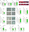
Fig. 1
Fig. 1. Supernatant of activated platelets reduces infarct size and improves cardiac outcome after myocardial infarction—this depends on platelet sphingosine kinase 1.
a Injection of cell-free supernatant of activated platelets (SNT+) prior AMI leads to decreased infarct size (INF/AAR) after 24 h of reperfusion as compared to supernatant of non-activated platelets (SNT−) and NaCl (ANOVA-analysis, nNaCl = 10, nSNT− = 13, nSNT+ = 13). b Area at risk per left ventricle (AAR/LV) did not differ between the groups. c Echocardiographic assessment 24 h post AMI showed improved cardiac function (EF = ejection fraction) in SNT+ treated mice (ANOVA-analysis, nNaCl = 10, nSNT− = 13, nSNT+ = 13). d Exemplary images of TTC-stained hearts. e This reduction of infarct size by SNT+ was associated with reduced neutrophil accumulation 24 h after AMI (unpaired t test, nSNT− = 7, nSNT+ = 8, scale bar left: 100 µm, scale bar right: 30 µm). f Apoptosis was reduced five days after AMI (unpaired t test, nSNT− = 7, nSNT+ = 7, scale bar left: 100 µm, scale bar right: 30 µm), and gj cardiac function was improved 21 days after AMI (SV = stroke volume, CO = cardiac output, HR = heart rate, unpaired t test, nSNT− = 6, nSNT+ = 7). k S1P concentrations in SNT+ was 25% higher as compared to SNT−. S1P concentrations in SNT− of mice lacking SphK1 were 70% lower and did not increase in SNT+ (two-way ANOVA, C57BL6/J nSNT− = 16, nSNT+ = 8, SphK1−/− nSNT− = 9, nSNT+ = 9). l Accordingly, SNT+ of SphK1-deficient platelets fails to reduce infarct size (two-way ANOVA, C57BL6/J nSNT− = 9, nSNT+ = 7, SphK1−/− nSNT− = 7, nSNT+ = 8). m No difference in the area at risk was observed. Error bars in each panel represent standard deviation.
Fig. 2
Fig. 2. Platelet-derived S1P exerts cardioprotection through the cardiomyocyte S1P receptor 1 (S1P1) and deficiency of platelet S1P transporter major facilitator superfamily domain containing 2B (Mfsd2b) leads to increased infarct size in vivo.
a SNT+ leads to decreased infarct size (IS [%LV]), b higher left ventricular pressure gradient (DP), and c lower left ventricular end-diastolic pressure (LVEDP) in a model of Langendorff-perfused hearts. This was blunted by pharmacological inhibition of S1P receptor 1 via W146 treatment (S1P1; two-way ANOVA, Vehicle nSNT− = 10, nSNT+ = 8, W146 nSNT− = 7, nSNT+ = 5). d Accordingly, genetic cardiomyocyte deficiency of S1P1 (S1P1αMHCCre−) abrogated beneficial SNT+ effects in Langendorff-perfused hearts in terms of infarct size, e left ventricular pressure gradient (DP) and f LVEDP (two-way ANOVA, S1P1αMHCCre− nSNT− = 6, nSNT+ = 5, S1P1αMHCCre+ nSNT− = 6, nSNT+ = 5). g In vivo AMI experiments in these animals revealed preserved cardioprotection in littermate controls but blunted reduction of infarct size (INF/AAR) in cardiomyocyte-specific S1P1 deficiency (Two-way ANOVA, S1P1αMHCCre− nSNT− = 14, nSNT+ = 14, S1P1αMHCCre+ nSNT− = 9, nSNT+ = 11). h No differences in area at risk (AAR/LV) were observed. i Time-series analyses of plasma and circulating cell S1P concentrations in mice undergoing in vivo AMI revealed an increase in plasma S1P (ANOVA-analysis, n = 4). j This was mirrored by reduced platelet S1P content (ANOVA-analysis, n = 3). k, l No changes in red blood cells (RBC) and myocardium were observed (ANOVA-analysis, n = 4). m In addition, SNT+ of Mfsd2b-KO mice did not decrease infarct size of treated C57BL/6 J mice (Two-way ANOVA, n = 6), n while now changes of area at risk were detectable. o, p Deficiency of platelet S1P exporter Mfsd2b showed increased infarct size during in vivo AMI compared to littermate controls (unpaired t test, nWT = 6, nKO = 8). q Cardiac-specific effects in Mfsd2b-deficient animals were ruled out by similar infarct sizes in ex vivo Langendorff experiments (unpaired t test, nWT = 5, nKO = 5). Error bars in each panel represent standard deviation.
Fig. 3
Fig. 3. S1P concentrations are associated with outcome of patients with STEMI - inhibition of platelet aggregation by GPIIb/IIIa but not P2Y12 antagonists preserves platelet S1P release and cardioprotection.
a Platelet S1P release during activation was reduced by P2Y12 inhibition (Can=Cangrelor) but preserved during GPIIb/IIIa inhibition (Tiro=Tirofiban, ANOVA-analysis, n = 9). b, c This translated to blunted cardioprotection by activated platelet supernatant (SNT+) of cangrelor-treated platelets but sustained cardioprotection during tirofiban treatment (ANOVA-analysis, n = 8). d As expected platelet aggregation was sufficiently inhibited by both cangrelor and tirofiban (maximum of aggregation (MoA), ANOVA-analysis, nVehicle = 6, nCan = 6, nTiro = 6). e, f In contrast, alpha-granule (e) and dense-granule (f) release were not affected by tirofiban (ANOVA-analysis nVehicle = 6, nCan = 6, nTiro = 6). g Plasma S1P concentrations were associated with cardiovascular death and h infarct size in patients with ST-elevation myocardial infarction (Pearson correlation, n = 38). Error bars in each panel represent standard deviation.
Fig. 4
Fig. 4. Main findings of this study.
Activated platelets release S1P during acute myocardial infarction (AMI) through the S1P exporter Mfsd2b; platelet-derived S1P during AMI is cardioprotective by engaging the cardiomyocyte S1P1; platelet S1P-mediated cardioprotection is preserved with GPIIb/IIIa antagonists, but not P2Y12 antagonists. The Figure was partly generated using Servier Medical Art, provided by Servier, licensed under a Creative Commons Attribution 3.0 unported license (https://creativecommons.org/licenses/by/3.0/).

Similar articles

Cited by

References

    1. Gawaz M, Neumann FJ, Ott I, Schiessler A, Schömig A. Platelet function in acute myocardial infarction treated with direct angioplasty. Circulation. 1996;93:229–237. doi: 10.1161/01.CIR.93.2.229. - DOI - PubMed
    1. Gidlöf O, et al. Platelets activated during myocardial infarction release functional miRNA, which can be taken up by endothelial cells and regulate ICAM1 expression. Blood. 2013;121:3908–3917. doi: 10.1182/blood-2012-10-461798. - DOI - PubMed
    1. Valgimigli M, et al. 2017 ESC focused update on dual antiplatelet therapy in coronary artery disease developed in collaboration with EACTS: the Task Force for dual antiplatelet therapy in coronary artery disease of the European Society of Cardiology (ESC) and of the European Association for Cardio-Thoracic Surgery (EACTS) Eur. Heart J. 2018;39:213–260. doi: 10.1093/eurheartj/ehx419. - DOI - PubMed
    1. Walsh TG, Metharom P, Berndt MC. The functional role of platelets in the regulation of angiogenesis. Platelets. 2015;26:199–211. doi: 10.3109/09537104.2014.909022. - DOI - PubMed
    1. Parsons MEM, et al. Platelet releasate proteome profiling reveals a core set of proteins with low variance between healthy adults. Proteomics. 2018;18:e1800219. doi: 10.1002/pmic.201800219. - DOI - PubMed

Publication types