. 2023 Apr 26;30(1):27.
doi: 10.1186/s12929-023-00921-7.

Degradation of neurodegenerative disease-associated TDP-43 aggregates and oligomers via a proteolysis-targeting chimera

Affiliations

Degradation of neurodegenerative disease-associated TDP-43 aggregates and oligomers via a proteolysis-targeting chimera

Yu-Ling Tseng et al. J Biomed Sci. .

Abstract

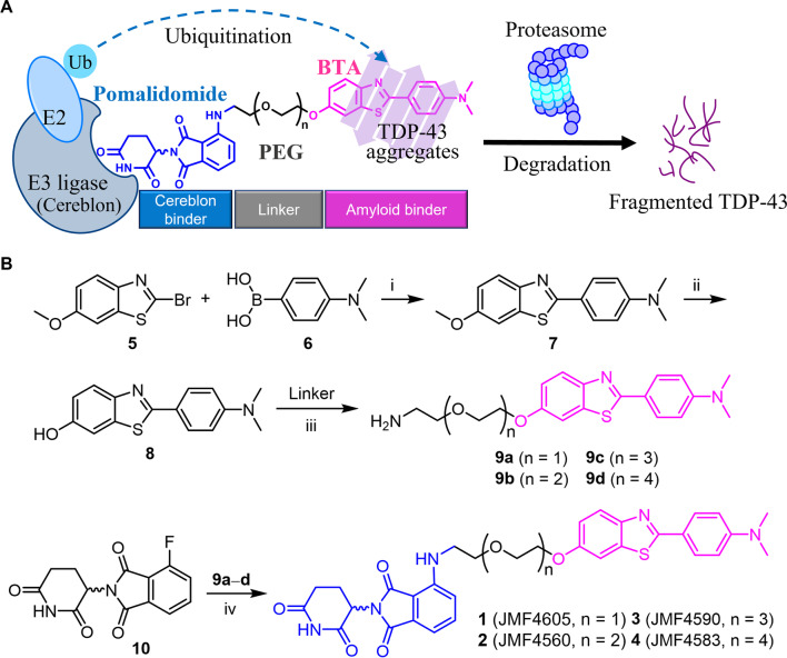
Background: Amyotrophic lateral sclerosis (ALS) associated with TAR DNA-binding protein 43 (TDP-43) aggregation has been considered as a lethal and progressive motor neuron disease. Recent studies have shown that both C-terminal TDP-43 (C-TDP-43) aggregates and oligomers were neurotoxic and pathologic agents in ALS and frontotemporal lobar degeneration (FTLD). However, misfolding protein has long been considered as an undruggable target by applying conventional inhibitors, agonists, or antagonists. To provide this unmet medical need, we aim to degrade these misfolding proteins by designing a series of proteolysis targeting chimeras (PROTACs) against C-TDP-43.

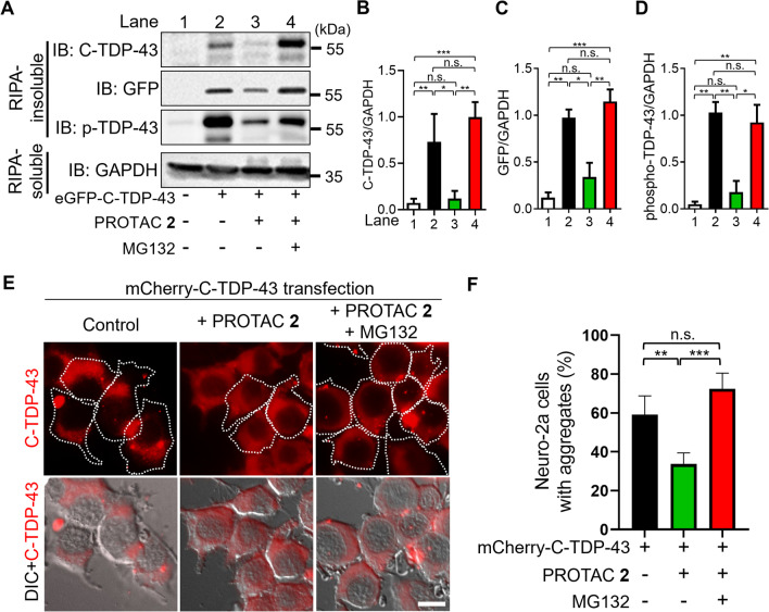
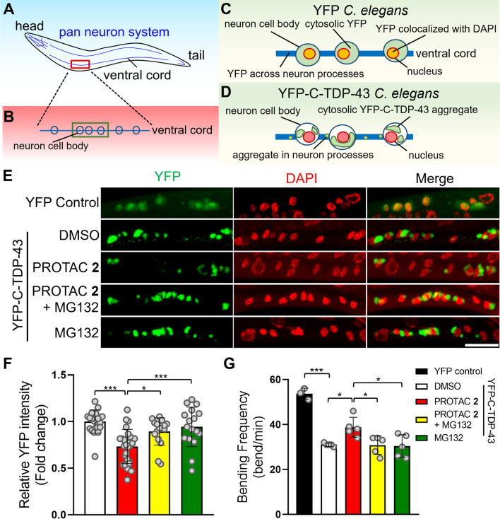
Methods: By applying filter trap assay, western blotting, and microscopy imaging, the degradation efficiency of C-TDP-43 aggregates was studied in Neuro-2a cells overexpressing eGFP-C-TDP-43 or mCherry-C-TDP-43. The cell viability was characterized by alarmarBlue assay. The beneficial and disaggregating effects of TDP-43 PROTAC were examined with the YFP-C-TDP-43 transgenic C. elegans by motility assay and confocal microscopy. The impact of TDP-43 PROTAC on C-TDP-43 oligomeric intermediates was monitored by fluorescence lifetime imaging microscopy and size exclusion chromatography in the Neuro-2a cells co-expressing eGFP-C-TDP-43 and mCherry-C-TDP-43.

Results: Four PROTACs with different linker lengths were synthesized and characterized. Among these chimeras, PROTAC 2 decreased C-TDP-43 aggregates and relieved C-TDP-43-induced cytotoxicity in Neuro-2a cells without affecting endogenous TDP-43. We showed that PROTAC 2 bound to C-TDP-43 aggregates and E3 ligase to initiate ubiquitination and proteolytic degradation. By applying advanced microscopy, it was further shown that PROTAC 2 decreased the compactness and population of C-TDP-43 oligomers. In addition to cellular model, PROTAC 2 also improved the motility of transgenic C. elegans by reducing the C-TDP-43 aggregates in the nervous system.

Conclusions: Our study demonstrated the dual-targeting capacity of the newly-designed PROTAC 2 against both C-TDP-43 aggregates and oligomers to reduce their neurotoxicity, which shed light on the potential drug development for ALS as well as other neurodegenerative diseases.

Keywords: Aggregate and oligomer; Amyotrophic lateral sclerosis; Neurodegenerative diseases; PROTACs; Protein degradation; TDP-43 cytotoxicity; Transgenic C. elegans.


Conflict of interest statement

The authors declare that they have no competing interests.

Figures

Fig. 1
Fig. 1
Mechanism for degradation of TDP-43 aggregates by PROTAC molecules. A The PROTAC binds E3 ligase and TDP-43 aggregates simultaneously to facilitate the transfer of ubiquitins to TDP-43 aggregates. As ubiquitin chains on TDP-43 aggregates are recognized by proteasome, TDP-43 aggregates are degraded. BTA: benzothiazole-aniline derivative. B The synthetic process of PROTACs 14. Reagents and reaction conditions: (i) Pd(dppf)Cl2, K2CO3, DMF, 80 °C, 18 h; 60%. (ii) BBr3, CH2Cl2, 0 °C, 20 h; 98%. (iii) For 9a, N3CH2CH2(OCH2CH2)OMs, K2CO3, DMF, 80 °C, 21 h; then PPh3, THF, rt, 24 h; 50% overall yield. (iv) i-Pr2NEt, NMP, 90 °C, 16–18 h; 32%. For the synthesis of 9b9d, see experimental section
Fig. 2
Fig. 2
Examination of the C-TDP-43 disaggregation and beneficial effects of PROTACs 1–4. A Representative images of eGFP-C-TDP-43-expressing Neuro-2a cells with or without PROTAC 14 (5 μM). Scale bar = 10 μm. B Filter trap assay of eGFP-C-TDP-43 expressed Neuro-2a cells in the presence and absence of PROTACs 14 (5 μM). The cell lysate was either loaded on cellulose acetate (CA) or nitrocellulose (NC) membrane probed with TDP-43 (C-terminal) antibody and β-actin antibody (loading control), respectively. C Quantification of blots in panel B. D AlamarBlue reduction assay of eGFP-C-TDP-43 expressed Neuro-2a cells treated with PROTACs 14 (5 μM). E Western blot of eGFP-C-TDP-43 transfected Neuro-2a cells treated with various concentrations of PROTAC 2. The RIPA-insoluble fraction and RIPA-soluble fraction of Neuro-2a lysate were further probed with GFP and GAPDH antibody, respectively. All the statistic results were quantified by ImageJ and shown as mean ± SD (n ≥ 3). Data were analyzed by one-way ANOVA with Dunnett post-hoc test (*P < 0.05, **P < 0.01, ***P < 0.001)
Fig. 3
Fig. 3
PROTAC 2 decreased insoluble C-TDP-43 aggregates via UPS. A Western blot of eGFP-C-TDP-43 harboring Neuro-2a cells with or without PROTAC 2 (5 μM) or/and MG132 (2 μM) treatment. The blots of RIPA-insoluble and RIPA-soluble cell lysates were demonstrated by SDS-PAGE and probed with C-TDP-43, GFP, phospho-TDP-43 (Ser409/410), and GAPDH antibodies. BD Quantification of blots of C-TDP-43 (B), GFP (C), and phospho-TDP-43 (D) in panel A. E Representative images of mCherry-C-TDP-43 expressed Neuro-2a cells with or without PROTAC 2 (5 μM) or/and MG132 (2 μM). To morphologically monitor the C-TDP-43 puncta upon drug treatment, the cells with mCherry-C-TDP-43 puncta larger than 0.1 μm2 were considered as the aggregate-positive cells (featured with dash line). Scale bar = 10 μm. F Quantification of the percentage of aggregate-positive Neuro-2a cells among total cells in panel E. All the statistic results were quantified by ImageJ and shown as mean ± SD (n ≥ 3). Data were analyzed by one-way ANOVA with Tukey post-hoc test (*P < 0.05, **P < 0.01, ***P < 0.001)
Fig. 4
Fig. 4
PROTAC 2 decreased the compactness of oligomeric intermediates C-TDP-43 and reduced the high molecular weight oligomers in Neuro-2a cells. A Schematic illustration of FLIM-FRET analysis on the 2FP-C-TDP-43 oligomeric intermediates in the cytoplasm of Neuro-2a cells with or without PROTAC 2. (2FP-C-TDP-43 represents co-expressing eGFP-C-TDP-43 and mCherry-C-TDP-43.) B The color-coded images of the EFRET distribution throughout Neuro-2a cells (“frame” fitting model, upper panel) and its per-pixel distribution histograms (lower panel). The palette (color coding on the upper-right) corresponded to the EFRET levels of overall C-TDP-43 species (monomers + oligomeric intermediates + aggregates). C, D The average EFRET (C) and the population (D) of C-TDP-43 oligomeric intermediates in 2FP-C-TDP-43 expressed Neuro-2a cells with or without PROTAC 2 (5 μM) by applying “highlighted-pixel” fitting model. Each cell was arbitrarily selected (n = 20) and calculated according to the region average lifetime on a pixel-by-pixel basis. Statistic results were shown as mean ± SD (n ≥ 3). Data were analyzed by two-tailed unpaired t-test (*P < 0.05, ***P < 0.001). E Neuro-2a cells expressing eGFP-C-TDP-43 with or without treatment of PROTAC 2 (5 μM) or MG132 (2 μM) were fractionated by applying FPLC on the size exclusion column (SEC). The elution of proteins was monitored by absorbance at 280 nm and fractions were collected every 1 mL. Fractions 9–14 were further loaded on NC membrane and probed with A11 antibody. The elution times of two standards, 670 kDa and 158 kDa, were marked as arrowheads
Fig. 5
Fig. 5
PROTAC 2 reduced C-TDP-43 aggregation and improved the motility of the neuronal YFP-C-TDP-43 transgenic C. elegans. A, B Schematic drawings of neuronally expressing C. elegans and its ventral cord. C, D Illustration of YFP (C) and YFP-C-TDP-43 (D)expression pattern within the region of interest in panel B. E Representative images of either YFP control or YFP-C-TDP-43 transgenic C. elegans with or without PROTAC 2 (5 μM) or/and MG132 (5 μM). While the cytosolic aggregates within the ventral cord were visualized in YFP channel (green), the nuclei of neuron cell bodies were monitored in DAPI channel (pseudo red color). Scale bar = 10 μm. F The relative YFP intensity of YFP-C-TDP-43 strain in panel E. G The bending frequency of YFP-C-TDP-43 transgenic C. elegans with or without PROTAC 2 (5 μM) or/and MG132 (5 μM) and YFP control. Each dot represents an independent experiment containing at least 15 worms with three repeat videos. All the statistic results were quantified by ImageJ and shown as mean ± SD (n ≥ 3). Data were analyzed by one-way ANOVA with Tukey post-hoc test (*P < 0.05, ***P < 0.001)
Fig. 6
Fig. 6
A schematic summary of the results. PROTAC 2 reduced C-TDP-43 aggregation and oligomerization, increased viability in the cell model, and improved the motility of the neuronal C-TDP-43 transgenic C. elegans

References

    1. Sweeney P, et al. Protein misfolding in neurodegenerative diseases: implications and strategies. Transl Neurodegener. 2017;6:6. doi: 10.1186/s40035-017-0077-5. - DOI - PMC - PubMed
    1. Hou Y, et al. Ageing as a risk factor for neurodegenerative disease. Nat Rev Neurol. 2019;15:565–581. doi: 10.1038/s41582-019-0244-7. - DOI - PubMed
    1. Taylor JP, Hardy J, Fischbeck KH. Toxic proteins in neurodegenerative disease. Science. 2002;296:1991–1995. doi: 10.1126/science.1067122. - DOI - PubMed
    1. Lamptey RNL, et al. A review of the common neurodegenerative disorders: current therapeutic approaches and the potential role of nanotherapeutics. Int J Mol Sci. 2022;23:1851. doi: 10.3390/ijms23031851. - DOI - PMC - PubMed
    1. Akhtar A, et al. Neurodegenerative diseases and effective drug delivery: a review of challenges and novel therapeutics. J Control Release. 2021;330:1152–1167. doi: 10.1016/j.jconrel.2020.11.021. - DOI - PubMed

MeSH terms

Substances