. 2023 Apr 26;22(1):139.
doi: 10.1186/s12936-023-04566-7.

Retinal imaging technologies in cerebral malaria: a systematic review

Affiliations

Retinal imaging technologies in cerebral malaria: a systematic review

Kyle J Wilson et al. Malar J. .

Abstract

Background: Cerebral malaria (CM) continues to present a major health challenge, particularly in sub-Saharan Africa. CM is associated with a characteristic malarial retinopathy (MR) with diagnostic and prognostic significance. Advances in retinal imaging have allowed researchers to better characterize the changes seen in MR and to make inferences about the pathophysiology of the disease. The study aimed to explore the role of retinal imaging in diagnosis and prognostication in CM; establish insights into pathophysiology of CM from retinal imaging; establish future research directions.

Methods: The literature was systematically reviewed using the African Index Medicus, MEDLINE, Scopus and Web of Science databases. A total of 35 full texts were included in the final analysis. The descriptive nature of the included studies and heterogeneity precluded meta-analysis.

Results: Available research clearly shows retinal imaging is useful both as a clinical tool for the assessment of CM and as a scientific instrument to aid the understanding of the condition. Modalities which can be performed at the bedside, such as fundus photography and optical coherence tomography, are best positioned to take advantage of artificial intelligence-assisted image analysis, unlocking the clinical potential of retinal imaging for real-time diagnosis in low-resource environments where extensively trained clinicians may be few in number, and for guiding adjunctive therapies as they develop.

Conclusions: Further research into retinal imaging technologies in CM is justified. In particular, co-ordinated interdisciplinary work shows promise in unpicking the pathophysiology of a complex disease.

Keywords: Cerebral malaria; Fluorescein angiography; Fundus photography; Malarial retinopathy; Optical coherence tomography.


Conflict of interest statement

The authors declare that they have no competing interests.

Figures
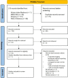
Fig. 1
Fig. 1
PRISMA flowchart
Fig. 2
Fig. 2
a Colour fundus photograph of the left eye of a child with cerebral malaria aged 28 months. Macular whitening is widespread (white circle and elsewhere) and there are retinal haemorrhages (black arrows). Cotton wool spots (white arrows) are whiter and more superficial than whitening. b Colour fundus photograph of the left eye of a different child with cerebral malaria aged 24 months. Extensive vessel discolouration (black arrows highlight one vessel segment) and white-centred haemorrhages (white arrows) are present
Fig. 3
Fig. 3
a Arteriovenous phase fluorescein angiogram of the left eye of a child with cerebral malaria aged 20 months shows macular (red outline) and peripheral (green outline) capillary non-perfusion. Note that haemorrhage (asterisk) masks fluorescence. b Venous phase angiogram of a different paediatric patient with cerebral malaria shows large focal leak (black arrow) and enlargement of the foveal avascular zone (white arrow), indicating capillary non-perfusion. Note again masking from haemorrhages
Fig. 4
Fig. 4
a SD-OCT machine: a low-coherence near-infrared beam is split such that it is directed at both the sample and a reference mirror. The beam is reflected from reflective surfaces (cellular interfaces) in the sample and from the reference mirror. b In the spectrometer, the recombined beams undergo Fourier transformation, and the resulting interference is detected by the detector array. Signals are processed to form OCT images. c) OCT scan of the left eye of a child aged 15 months. Hyperreflective capillaries (pale yellow arrows), hyperreflective areas (long white arrows) and haemorrhage (black asterisk) are visible
Fig. 5
Fig. 5
Statistical tests in image analysis may be applied at the pixel or region level. TP true positive; TN true negative; FP false positive; FN false negative; AUC area under the curve; ROC receiver operating curve, 1-specificty against sensitivity; DC dice coefficient, |X| number of elements in X, X ∩ Y elements that are similar in X and Y

Similar articles

Cited by

References

    1. WHO . World malaria report. Geneva: World Health Organization; 2021.
    1. WHO . Guidelines for malaria. Geneva: World Health Organization; 2022.
    1. Beare NAV, Taylor TE, Harding SP, Lewallen S, Molyneux ME. Malarial retinopathy: a newly established diagnostic sign in cerebral malaria. Am J Trop Med Hyg. 2006;75:790–797. doi: 10.4269/ajtmh.2006.75.790. - DOI - PMC - PubMed
    1. Beare N, Southern C, Chalira C, Taylor T, Molyneux M, Harding S. Prognostic significance and course of retinopathy in children with severe malaria. Arch Ophthalmol. 2004;122:1141–1147. doi: 10.1001/archopht.122.8.1141. - DOI - PubMed
    1. Harding SP, Lewallen S, Beare NAV, Smith A, Taylor TE, Molyneux ME. Classifying and grading retinal signs in severe malaria. Trop Doct. 2006;36(Suppl 1):1–13. doi: 10.1258/004947506776315781. - DOI - PubMed

Publication types

MeSH terms